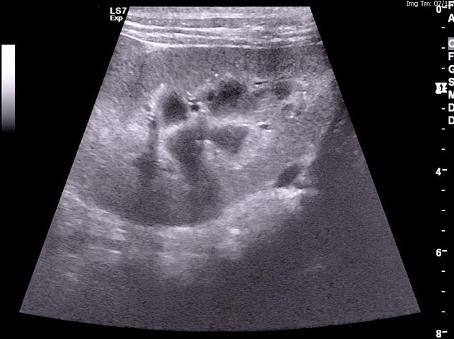
Figure 1d. An ultrasound image of the kidney of a dog with cutaneous and renal glomerular vasculopathy. The kidney is mildly hyperechoic. Some cases have had reduced corticomedullary definition, but generally any changes are mild. Image: Anderson Moores Veterinary Specialists.
Leave a Reply