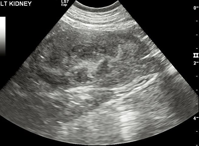
Figure 1b. An ultrasound image of a kidney affected by chronic kidney disease. The kidney is slightly small and irregular, with reduced corticomedullary definition. Image: Anderson Moores Veterinary Specialists.
Written by
in
Figure 1b. An ultrasound image of a kidney affected by chronic kidney disease. The kidney is slightly small and irregular, with reduced corticomedullary definition. Image: Anderson Moores Veterinary Specialists.
Leave a Reply