Tag: x-ray

  • Presurgical treatment of acute gastric dilatation-volvulus

    Presurgical treatment of acute gastric dilatation-volvulus

    Resolution of the hypovolaemia is the primary concern. Two large bore catheters are placed in the cephalic veins. If the cephalic veins are not available, the jugular vein is used. Fluid resuscitation through the saphenous veins is unlikely to be successful because of the caudal vena caval obstruction.
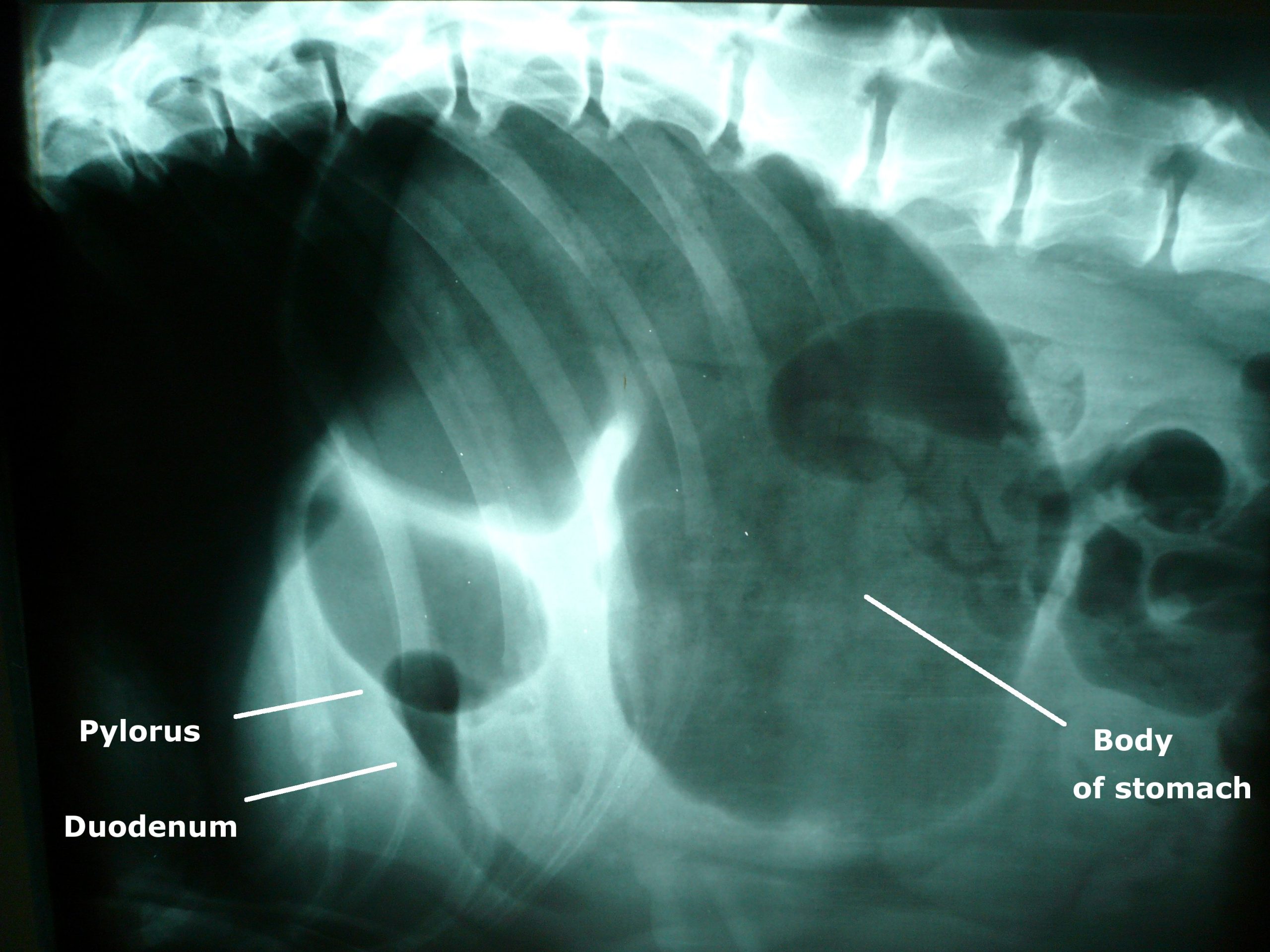
    Photo of an x-ray showing gastric dilatation and volvulus in a large mixed-breed dog. The large dark area is the gas trapped in the stomach. The pylorus and duodenum are in an abnormal position cranial to the stomach and are separated by a fold in the stomach, creating a "double bubble" appearance. By Joel Mills (Own work) [GFDL (http://www.gnu.org/copyleft/fdl.html), CC-BY-SA-3.0 (http://creativecommons.org/licenses/by-sa/3.0/) or CC-BY-SA-2.5-2.0-1.0 (http://creativecommons.org/licenses/by-sa/2.5-2.0-1.0)], via Wikimedia Commons
    Photo of an x-ray showing gastric dilatation and volvulus in a large mixed-breed dog. The large dark area is the gas trapped in the stomach. The pylorus and duodenum are in an abnormal position cranial to the stomach and are separated by a fold in the stomach, creating a “double bubble” appearance.

    Image by Joel Mills [CC-BY-SA-3.0], via Wikimedia Commons.

    Either isotonic crystalloids (90ml/kg in the first hour) or hypertonic saline (7% NaCl in 6% Dextran: 5 ml/kg given over five minutes) followed by crystalloid are administered.

    Controversial treatments

    The use of corticosteroids remains controversial. They have many theoretical benefits but have not been unequivocally demonstrated to improve survival in cases of gastric dilatation-volvulus (GDV).

    Prophylactic antibiotics are also somewhat controversial, but rational arguments are made for their use. GDV dogs do have increased levels of circulating endotoxin, perhaps indicating increased GI mucosal permeability. Poor perfusion to the liver could inhibit reticuloendothelial function.

    Improving tissue perfusion by fluid resuscitation and subsequent gastric decompression and de-rotation can potentially result in the production of damaging, highly reactive oxygen free radicals. These radicals can cause significant reperfusion injury that may be as damaging as the initial hypoperfusion episode.

    It is possible treatment to prevent free radical generation may be beneficial in dogs with GDV. Of the drugs trialled in experimental models, deferoxamine, an iron chelator, shows the most promise for clinical application.

    Gastric decompression

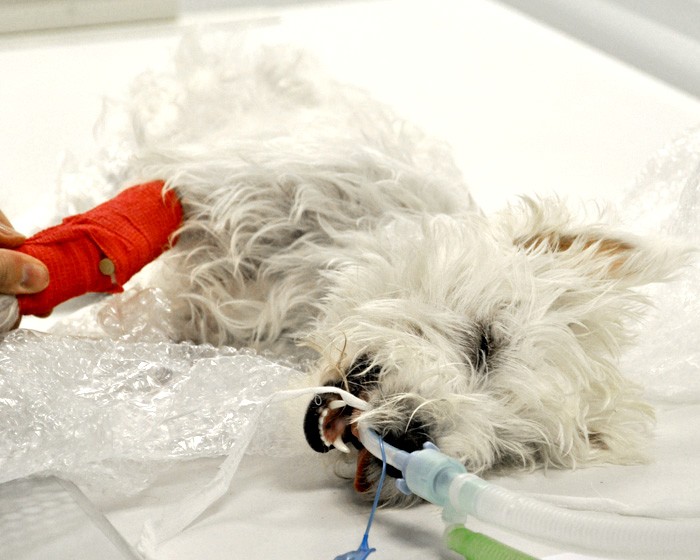
    Ideally, a continuous electrocardiogram is connected. Once the animal has been stabilised, gastric decompression is attempted using a silicone or rubber tube. The tube is pre-measured to the level of the stomach and marked. A 2in roll of tape is placed in the dog’s mouth and the tube passed through the tape and slowly into the oesophagus and stomach.

    If resistance is encountered at the level of the cranial oesophageal sphincter, the tube must not be forced, as this could cause rupture of the caudal oesophagus.

    In some fractious animals, sedation and intubation is necessary for gastric decompression. If orogastric intubation is unsuccessful, the stomach is decompressed by trocarization. The abdomen is carefully palpated, and the enlarged spleen is avoided. A large gauge catheter (10-12F) is placed into the stomach percutaneously to relieve pressure.

  • Decubitus lateral view of chest

    Decubitus lateral view of chest

    Westie
    West Highland terrier being prepared for-x-ray. Image: Jackie Morrison

    This view is particularly useful for demonstrating small pneumothoraces, emphysema or loculated pleural fluid.

    The animal is placed in lateral recumbency. To demonstrate trapped pleural fluid, the affected side should be uppermost; to demonstrate emphysema, the affected side is placed down.

    The x-ray cassette is positioned perpendicular to the spine on the table with a block – for example, a foam wedge or sandbag behind the cassette – to stop it from tipping backwards. The x-ray tube head is rotated so that it is parallel with the table and the x-ray beam is horizontal.

    The x-ray beam should be directed towards an external wall and care should be taken that no people are on the other side of the wall.

  • Buy a dental x-ray machine

    Dental radiograph showing periodontal disease in a 2-year-old cat.
    A dental radiograph showing periodontal disease in a 2-year-old cat (note bone loss), by mariposavet. Licensed under CC BY 2.0 via Flickr.

    If I am being perfectly honest, dentistry has never filled me with excitement.

    That said, attending a number of sessions on dentistry at the North American Veterinary Conference this year was fascinating and convinced me of the value of buying a dental x-ray machine.

    Goldstein (2015), to name but one speaker, evaluated the use of radiography in assessing dental conditions in cats and convinced me of the need.

    In another talk the value in assessing canine lesions was discussed (Lewis, 2015) – definitely something to bring up at our practice’s next clinical effectiveness meeting.

    References
    Goldstein, G.S. (2015). Dental pathology case presentations, clinical and radiology: interactice discussion of what’s new, North American Veterinary Conference, Orlando 2015.

    Lewis, J.R. (2015). Interpreting canine dental radiographs: learning what you were never taught, North American Veterinary Conference, Orlando 2015.

  • I blame the NHS

    "Those detached from the veterinary world still uphold the perception that vets are rich and set high prices to rip off the unsuspecting public."
    Original image ©iStockphoto.com/hidesy

    A couple of articles have been brought to my attention this week regarding the public perception of vets versus the reality.

    When somebody finds out that you want to be a vet or are studying veterinary medicine, there are a few arduous questions that usually follow:

    • “Is it seven years at university for that?”
    • “It’s hard to get into isn’t it?”

    And, of course:

    • “Well, vets earn loads of money don’t they?”

    Unfortunately, only one of those three assumptions is true. However, those detached from the veterinary world still uphold the perception that vets are rich and set high prices to rip off the unsuspecting public. What these people don’t understand is that a new veterinary graduate can expect to start on a salary of around £20k while working all living hours of the day, plus being on call.

    While each individual job varies, the reality is that we can expect to earn very little considering the length of intense training required and the high levels of stress and responsibility that come with the job.

    With experience and promotion to more superior roles – such as becoming a partner in a practice – the salary will increase, but often to less than half the average wage of a GP doctor. In general, vets accept this and are highly dedicated to the health of the animal they work with. If they were after a high salary, they’d be better off in a different career field entirely.

    But the public can’t be entirely blamed for their own ignorance – I think a lot of the problem lies with the National Health Service

    There is no NHS for pets...
    “There is no NHS for pets, and I think many people would do well to remember this.”

    We are blessed with a “free” health service in the UK, but this means those of us outwith the field of human medicine have very little understanding of how much treatments, operations or drugs cost.

    Perhaps if people knew how much these things would cost if they had to fund them privately, they’d have a greater appreciation of both the NHS itself and the veterinary care they pay for for their animals. After all, there is no NHS for pets, and I think many people would do well to remember this.

    My American friends tell me that clients in the US do seem to have a more grounded understanding of the cost of healthcare and are able to apply this to veterinary care without quite so much complaining.

    It’s also worth noting that the money people spend on their animals’ treatments does not go directly into the pocket of the surgeon, but contributes to the cost of anything required for the procedure, including medication, electricity, needles and syringes, catheters, x-ray plates, bedding, food, anaesthetic, licensing… this list goes on – and somewhere at the bottom of that list sit the wages of the hardworking and dedicated vet, who often only receives a grumbling about the expense in “thanks”.

    On a recent EMS placement at a small animal veterinary surgery, I witnessed a lot of this grumbling, and sometimes even full blown arguments about cost. Luckily it’s the few clients that are truly grateful and would do anything for their animals that make it all worth it.

    I find it highly offensive and disrespectful when I hear remarks that vets are “only in it for the money” because, if that were true, then we are not as intelligent as our education might suggest.

  • The German attitude to learning

    A veterinarian placing a syringe in the vein of a horse. © iStockcom/Jan-Otto
    A veterinarian placing a syringe in the vein of a horse. © iStockcom/Jan-Otto

    The vet handed me the needle and vacuum tubes and, at the slightly bewildered look on my face, asked if I’d ever taken blood from a horse before. Upon my answer of “no”, he shrugged and said: “I’ll show you the first one, instruct you for the second, then you can do it by yourself.”

    Having started at 8am on my first morning, he had me taking blood samples from broodmares used to produce top class racehorses by 8:05 – not something I would expect to be allowed to do as a second year vet student anywhere in England.

    I spent the rest of that morning with Neils, the vet, driving to different yards and observing while he performed rectal ultrasound scans on mares, assessed an ongoing case of RAO (Recurrent Airway Obstruction) and extracted a tooth from a very old and very hairy pony, alternating between being utterly flummoxed by his exchange of German conversion with clients and him then explaining things to me in perfect English. I then returned to the stud yard I was based at (between Hannover and Hamburg) to groom, feed and bring in the mares.

    Although I was technically supposed to be on pre-clinical EMS at the stud, Neils was eager for me to learn from him, in addition to the more husbandry-based experience I was gaining from being on the yard. Some days were spent entirely on the yard, and others were spent partially with him, gaining bonus clinical experience. Neils was a “one-man-band”, running a mobile equine practice by himself – an alien concept, compared to the practice based vets that are the norm at home.

    About halfway through my first week, I spent an entire day with Neils and, having watched him scan (via rectum) more mares than I could count, he decided there were a few safe candidates for me to try my hand on (or, rather, arm in). After a few minutes of fumbling around, I managed to orientate myself and understood far more clearly what the grey and black mush on the ultrasound screen represented.

    Creme egg

    We then went on to x-ray a horse with a fractured radius and I assisted in applying its Robert Jones bandage. I took a few more blood samples and we called at other horses to drop off medication, vaccinate, assess lameness and rasp some teeth.

    I felt like I’d had a taste of what it would be like to be a qualified vet – not from the practical and clinical things I got to see and do that day, but from the 14 hour day, having had nothing but a Creme Egg to eat and not stopping for breath…

    However, arriving back to the yard that evening just after the arrival of a new foal made it worth every second. Between them, Neils and the yard manager explained everything that was done and needed to be done just after a foaling; we examined the afterbirth to ensure none had been retained, assisted the foal while it began to suckle and kept an eye on both the mare and foal for the next few hours.

    The end of my two weeks in Germany came around all too soon and was quite sorry to have to leave. I was taken aback by their hands-on attitude and desire for me to get as much out of my placement as possible, and not just be another pair of hands for mucking out.

    The generosity I experienced from everyone I worked with is something I’m extremely grateful for, and will never forget.