Tag: Urine

  • Urinalysis: dipstick tips

    Urinalysis: dipstick tips

    Following on from July’s post entitled Urinalysis: the neglected test, let’s have a look at the dipstick – it’s a very easy part of a urinalysis and essential to perform.

    Here are some of my tips in regards to using dipsticks:

    Poli_dipstick
    Dipstick: despite the name, DON’T DIP!
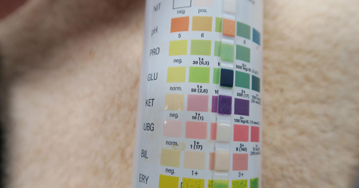
    • It may sound obvious, but you should always use veterinary-specific dipsticks. Human-specific dipsticks include panels for urobilinogen, nitrates and leukocytes, which we often do not interpret in small animal patients, as they are neither sensitive nor specific.
    • DON’T DIP! Use a syringe and drop samples on to each square, leave for 10 seconds, then flick off the excess.
    • Any amount of protein in dilute urine should raise suspicion. A reasonably large amount of protein has to be present in the urine for it to be positive on a dipstick. A urine protein to creatinine ratio may be the only way to quantify the amount of protein present, but first you must rule out evidence of inflammation or haematuria via a sediment examination.
    • The ketone panel on the dipstick test is only for acetoacetate (and not beta-hydroxybutyrate), although it is extremely rare for diabetic ketoacidosis patients to not produce any acetoacetate.
    • Trace blood can be a common artefact finding, especially during a cystocentesis where needle trauma can contaminate the sample with blood.
    • In our feline patients, any hyperbilirubinuria is abnormal, but this may be normal in a dog depending on urine concentration.
  • Handling an Addisonian crisis – part 1

    Handling an Addisonian crisis – part 1

    Addison’s disease (hypoadrenocorticism) is one of those annoying diseases that does not always play by the rules.

    One of the main reasons is the clinical signs of Addison’s disease can be frustratingly non-specific and we don’t often see the classic “low sodium, high potassium” electrolyte changes we are attuned to noticing. Therefore, it is important to recognise the early signs, or have a set of clinical signs, history and biochemistry changes that trigger the Addison’s disease alarm bells.

    Once diagnosed, we can look at the approach to stabilising a patient in an Addisonian crisis.

    Clinical signs

    The clinical signs of Addison’s disease can be vague and non-specific, these include:

    • anorexia
    • lethargy
    • weakness
    • gastrointestinal signs
    • polyuria and polydipsia

    A history of chronic intermittent vomiting and/or diarrhoea that resolves with symptomatic management would be one of the triggers.

    Another trigger is the subtle changes in blood tests. These include:

    • the absence of a stress hyperglycaemia in a sick patient (normal glucose or even a low blood glucose, for example)
    • hypercalaemia (ionised) of any degree
    • absence of stress leukogram

    These changes are often seen in isolation, so don’t expect them to all be there at the same time. We see patients all the time with inappropriately normal or low blood glucoses with gastrointestinal signs that we later diagnose with Addison’s.

    Hypercalcaemia – what’s the big deal?

    Why hype up and down about normal or low blood glucoses? Because low blood glucoses are rarely caused by inappetence or gastroenteritis alone, and the body is pretty good at maintaining glucose within normal levels (except, maybe, for very young patients and some toy breeds). Any low glucose Addison’s should be considered, and a stress hyperglycaemia is so common in ill patients that its absence makes me concerned.

    What is the big deal about hypercalcaemia? It is so tightly regulated that any elevation Addison’s is on the differential list. How does Addision’s cause these changes? Corticosteroids are released during stress or ill states, which result in a stress hyperglycaemia. They also play a role in the excretion of calcium in the urine, so preventing a hypercalcaemia.

    What about the electrolyte changes, the hyponatraemia and hyperkalaemia? When these changes are present they make diagnosis much easier, but they are not always present, such as with “atypical hypoadrenocorticism”, so the absence of that classic change does not rule out Addison’s.

    Next, we will cover the management of the Addisonian crisis.