Following on from parts one and two in this coagulation series – in which we described how to use your history, signalment and clinical signs to help determine if your patient has a coagulopathy and narrow down the list of differentials – we now look at the diagnostic tests you can use to confirm your suspicions.
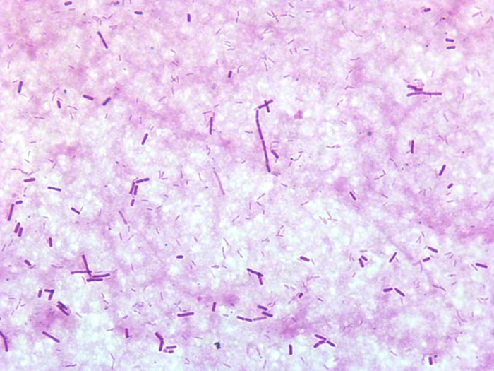
In the initial stages, you only need two simple tests – a blood smear and an activated clotting time.
Less than between 35,000 platelets per μL and 50,000 platelets per μL – equivalent to less than two to three platelets per 100× field – is required for a spontaneous bleed and you should consider that your patient has a clinically relevant thrombocytopenia.
If you see more than 10 platelets per 100× field, you do not have a thrombocytopenia causing bleeding.
Pro Tip #1
Anytime you are collecting blood, keep one drop for a smear – it saves you from having to stick the patient again, and you can easily throw it away later. You can also perform an ear prick if you don’t want to ruin a vein.
Pro Tip #2
If you can‘t find a platelet in the monolayer, check the feathered edge. If you see platelet clumps then you are all good.
If platelet numbers are normal then you either have:
a platelet disorder (thrombocytopathia)
a problem with secondary haemostasis (clotting factors)
Activated clotting time – tests secondary coagulopathies
Normal activated clotting time (ACT) is generally less than 120 seconds, or 90 seconds with some types of tubes. The issue with this test is that it requires a significant deficiency in clotting factors (less than 5% normal factor activity) for prolongation to occur. However, this is usually not a problem if they are clinically bleeding.
As a rough rule, if the ACT is greater than 25% of normal then it can be considered abnormal and you have a secondary coagulopathy. The ACT is an inexpensive and quick bedside test; however, partial thromboplastin time (PTT) and activated partial thromboplastin time (aPTT) are more sensitive, which will be further discussed in the next part.
Pro Tip #3
ACT can be prolonged with severe thrombocytopenia, so check your platelets.
Pro Tip #4
PT and aPTT are not affected by platelets like the ACT.
If your ACT is normal and your platelets are normal:
Consider thrombocytopathia. However, testing to see if PTT and aPTT are normal as well helps rule out a secondary coagulopathy.
Thrombocytopathia is assessed by performing a buccal mucosal bleeding time (BMBT), which will assess for platelet disorders like von Willebrand disease.
A BMBT of more than four minutes is abnormal.
Pro Tip #5
Avoid using a scalpel blade for a BMBT due to a large amount of error. Use a specific “surgicut”.
In the next part, we delve deeper into secondary coagulation disorders.
Last week we discussed the causes and diagnostic pathway for investigating immune-mediated thrombocytopenia. This week we will go through the management of this condition.
Despite the fact red blood cells are not actually being destroyed, a severe anaemia can develop from blood loss due to coagulopathy – a common reason for why they present to emergency practices. The management of these patients is broken down into three main areas:
improving oxygen delivery
commencing immunosuppression
management of the underlying cause (if identified)
Optimising oxygen delivery in the acute phase is going to keep them alive long enough for immunosuppression to work. This is achieved through IV fluids to help improve perfusion and blood transfusions to replace red blood cells. If fresh whole blood is available, it can assist in increasing platelet numbers, but generally it is not very effective.
Platelet transfusions using platelet-rich plasma can be considered if it is available. Plasma transfusion is not effective at managing the coagulopathy as it is due to a loss of platelets, not a loss of coagulation factors.
Immunosuppression
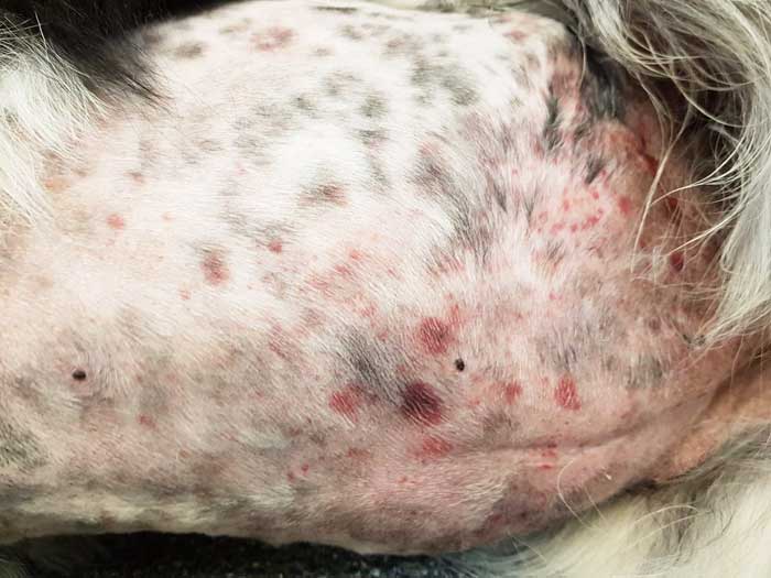
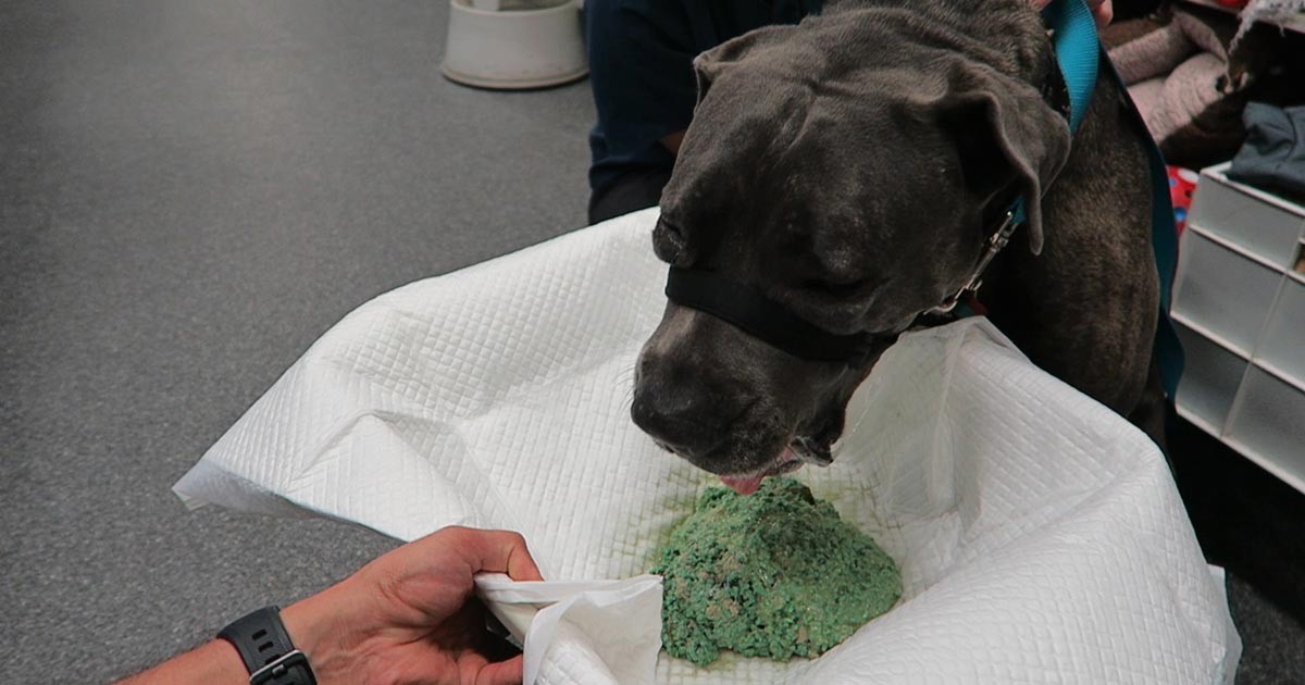
Large areas of ecchymotic haemorrhage on the skin are a quite obvious sign of thrombocytopenia.
Immunosuppression therapy is often commenced concurrently as the patient is being stabilised.
The first choice is either dexamethasone 0.5mg/kg IV every 24 hours if the patient is not stable enough for oral medications; otherwise, once stable, start prednisolone at 2mg/kg by mouth per day divided every 12 hours.
Other immunosuppressive agents include:
Azathioprine – 2mg/kg by mouth every 24 hours then 0.5mg/kg by mouth every other day. The main concerns are bone marrow suppression and hepatoxicity – also, it is very toxic in cats.
Ciclosporin – 5mg/kg to 10mg/kg by mouth divided twice a day; cats 5mg/kg by mouth every 24 hours.
Chlorambucil could also be used at a dose of 0.1mg/kg/day to 0.2mg/kg/day by mouth if the response to prednisolone is insufficient.
Management
Management of the underlying cause should be commenced if a cause is identified, but this is often not the case.
Other management options include:
Vincristine can be trialled to increase platelet number as it stimulates the release of platelets from the bone marrow.
Gastroprotectants can be considered if gastrointestinal bleeding has occurred – these include proton pump inhibitors and sucralfate.
Strict confinement, potentially sedatives and minimal blood sampling are important to minimise injury that may result in further bleeding and blood loss.
Antithrombotic therapy is not part of standard management as, unlike immune-mediated haemolytic anaemias, thrombotic events rarely occur.
When it comes to monitoring, platelet counts are performed daily until more than 40 × 109/L – this can take up to two weeks to occur.
Once above this level, take weekly counts until the numbers have normalised. Once they have, taper immunosuppressive medications over four to six months, with 20% dose reduction every couple weeks, generally with the adjunctive immunosuppressants first and prednisolone last.
Thrombocytopenia is a condition characterised by a decrease in platelet numbers, which is often caused by increased destruction of platelets or a decrease in production.
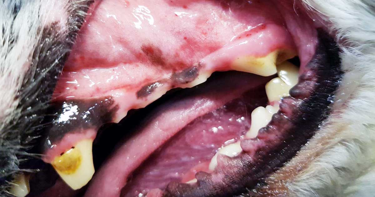
Thrombocytopenia can manifest in many ways – the signs can be subtle and easily missed, such as small petechiae on gums, or quite obvious signs, such as large areas of ecchymotic haemorrhage on the skin.
Ecchymotic haemorrhages are often attributed to disorders of secondary haemostasis, such as rodenticide intoxication, but it can also occur with thrombocytopenia depending on the severity and chronicity.
Other common clinical signs include:
epistaxis
blood in stools, urine or vomit
pale mucus membranes
lethargy
weakness
Therefore, the first step to managing a patient with a severe thrombocytopenic episode that has resulted in significant blood loss is to manage shock, if present, with IV fluids, then administer a red blood cell transfusion.
Patient handling
Careful patient handling is critical as these patients can bleed easily, leading to blown veins, large bruises that contribute to the development of anaemia and significant patient discomfort. Beyond initial patient stabilisation, the next step is to determine the underlying cause.
Diagnosing thrombocytopenia is relatively straightforward with the demonstration of low platelet counts. Generally, bleeding does not occur until the platelet count drop below 40,000 thousands per cubic milliliter (k/uL). This can be determined by either a haematology machine or manually via blood smear analysis.
When assessing blood smears, the general rule is one platelet per high-powered field on the monolayer is equal to 15,000k/uL. With either method, you must assess for platelet clumping on a blood smear as this can artefactually drop platelet numbers, leading to a false diagnosis.
The diagnostic pathway should not stop there. It needs to continue to determine the underlying cause.
The most common cause of thrombocytopenia is immune-mediated destruction. This can be either a primary (diagnosis of exclusion) or secondary cause (such as Rickettsia infection, and drugs such as sulphonamides, toxins and neoplasia). Other less common causes include:
splenomegaly, which can lead to platelet sequestration
disseminated intravascular coagulation and acute blood loss, leading to platelet consumption
bone marrow disease, which results in reduced platelet production
Signalment and history will refine such a diagnosis, as certain breeds are more prone to developing thrombocytopenia than others – for example, grey collies due to a defect in haematopoietic stem cells, and whippets and greyhounds, which traditionally have a lower platelet count than other breeds.
The generally diagnostic pathway continues to include haematology and biochemistry, thoracic radiographs, abdominal ultrasound and depending regional prevalence testing for infectious organisms with PCR and ELISA assays.
Next week I will cover the management principles of the thrombocytopenic patient.
Idiopathic acute haemorrhagic diarrhoea syndrome (AHDS) – previously known as haemorrhagic gastroenteritis – remains the one disease where constant debate exists as to whether antibiotics should be used as part of the standard treatment.
The logic behind using antibiotics to prevent bacterial translocation is sound, and if AHDS is truly initiated by Clostridium species or their toxins then the use of antibiotics can be justified.
However, no knowledge exists of the true frequency of bacterial translocation in AHDS patients and conflicting evidence supports Clostridium being the initiating cause of AHDS in dogs.
Together with new data indicating the use of antibiotic therapy in aseptic AHDS patients did not change the case outcome or time to recovery, the benefit of using antibiotics must be weighed against the very real risk of selection of antibiotic resistance and other complications associated with inappropriate antibiotic use.
In this blog, we will explore the evidence against the use of antibiotics in AHDS.
Cause unknown
AHDS is characterised by an acute onset of vomiting (of less than three days’ duration) that can quickly progress to haemetamesis, and severe and malodorous haemorrhagic diarrhoea, accompanied by marked haemoconcentration that can be fatal if left untreated.
AHDS is a diagnosis of exclusion; other diseases (such as canine parvoviral enteritis, thrombocytopenia, hypoadrenocorticism, azotaemia, hepatopathy, neoplasia, intussusception, intestinal foreign body and intestinal parasitism) must be ruled out by a combination of medical history, vaccination status, complete blood count, serum biochemistry, coagulation times, diagnostic imaging and faecal testing.
Small breed dogs – in particular, the Yorkshire terrier, miniature pinscher, miniature schnauzer and Maltese – have been found to be particularly predisposed. On average, the affected dogs were young (a median of five years old).
The cause of AHDS is still unknown. Clostridium perfringens and its toxin has been incriminated as being the initiating cause; however, conflicting studies have refuted this claim. It is also difficult to determine whether overgrowth of Clostridium speciesis primary or secondary to the intestinal injury.
Virus theory
Another theory is viruses may have a role in AHDS’ aetiology. At this stage, only single agents had been investigated. It is possible a novel agent not yet been tested is the cause of this syndrome, or possibly the syndrome is the result of a very complex interaction between multiple organisms or their toxins.
For the aforementioned reason, no indication exists for the use of antibiotics to treat for the underlying cause.
Another argument behind the use of antibiotics lies in the fact most idiopathic AHDS patients have several risk factors for bacteraemia.
Necrosis of intestinal mucosa, leading to the disruption of the gastrointestinal mucosa-blood barrier; adherence of significant numbers of bacteria to the necrotic mucosal surfaces that increases the risk of bacterial translocation; significant hypoalbuminaemia indicating substantial loss of mucosal epithelial layer; splanchnic and intestinal hypoperfusion, leading to reduced intestinal barrier function; and microbial dysbiosis all contribute to an increased risk of bacterial translocation.
Although bacterial translocation has the potential to lead to sepsis, the true incidence of bacterial translocation needs to be established in idiopathic AHDS patients, as well as their influence on the outcome of the patients.
Antibiotic requirement
Use of unnecessary antibiotics not only disrupts the protective mechanisms of a normal intestinal microflora, but also the real risk of post-antibiotic salmonellosis and Clostridium difficile-associated diarrhoea.
Multiple studies have suggested antibiotics are not required in the treatment of aseptic idiopathic AHDS patients.
In a prospective study of bacteraemia in AHDS dogs by Unterer et al (2015), the incidence of bacteraemia of patients with idiopathic AHDS was 11%, compared to those of healthy controls, where it was 14%.
Transient bacterial translocation to mesenteric lymph nodes occurred in 52% of dogs undergoing elective ovariohysterectomy (Dahlinger et al, 1997), and confirmed in studies by others (Harari et al, 1993; Howe et al, 1999; Winkler et al, 2003), where portal and systemic bacteraemia was reported in clinically normal dogs.
As long as the immune system is competent, and the functional capacity of the hepatic reticuloendothelial system is not overwhelmed, the body is usually effective at eliminating organisms from the blood.
This is reflected in the Unterer et al (2015) study result, where – regardless of the bacteraemia status – all idiopathic AHDS dogs survived to discharge.
In another prospective, placebo-controlled, blind study by Unterer et al (2011), idiopathic AHDS patients were either treated with amoxicillin/clavulanic acid for six days or a placebo, and no significant difference occurred between the treatment groups concerning mortality rate, duration of hospitalisation or severity of clinical signs.
They concluded, without the evidence of sepsis, antibiotics do not appear to change the case outcome or shorten the time to recovery.
Negative impacts
The negative impacts of inappropriate antibiotic use are undeniable – especially in a time where resistance has become a worldwide public health concern.
Use of unnecessary antibiotics not only disrupts the protective mechanisms of a normal intestinal microflora, but also the real risk of post-antibiotic salmonellosis and Clostridium difficile-associated diarrhoea.
With evidence all pointing against the use of antibiotics as routine treatment of aseptic idiopathic AHDS, next time you are about to reach for antibiotics, I urge you to reconsider. Although it has taken some time to adopt and requires clear communication with clients, all vets should feel comfortable not using antibiotics for AHDS patients.
Until I started researching this Tip of The Week, I did not know the medical profession has abandoned the routine use of emesis in oral poisoning.
This is based on multiple medical literatures that have proven emesis induction does not influence the clinical severity of poisoning, the length of hospitalisation and the clinical outcome or mortality.
Although the rationale for inducing emesis is obvious, it is not necessarily evidence based. It is also dependent on satisfying a few large assumptions, all of which are untrue:
Emesis is a very effective way of removing gastric contents.
No separation exists of poison from its vehicle while inside the acidic environment of the stomach.
Poison is not absorbed through the stomach wall.
Ineffective method
Snail bait ingestions: this patient ate 500g of snail bait containing metaldehyde.
Emesis induction is an ineffective way of clearing stomach contents. A review of the effectiveness of induced emesis, with both human and canine participants, showed at 30 minutes post-ingestion of non-absorbable markers, the recovery rate averaged between 17.5% and 52.1%, but never exceeded 62%.
In fasted puppies, this was even lower at 2% to 31%, despite inducing emesis immediately after marker administration. These have been confirmed by the presence of poisonous materials in the stomach of dead patients, despite effective emesis induction until clear fluid was brought up.
The clinical outcome only improves if the systemic exposure of a toxicant is reduced by more than half. However, considering animals rarely practice restraint, the ingested amount is unlikely to be exactly the lethal dose and no more. Therefore, even reducing the ingested toxic dose by 62% is unlikely to make a clinical difference.
Furthermore, most patients rarely present within 30 minutes of ingesting a toxicant, thus further reducing its efficiency.
The absorption conundrum
Some may argue the retrieval of metaldehyde or anticoagulant rodenticide granules from vomitus is indicative of reducing the toxicant dose. This could be true, but only if emesis was induced immediately after ingesting the poison.
The poison itself is colourless and has a different absorption characteristic to the coloured vehicle (granule); therefore, the presence of granule only serves to confirm ingestion, but is of no indication whether the poison has already been absorbed.
Contraindications
Many well-recognised absolute contraindications also exist to inducing vomiting:
Ingestion of oils, which includes waxes that melt to oil in the internal body environment, as this poses a high risk of lipoid and bacterial pneumonia. This is of significant veterinary significance, as wax is routinely used in rodenticide baits.
Ingestion of hydrocarbons and other volatile substances, or caustic or corrosive substances.
When the mental status is altered – for example, hyperexcitable or depressed mental state.
Where the patient is at risk of seizures (seizures can be induced by emesis).
Increased intracranial pressure.
Risk of intracranial or cerebral haemorrhage – for example, thrombocytopenia or abnormal clotting parameters.
Other less severe, yet important, reasons include:
delays administration of more effective treatment, such as activated charcoal, antidote or other treatments
risk of aspiration pneumonia
hypochloraemia in recurrent emetic patients
significant CNS and respiratory depression from apomorphine
rare, but reported, complications such as cerebral haemorrhage, oesophageal tear/ rupture, hiatal hernia, gastric rupture, pneumothorax and pneumomediastinum
legal implications – for example, if the product information clearly states emesis should not be induced
A place for everything
Emesis induction is not a benign procedure. It still has its place in certain circumstances, but its use in the routine management of oral poisonings may need to be reconsidered – especially if it means delaying administration of a more effective treatment, such as activated charcoal.
So, after all this, how do I tackle this information? It is a bit hard to swallow. My clinical experience is emesis is generally safe, especially in canine patients using apomorphine. So, I still feel some merit exists in reducing the amount of toxicant in the stomach if you have a chance – and in some situations, you don’t know until you try.
Emesis after ingestion of a toxic dose of chocolate can be incredibly rewarding, even six hours after ingestion, leading to patients not developing clinical signs at all.
Overall, I am biased by my personal successes with emesis, so still feel a time and place exist for emesis induction. But I now stop and question my decision to induce emesis, whereas I did not hesitate before.
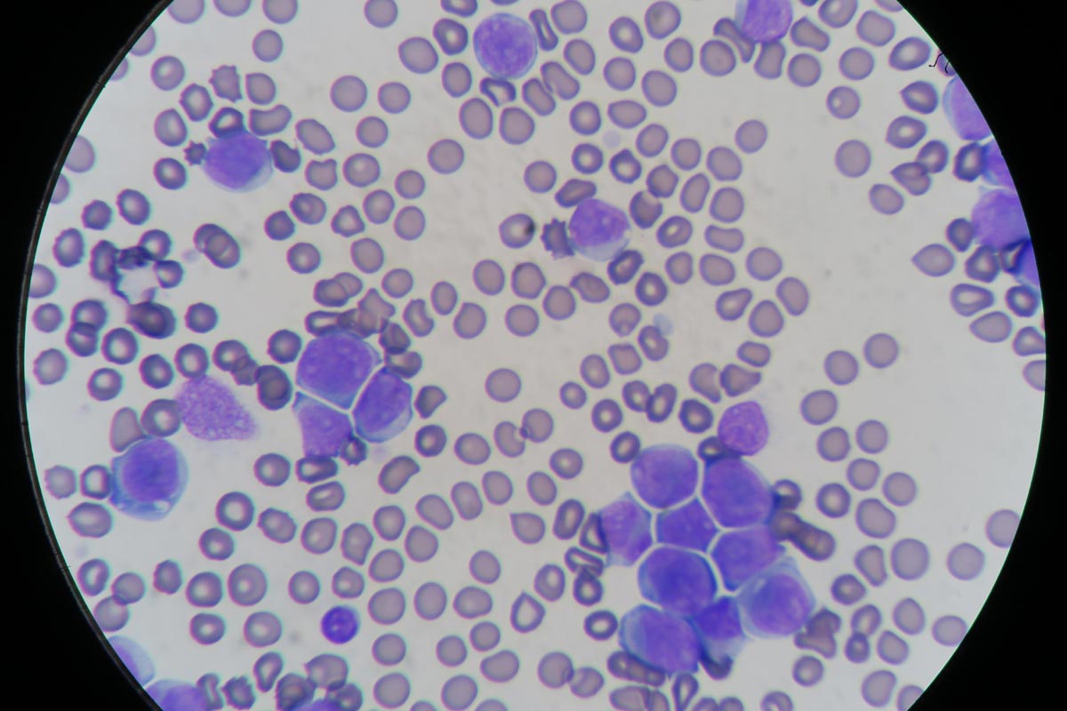
Blood smear evaluation is an often overlooked, but very important, aspect of an in-house haematology.
With the advancement in haematology analysers that can now detect reticulocytes and even band neutrophils, some practitioners are beginning to rely solely on the numerical data alone in evaluating the patient’s blood.
Patient with elevated white blood cells caused by leukaemia (click to zoom).
The art of blood smear interpretation is on the decline. However, it is an extremely valuable skill that must be practised and perfected and really should be part of every in-house haematology.
Plus points
What are the benefits of being good at blood smears?
Identifying a regenerative response, looking for reticulocytes (polychromatophils).
Looking for other possible causes of an anaemia – such as Heinz bodies, infectious microorganisms or spherocytes, which can indicate an immune-mediated haemolytic anaemia.
Confirming thrombocytopenias, as frequently platelet clumping can be reported as a thrombocytopenia.
Assessment of the nature of a leukocytosis. High leukocyte counts do not always mean infection. Neutrophilia can be caused by both elevated immature and mature neutrophils. Determining the nature of neutrophilia can provide crucial information in identifying the underlying cause and if the patient is coping or not. Apart from infection, other causes can include stress, corticosteroids and neoplastic leukaemias.
Normal leukocyte counts do not always mean the patient is okay. Patients can have severe left shifts, but normal leukocyte counts.
Practice makes perfect
Blood smear evaluation begins with becoming accomplished at producing great diagnostic smears. This takes practice; poorly performed smears can be non-diagnostic and frustrating to assess for both yourself and an external pathologist.
A few tips on the technique:
Use a very small drop of blood. If you have picked up too much blood with the “spreader” slide, lift off and start the smear away from that drop of blood.
Angle the “spreader” slide about 30°. The bigger the angle, the shorter your smear.
Push the “spreader” slide forward.
The smear should end at about half to three-quarters of the way down the slide and must have a “feathered edge”.