Rupture of the cranial cruciate ligament in an English Bulldog by Uwe Gille, licensed under CC-BY-SA-3.0
For dogs weighing less than 15kg, cranial cruciate disease can be managed conservatively – weight loss until an appropriate Body Condition Score (BCS) is achieved, exercise restriction for 3 to 6 weeks, and possibly physical therapy and pain medication – allowing acceptable comfort and function.
In dogs weighing more than 15kg, cruciate disease will eventually cause significant arthritis, and dysfunction is inevitable without surgical treatment.
No single surgical technique is clearly superior, so the choice of surgical repair should be decided by the surgeon and the needs of the owner.
An experienced vet could complete the entire procedure easily within 10 minutes. We “tentatively ambled” through our surgeries in 20.
Having finally settled in one place in Jaipur, India, my friend and I were able to relax a little, safe in the knowledge we had two weeks of neutering for population control ahead of us.
Being in an unfamiliar environment, and with our patients mainly being strays, we were prepared for very different methods of anaesthesia, variations on drugs we’re used to at home, and potentially questionable sterility. Even so, when the vet, stood with his scalpel at the ready, said “oh yes, we use the right flank method” as if it were the norm, we were a little surprised.
At home, we’re so used to seeing flank cat spays and midline bitch spays, my gut reaction was “is that even anatomically possible?”. As it turns out, it is.
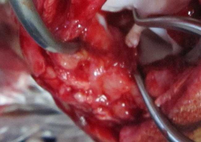
The method
A small incision (<2cm) is made on the right flank, first through the skin and then each of the 3 underlying muscles (transverse abdominis, external abdominal oblique and internal abdominal oblique). A spay hook is then used to exteriorise the right uterine horn.
Once identified, the surgeon follows the horn to the ovary and applies tension caudally to break the suspensory ligament. A ligature (note single) is placed around the blood vessel and the ovary cut from it using the three clamp method in the same way as spays in the UK. The surgeon then follows the uterus to the cervix and along the left horn to the left ovary, where the procedure is repeated. A ligature is placed just above the cervix (again using the triple clamp method) and the uterus removed.
Closing the incision comprises placing a horizontal mattress suture in each of the muscle layers, a cruciate suture in the subcutaneous fascia, and intradermal sutures in the skin.
The positives
While the very idea of flank spays in the bitch just seemed alien, this method seems to be successful and works well in a charity environment in a country where certain resources are unavailable.
The reasons for choosing this method include easier wound checking, a shorter wound healing time (meaning the dogs can be re-released sooner) and less tension at the incision site, decreasing the risk of wound breakdown – essential for animals that, once released, are unlikely to be seen again.
Despite her initial surprise at the method used, Jordan admits the flank approach is the best compromise, considering the resources available.
The surgeons at the charity have found, over the years, the single horizontal mattress suture seems to be the least aggravating to the body wall muscles, and intradermals are the closure of choice in any stray or vicious animal that would be difficult to get near to remove sutures.
Another key advantage to the flank approach is speed; important for two reasons:
The sheer number of stray dogs to neuter to reach an adequate level of population control means faster surgery is required to reach the target numbers.
The surgical time under IV anaesthesia should be kept to a minimum to avoid prolonged or rocky recoveries and minimise side effects.
The experienced vet could complete the entire procedure easily within 10 minutes (in a normal young bitch, opposed to a pregnant or in season girl), and we, tentatively ambling through our surgeries, could complete within 20.
The negatives
Disadvantages to this method include more potential bleeding due to incising through the three muscle layers, a possibility of more postoperative pain and increased difficulty in extending the incision if there are complications. The most important, however, is that recovery of a dropped or bleeding ovarian stump is extremely difficult (or near impossible).
The anaesthesia protocol used is premed: xylazine, induction/maintenence; IV ketamine and IM meloxicam as pain relief. Hence, the speed of the flank approach will also minimise the number of top ups needed and reduce the anaesthetic hangover comparing to a technique (such as midline) that is more time consuming.
Compromise
The method seems to be the best compromise, considering the resources available. I think the overruling disadvantage is that, if you were concerned about a slipped ligature, the ovarian and uterine stumps would be virtually impossible to find again via the original incision.
However, that said, the only postoperative death we saw during our time on postmortem had all ligatures intact.
It was eye-opening to see an entirely different approach to a bitch spay, and while it may not be the same as the routine at home, I still felt that we gained a lot of surgical experience and developed transferable skills.
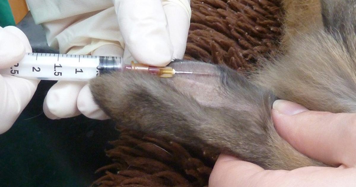
A venipuncture site should be chosen where the skin is clean and has no obvious inflammation or infection. The marginal ear vein or the lateral saphenous vein are usually good choices.
The fur should be clipped and the skin cleaned. EMLA cream can be applied over the site 45-60 minutes prior to venipuncture and covered with a dressing or cling film. The site should then be wiped with surgical spirit.
The vein is raised and the needle should be inserted at a very acute angle, almost parallel to the skin.
Gently does it…
Use only very gentle pressure to draw back on the syringe plunger, to prevent the vein from collapsing.
Too great a pressure may also damage the blood cells, especially when using a small gauge needle. Rotating the needle around its long axis so the bevel faces sideways or downwards may improve blood flow if the vein appears collapsed.
After taking the blood, pressure over the venipuncture site should be applied as rabbit veins are prone to haematomas.
Have you ever noticed that, sometimes after starving, the haematocrit (Hct) and haemoglobin (Hgb) levels appear a little high on pre-anaesthetic bloods prior to a surgical procedure that morning?
If so, make sure you look at the figures.
If, like us, your laboratory machine produces a band with a red marker in the middle indicating some elevation, make sure you also look at the absolute figures.
I recently saw a nine-year-old boxer that, after a surgical procedure, developed redder and redder skin and mucous membranes. She had high Hct and Hgb on pre-anaesthetic bloods and subsequently turned out to have polycythaemia vera with, eventually, a Hct of 84.9.
Four episodes of venipuncture (taking 200ml to 300ml of initially very viscous blood on each occasion), plus treatment with hydroxycarbamide, and she was feeling much better.
I spent a couple of weeks at a mixed practice on extramural studies. It was a placement of firsts – first experience at a very “young” practice (lots of newish graduates), first clinical farm experience and first surgery experience.
It gave me an insight into the difference between older, more experienced vets and new grads – from the way they approach consultations and cases to the time/skill difference in surgical procedures. Obviously, a lot of this comes with experience, but it was reassuring to know I’m not quite as lost as I thought I was (and it also helped me recognise areas to work on).
I did work experience at farm practices before uni, but hadn’t been to one as a proper student. It entailed the bread and butter of farm practice – being shoulder deep in a cow’s rectum.
My younger brother found it particularly fascinating, always asking me when I got home each night how many cows I’d seen. Appealing as it sounds, I found reproductive physiology and pharmacology began making a lot more sense.
One day we were called out to castrate 34 calves, which turned out to be a day of avoiding hailstones while the vet cut, twisted, pulled and I injected. Nearing the end of the never-ending stream of beef calves, the vet invited me to have a go. A few minutes later, covered in blood, cow poo and sweat (it required a surprising amount of elbow grease), I had performed my first surgical procedure.
The following day I found myself being told to scrub up, then was guided through a cat castrate. It was a bit surreal, because the vet started off with “cats are a bit like calves…” I couldn’t help wondering how many students castrated a calf before a cat…
A few days later, the same vet supervised my second unassisted castrate. I had a real sense of achievement, having been able to perform the procedure without being told what to do. I know it’s a simple surgery and may seem like peanuts to a qualified vet, but it was quite a step for me – and everyone has to start somewhere.
I also scrubbed in on an exploratory laparotomy on a ferret, which was unusual to say the least, especially when the huge mass we were investigating in the abdomen turned out to be fluid filled and exploded slightly after a needle was stuck in. Since it was attached to the uterus, the vet decided to spay the ferret, which sparked the conversation of why ferrets aren’t often spayed and the resulting hormonal changes involving the adrenals.
Overall, I had a tiring couple of weeks, but felt the vets were eager to get me involved and my clinical skills certainly progressed further than I expected.
“Why diagnose early? Obtaining a definitive diagnosis with cytology or biopsy early and before excision will lead to improved patient outcomes for superficial masses. Surgery is likely curative for the majority of superficial tumours when detected early, when they are small – especially benign lesions and locally invasive tumours with a low probability of metastasis. If tumours are removed with complete surgical margins, the prognosis is often good with no additional treatments needed.
“Pet owners need to be aware of the ‘pea’ size requirement to have masses evaluated, and veterinarians must measure and document the size of the mass to compare growth.
“If more than 1cm (or the size of large pea) and present for a month, the mass should be aspirated or biopsied.
“Knowing the tumour type prior to the first surgery will increase success of a curative-intent surgery.”
In all honesty, I rarely do this for masses booked in for surgery and I suspect as lipomatosus, and for masses that visually appear consistent for histiocytoma – but there are tips here I will follow in the future.
Reference Ettinger S (2015). Top Ten Oncology Mistakes and How to Avoid Them, North American Veterinary Conference: Small Animal & Exotics Proceedings, Gainsville, Florida.
As a student on placement, I’m often in awe of the vets I’m working with. The ability to take a history, examine an animal, run through differentials and come up with a diagnosis or action plan within 10 minutes – all while listening to an owner commenting on the weather or traffic – seems superhuman.
This may seem an exaggeration (after all, vets aren’t superheroes), but when considered like that, it is pretty impressive.
While seemingly intangible at the moment, I know the ability to do this with such ease comes with practice – and clearly some presentations are far more complex than that.
However, while I find this impressive, others have a different opinion…
A family friend recently commented on their own vets, claiming they would avoid seeing the partners if possible because – in their opinion – they see an animal for five minutes and see it as a money making exercise, whereas the younger vets spend a bit more time with the clients.
Obviously I can’t comment on the vet/client rapport, which may have a huge influence on this opinion, but I can’t help but think that a younger, newly qualified vet would spend more time during consultations purely due to experience, or lack thereof.
It has become evident recently that the profession has an image problem and we must try to change that for the better. But what do the public consider as a “good vet”? Apparently the opinion differs depending which side of the table you’re on.
This is just one example, but in general, do clients want the vet to spend more time with their animal? They probably do – but, at the same time, they don’t want to be kept waiting and they want to be able to get an appointment. There has to be a balance between the three.
As for cost, I’ve seen some vets charge meticulously, whereas others would try and keep prices as low as possible to please customers. In the clients’ eyes, the cheaper the better. But a vet practice has to function. It’s no good offering neutering for £10 because the practice would be bankrupt within a week.
“The most highly qualified and experienced surgeon in the practice might not be the best at client communication,” claims Jordan.
Surgical skills and experience are perhaps something that the client will never fully appreciate. For a start, the vet seen in the consultation room may not be the same one who performed the operation, particularly if it’s something fairly routine. Also, the most highly qualified and experienced surgeon in the practice might not be the best at client communication.
A vet can have such a diverse set of skills and knowledge that it is difficult to pinpoint which of these defines a “good vet”. Many vets have certain areas of expertise and will be better than others in certain situations, but not all.
The key to time and money is striking the balance between what the client desires and what is realistic.
Communication, however, doesn’t need to be compromised and can be the difference that alters the client’s opinion. For example, the manner in which an examination is conducted and the attitude of the vet during a 10-minute consult could leave the client feeling rushed, whereas a different vet with a different approach could leave the client with a far more positive impression.
Client opinion is important, but at the end of the day, the welfare of the animal in front of you is your priority, whether or not the client values you highly.
While the profession as a whole should take heed of what clients want, the customer is not necessarily always right, and at the end of the day, it is the welfare of the animal in front of you that should be paramount.
Fine-needle aspiration (FNA) of a mass. Image source: BSAVA ’09 Congress Times.
We recently had a case where a freely mobile, soft mass on the ventral abdomen, which had been present for a number of years, had started to get larger.
We carried out a fine-needle aspiration (FNA) biopsy, and I fully expected this to confirm the presence of a lipoma (a benign fatty tumour common in dogs).
However, I was really surprised when the cytology revealed the presence of a mast cell tumour, with a surgical procedure to follow.
My tip would be that it is definitely worthwhile checking out those seemingly innocuous “lipomas” with FNAs.
Argentinian Tango dance duo German Cornejo and Gisela Galeassi. Image: Fuentes/Fernandez
Before the full force of third year hit, the first week back at vet school started with everyone catching up on tales from their summer holidays.
Before long, it was like we’d never left and the four months of freedom seemed to fade into a distant memory. However, one particular topic of holiday gossip that I have been dwelling on is extramural studies (EMS).
Everyone had undertaken some form of EMS over the summer, whether it was just a week or two, a solid two months, clinical, preclinical, large or small animal – there is a lot of room for variation in our placements, but I was still surprised to hear of how different some of my friends’ experiences had been, despite doing theoretically similar placements.
A number of us had embarked on our first clinical placements, and although we’re all at the same stage of our studies and therefore should be able to get involved during veterinary placements to a similar extent, the truth is somewhat different.
Just among my friends, there were experience levels at both ends of the scale, with some students having been simply told to observe consultations and others being allowed to scrub into surgical procedures.
This wide range of experiences can be attributed to many factors, including:
the veterinary practice
how well the vet knows the student (either from previous experience or length of placement)
how well the staff have judged the student’s knowledge and ability based on stage of the veterinary course
attitude and competency of the vet
the individual student’s skills, experience and attitude
I was advised by a final year student last year to undertake the majority of my clinical EMS at one single practice if possible, because by getting to know the vets well (and vice versa), they’ll be able to judge your level of competency better and encourage you to get more involved. I can now begin to appreciate this advice more, having listened to the anecdotes from my friends.
The practical teaching we receive at vet school is just not enough to be able to adequately develop and refine essential clinical skills that will be needed everyday in general veterinary practice. The solution to this is EMS, and we are constantly being told that we, as students, need to take responsibility for our own learning and ensure that we get the most out of EMS by getting involved. And I whole-heartedly agree – we can’t be spoon-fed forever and need to be proactive in gaining the right type of experience.
However, you could be the most enthusiast student in the world and read up on cases every night, and yet still be very limited in what you are allowed to do. While getting the most out of a placement is up to us, it takes two to tango, and we need the vets’ support too in order to enable us to do this.
I know taking on students and teaching or letting them practice techniques can be time-consuming and inconvenient, but we need to gain experience somehow. At some point during their training, all vets would have had to see practice and learn in the same way, so is it not just a way of giving back to the profession?
I can also appreciate that some people are just not natural teachers (after all we’re training to work in a vet clinic, not a school), but a little bit of patience and some advice can go a whole lot further than just ignoring a student.
It may sometimes be inappropriate for a student to be asking questions or trying things out – in the consultation room in front of the client, for example – but these situations can be fine when approached the right way. I was lucky enough to stand in with vets that would always try and get me to see/hear/feel things. If they found something interesting in the consultation room, they’d always explain to the client that I was a student and ask if they minded me having a look. This seems far more reasonable to me than telling a student they are to observe only.
Another approach I experienced myself was the vet taking the animal to the surgery room to take blood samples and allowing me to perform my own clinical examination (having not actually been in the original consultation).
As mentioned previously, there can be many factors involved in getting a “good” clinical placement. It also depends how busy the surgery is – if there are four clients waiting to see the same vet, it’s understandable for the vet to whizz through them without having much time for questions or explanations (whenever this happened to me, the vet apologised for not explaining, even though she really didn’t need to!).
I have to agree there are advantages to going back to a veterinary practice you know. I did work experience for three years before university at the practice I did my EMS at this summer, and definitely felt welcomed as part of the team, which can be difficult at an entirely new practice.
Yes, it is our responsibility to find the balance between getting involved to gain experience and not interfering with consults, but we also need vets to help us a bit too. Undertaking EMS is the only way we will prepare ourselves for the future, and we’re extremely grateful for the vets that encourage and help us every step of the way (partly why most vet students are pretty good at baking). I think it’s just a case of finding the right practice for both you and the vets you’ll be learning from.
Vet school doesn’t prepare you for making a complete idiot out of yourself.
At vet school, you learn some basic clinical skills and are taught how to conduct a general clinical examination to prepare you for EMS placements in veterinary surgeries. What they don’t prepare you for is making a complete idiot out of yourself.
Before my first clinical placement I told the vets I would be working with that I had only just finished second year and had no pharmacological knowledge as of yet, non-existent surgical experience and very little understanding of small animal medicine in general.
Luckily, all the vets in the practice were very good at judging the level of my understanding and seemed to find the right balance between patience and pushing me for answers.
Things seemed to be going OK. I’d successfully taken blood samples and started to make sense of abdominal palpation. However, applying clinical skills taught at vet school isn’t necessarily straightforward – cadavers have a distinct lack of weapons in the form of claws and teeth, but I was coping with that reasonably well and taking note of the vets’ advice on particular techniques.
This was until a few days in, when I found myself working with the head vet…
In the same morning, I managed to spray penicillin all over my face while trying to administer an injection, incorrectly insert an endotracheal tube despite being 99% sure it was OK, and cover myself in guinea-pig blood while clipping nails, leaving me to wear the stained tabard for the rest of the day.
To add insult to injury, I later misread the scales and recited the incorrect weight without thinking (it didn’t occur to me that there’s no way a fully grown border collie could weigh 10kg).
Isolated, these incidents might not seem like the end of the world, but when they all happen in the same day in front of the head vet and when one of the clients involved is your neighbour, you do feel like shouting “I am a vet student – honest”, despite feeling like a complete moron.
This was, however, followed by days of mini-triumph, such as inserting an IV catheter correctly for the first time or scaling and polishing a dog’s teeth myself.
The important thing to remember is that you are inexperienced, and you just have to accept there will be days when nothing seems to go your way, get past them and carry on with your head held high – even if it is covered in yellow spots of penicillin.