Tag: Stress

  • Triage, pt 1: primary survey

    Triage, pt 1: primary survey

    The art of triage takes time to master – particularly in emergency hospitals, where critical patients arrive in quick succession to the crash area.

    Patients need to be examined quickly and effectively to ensure the most critical issues are identified and stabilised. To do this, I break triage into two categories – primary survey and secondary survey.

    In part one I will discuss the primary survey process. The second part will go into the details of the secondary survey.

    ABCDE approach

    Primary survey refers to the initial stabilisation stage, where the aim is to preserve life, manage life-threatening injuries and re-establish tissue perfusion with oxygenated blood.

    The approach to all deteriorating or critical patients is the same: ABCDEAirway, Breathing, Circulation, Disability and External.

    ABC – Airway, Breathing, Circulation

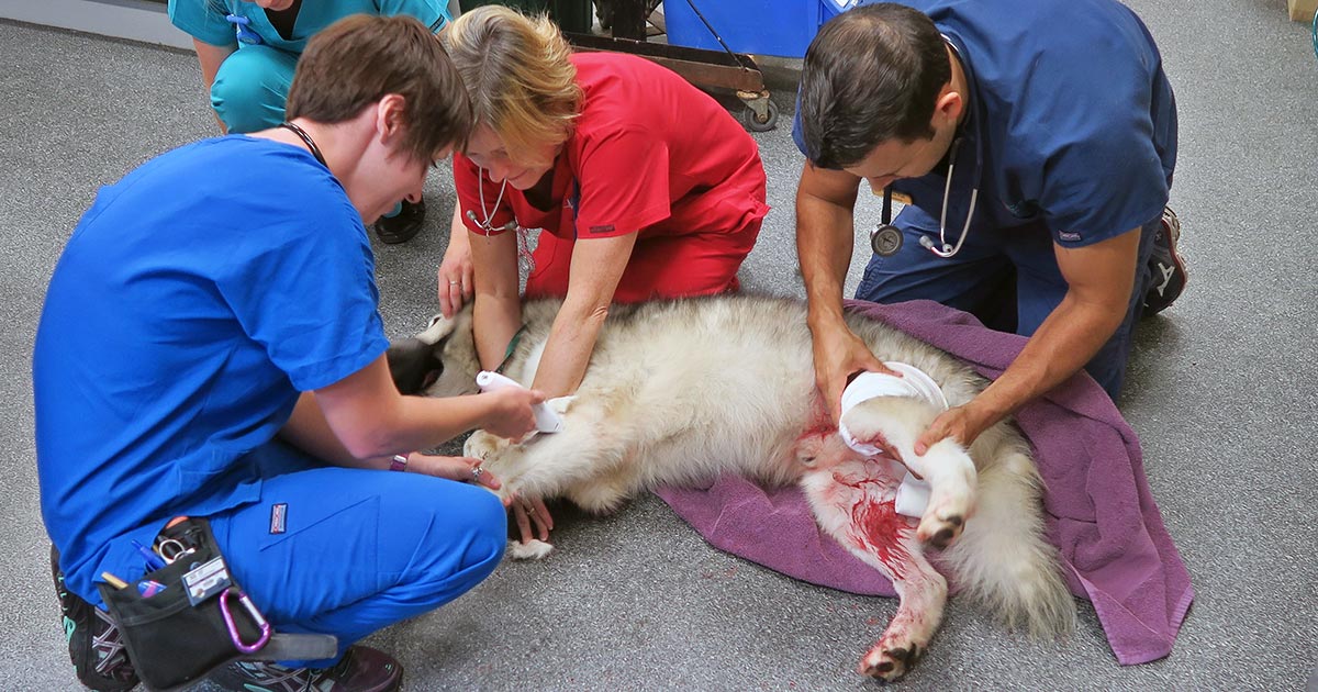
    Critically ill patients need to be examined quickly and effectively.
    Critically ill patients need to be examined quickly and effectively.

    Assess airway

    • Is it patent?
    • Does it require suctioning?
    • Does evidence exist of upper airway obstruction?
    • Intubate if you suspect the patient may need resuscitation.

    If you suspect cardiopulmonary arrest, begin basic life support immediately. This involves chest compressions, and intubation and ventilation in:

    • loss of consciousness
    • absence of spontaneous ventilation
    • absence of heart sounds on auscultation
    • absence of palpable pulses

    Assess breathing

    • Is the patient hyperventilating or hypoventilating?
    • How is the respiratory effort? Is it sustainable?
    • What is its oxygen saturation, or does the patient looks like it needs oxygen? If yes, provide supplemental oxygen therapy.
    • Does the patient need to be ventilated? Ventilation is required if any of these criteria are satisfied:
      • hypoxaemia (partial pressure of oxygen lower than 60mmHg or blood oxygen saturation level lower than 90%) and unresponsive to oxygen supplementation
      • hypercapnia or hypoventilation (partial pressure of carbon dioxiden greater than 60mmHg)
      • unsustainable respiratory effort?
    • Consider sedation. Reducing stress can make a dramatic difference in stabilising dyspnoeic patients. Butorphanol or low-dose acepromazine (try to rule out cardiac disease first) can be used in these situations.

    Assess cardiovascular system

    • Mucous membrane colour, capillary refill time, heart rate and rhythm, pulse pressure, and temperature.
    • If a heart murmur or arrhythmia is present, I try to rule out cardiogenic shock before resuscitation therapy.

    At the same time, draw enough blood to run baseline blood work and begin IV fluid resuscitation if signs of shock are present. Fluid boluses should be considered if any of these exist:

    • pale mucous membranes
    • slow capillary refill time
    • tachycardia
    • poor pulse pressures
    • hypothermia

    I generally start with 10ml/kg Hartmann’s Solution over five minutes and reassess.

    D – Disability assessment

    Any abnormalities in:

    • Mentation? Seizures?
    • Level of consciousness?
    • Altered pain sensation?

    E – External assessment

    • assessment for wounds or injuries
    • control any obvious bleeding, apply direct pressure – possibly tourniquets, but only for less than 30 minutes unless life-threatening bleeding
    • initial medications:
      • pain relief – generally opioids are safest with unstable patients
      • antiepileptics – diazepam
      • sedation for dyspnoeic patients – butorphanol or low-dose acepromazine IV

    Once you have finished your primary survey and performed the required intervention, you repeat your primary survey until the patient is appropriately stabilised.
  • Don’t rush: a systematic approach to x-rays

    Don’t rush: a systematic approach to x-rays

    One of my responsibilities in our emergency hospital is the training and mentoring of vets new to the field of emergency and critical care.

    First look
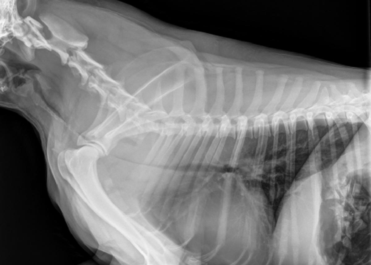
    FIRST LOOK: The patient was bitten by another dog and presented with multiple puncture wounds and difficulty breathing. Radiographs were taken to assess for thoracic injuries (click to zoom).

    A common area I have found where clinicians request more training is radiographic interpretation.

    When I review radiographs and find pathology that was missed, it Is more often due to a lack of systematic approach to reviewing the radiograph than the clinician’s lack of experience or knowledge.

    There is, of course, no one set way you should go about interpreting a radiograph – but whatever the method, the entire radiograph should be assessed, not just the area of interest.

    Radiograph tips

    1. Try not to struggle with your patient. If possible, appropriately sedate your patient (or anaesthetise if safe to do so). This reduces stress for everyone involved and improves your chance of getting a good radiograph. For musculoskeletal radiographs, you often need to manipulate painful joints and limbs to get diagnostic images.
    2. Take appropriate views. For example, I aim to get three plane projections for thorax and abdominal radiographs – i.e. left and right laterals and VD (or DV). Three views are critical for the assessment of both lung fields, and also to help interpret abdominal gas patterns more effectively.
    3. Collimate, rotate, crop, label and adjust the image appropriately. Displaying radiographs in a standardised format is important for proper assessment. Reviewing anatomy in the same way each time helps develop an understanding of what is normal, and makes identifying abnormalities easier.
    4. This is my top rule: At first, IGNORE the area you are interested in. This means, if you are interested in looking at the GI tract in a vomiting dog, try not to focus – albeit initially – on the stomach and intestines on your radiograph.
    5. Start at the periphery. This means things like the spine, subcutaneous tissue, etc – you would be surprised how often lesions are missed in these areas.
    6. reveal
      SECOND LOOK: This fracture was missed on initial review of the radiographs (click to reveal).

      Now take a look at the cavity space (the pleural and abdominal space, for example). You should not be able to see the pleural space, and you should see no evidence of air or fluid in the abdominal space. When it comes to musculoskeletal radiographs, don’t forget to consider joint cavities and soft tissue structures such as ligaments and tendons.

    7. Lastly, make sure you assess every organ (again leaving your organ of interest until last). Things often overlooked include the prostate, kidneys and mediastinal region. At the end of this, I always ask myself, have I missed an organ?

    Tunnel vision

    Another thing I like to do sometimes is take a step back and assess the radiograph again. I find this gives a better global view of the projection, as opposed to staring at it closely.

    This is because when we focus on our area of interest, we start developing the habit of tunnel vision, introducing the potential for missing lesions.

  • Oh, CR*P! Using point-of-care C-reactive protein tests

    Oh, CR*P! Using point-of-care C-reactive protein tests

    Few companies now offer affordable point-of-care tests for canine C-reactive protein (CRP). As we did when we recently received our new box of CRP slides, you might soon be asking the question: what do we even do with this stuff?

    Here’s what we’ve learnt…

    CRP is one of the acute phase proteins produced by the liver in response to inflammation. Healthy patients have very low levels of CRP, but a systemic inflammatory condition will cause an increase in CRP within four to six hours. Conversely, increased levels will decrease rapidly on resolution of inflammation. This provides an almost real time measure of inflammation that is more responsive and reliable than the white blood cell response.

    In other words, CRP can indicate the presence of inflammation before the patient’s white blood cell count gives any clues, or before it becomes pyrexic – and, unlike the white blood cell count, stress and steroids do not affect CRP levels.

    Uses

    So, how do we use it?

    • I love it for early pickups of problems in those grey area cases: the dog seems okay on clinical examination, but something about it bothers me. A normal or mildly increased CRP test will make me sleep more easy, while a surprise high reading will prompt me to admit for full diagnostics, or at least get the patient in for a follow-up CRP the next day. Conversely, a localised problem – such as an abscess – combined with a normal CRP test might mean you can hold off on antibiotics and just recheck CRP in 24 hours.
    • It’s great for monitoring response to treatment. If my plan is working then I’d expect CRP to show a significant decrease by day two or three. If it’s not dipping by then, I need to reassess my treatment plan. Do I need to change antibiotics? Scan it again? Maybe we need to consider surgery? It can also be a good prognosticator. Research has shown failure of CRP to decrease significantly (around a 3× decrease) by around day three is generally bad news for patients with inflammatory conditions such as pancreatitis and immune-mediated haemolytic anaemia.
    • We are starting to play with it for post-surgical monitoring. Any surgery will cause inflammation with an increase in CRP levels, but in an uncomplicated postoperative period, you should expect levels to start decreasing by day three to five. A base line CRP 24 hours after surgery with a recheck on day three should pick up early signs of postoperative problems such as infection, and prompt investigation or intervention.
    • A potentially nifty use for it that we haven’t yet had the opportunity to use is in differentiating inflammatory lamenesses (arthritis, infection, injury) from a neurological causes – that is, is it arthritis or a nerve problem?

    Limitations

    • Remember, it’s very sensitive, so will increase with almost any inflammation. A mild upper respiratory infection or a bad gingivitis will likely induce some changes, so it’s important not to over-interpret (keep in mind that the magnitude of the increase in CRP does generally correspond with the severity of the inflammatory response). A pancreatitis case where the CRP fails to drop does not always mean death is looming – you may have just missed the concurrent skin disease. Always interpret CRP values in concert with your clinical examination.
    • Be aware that pregnancy and intense exercise can increase CRP values.
    • Not all serious conditions have an inflammatory component. CRP will be unchanged in most veterinary cases of heart disease; in common hormonal disease, such as adrenal disease and uncomplicated diabetes; urinary obstructions; many localised cancers; epilepsy and many others. Don’t presume that just because CRP is normal, everything is fine.
    • No similar test exists for cats.

    Sit up and say…

    My favourite way to explain how to use this test is by using its highly appropriate acronym – any unexpected increase should make you sit up and say: “Oh CR*P! What am I missing?”

  • Rat bait’s sneaky trick: bleeding into the dorsal tracheal membrane

    Rat bait’s sneaky trick: bleeding into the dorsal tracheal membrane

    Most of us are familiar with anticoagulant rodenticide toxicosis and the range of clinical signs it can present with, but there is one potentially fatal manifestation of coagulation pathology that is perhaps not as widely known…

    Dogs with severe clotting problems will occasionally bleed into the dorsal tracheal membrane. This causes collapse of the thoracic trachea and can lead to severe respiratory distress.

    Presenting signs

    These cases can present with none of the other signs of bleeding normally associated with coagulopathies, so rat bait poisoning may not come to mind as a differential diagnosis if you are not aware of this syndrome.

    The typical case will present as an otherwise healthy dog that develops acute respiratory problems. Early signs can be as mild as a persistent cough, but it can quickly escalate into a life-threatening respiratory crisis.

    Severe cases will have an obvious stridor on both inspiration and expiration, cyanotic mucous membranes, and patients may be very distressed.

    It will look very much like:

    • a dog that is choking from a tracheal foreign body
    • an old dog with tracheal collapse
    • the end stages of laryngeal paralysis – except the stridor will come from much lower in the respiratory tract than it does in laryngeal paralysis

    So, what do you do?

    On initial presentation you would approach it as any respiratory distress case: oxygen, oxygen, oxygen, calm and stress-free handling, and light sedation (butorphanol, for example).

    bleeding_dorsal-tracheal-membrane

    Once it is safe to do so, you should take chest rads to look for what you’ll probably suspect is a tracheal foreign body, and you’ll get an image like the one above (although it may not be this severe). Then you’ll remember this article, have an “aha!” moment and run a clotting profile (but if it’s as bad as this case, you’ll obviously first save the animal’s life by passing an ET tube).

    Once a clotting problem is confirmed you’ll need to stop the bleeding with standard therapy for anticoagulant rodenticide toxicity: plasma and vitamin K.

    Severe cases

    In a severe case you may need to keep the dog intubated for several hours, until the clotting times have normalised, before cautiously attempting to extubate.

    If the patient is unable to stay well oxygenated without an ET tube (mucous membrane colour, pulse oximetry, blood gas), consider placing a long oxygen catheter past the narrowing – either via a tracheostomy or a nasal O2 catheter.

    If these cases are quickly recognised for what they are, and an open airway can be maintained, the prognosis should be good. These are potentially very satisfying cases with great potential for you to be a total hero.

  • The why of veterinary science

    The why of veterinary science

    In one of the last decade’s most influential books on motivation, Drive: The Surprising Truth About What Motivates Us, author Daniel Pink argues that the traditional motivators of the previous century – reward and punishment – mostly fail to deliver when it comes to keeping people engaged, fulfilled and happy in their careers.

    According to the research that Mr Pink cites, the things people crave most are:

    • autonomy
    • mastery
    • purpose

    If those are the goals, then we should have it pretty good in veterinary science: for the most part vets have a large amount of autonomy (the freedom to make our own decisions) and opportunities for achieving mastery in veterinary science are just about limitless – but purpose, defined in this book as “working in the service of something larger than ourselves”, can be less obvious.

    Autonomy,_Mastery,_Purpose_(11134670423)(1)
    Autonomy, Mastery, Purpose by Paul Downey / CC BY 2.0

    Serving what purpose?

    The question as to whether what we, as vets, do for a living serves a larger purpose is one you’ll often hear – and it’s one I’ve certainly asked myself throughout my career.

    And it’s a valid question: is it really that important, in the greater scheme of things, that I treat all those itchy dogs, while others are changing the world and amassing fortunes?

    Even within the veterinary profession we see individuals pioneering new science, teaching and inspiring thousands of people, or reaching positions of authority and influence. Am I wasting my time and talents in practice, treating one sick animal at a time? Do my days have purpose?

    The answer

    driveI’ve found the answer – or at least my answer – in the simple realisation that, on multiple occasions throughout the average working day, we have opportunities to relieve suffering, show compassion and provide some peace of mind.

    Reminding yourself of this can provide a protective shield against the forces of compassion fatigue (or just simple fatigue), high stress, and sometimes just sheer boredom that relentlessly try to chip away at motivation.

    When you can turn work into service, chores turn into privileges.

    This is my truth

    Here’s another quote from Drive I think applies beautifully to vets and our purpose: “The question is not how to be successful, or how to survive. It’s how to be useful.”

    Don’t forget how useful you are.

    And remember that every time you work towards relieving suffering in your patients or your clients, however minor or imagined that suffering may seem to you, you are doing something that transcends your own needs. In that simple truth lies purpose.

  • Dystocia, pt 2: diagnostics

    Dystocia, pt 2: diagnostics

    Part one of this series covered the stages of labour and indications dystocia is present.

    Once the bitch presents to the clinic, a few basic diagnostic checks need completing to determine the status of the bitch/queen and the fetuses.

    Physical examination

    The first is a thorough physical examination, starting with the bitch or queen:

    • Demeanour, hydration status, vital signs, mucous membrane colour, capillary refill time and temperature are important.
    • Pregnancy anaemia is not uncommon; however, for patients with a haemorrhagic discharge, it is important to know their cardiovascular status.
    • A thorough abdominal palpation should be carried out to assess comfort level and palpation for the presence of fetuses. Palpating fetuses can be difficult and cannot confirm if no fetuses are present.
    • A digital vaginal examination should be performed. Feathering response – also known as the Ferguson reflex in human medicine – is the neuroendocrine reflex where the self-sustained cycle of uterine contractions is initiated by firm pressure on the dorsal aspect of the vestibulovaginal wall. If this is absent, the patient is unlikely to progress with the parturition unaided.
    • Palpation of fetuses in the canal can help decide whether surgical management is required. Obvious fetal malposition, malposture or malpresentation, or fetopelvic disparity, will be indications of caesarean. Abnormal pelvic diameter is also another reason to not proceed with medical management. To confirm these suspicions, abdominal radiography is required.
    • Radiographs will also help determine the number of fetuses to be expected, the signs of fetal death (presence of gas surrounding the fetus) and aforementioned fetomaternal abnormalities. I always repeat radiographs after the expected number of neonates is passed, to make sure I have not miscounted at the start.

    Ultrasound

    Panel 1. Heart rate ranges to help indicate stress of fetuses

    Dogs:

    • normal – 180 to 220 beats per minute (bpm)
    • Stressed – 160bpm
    • Real concern – less than 160bpm

    Cats:

    • normal – more than 220pbm
    • fetal stress – less than 180bpm

    The second important diagnostic tool is ultrasound.

    Fetal heart rates are good indicators of fetal stress. Some heart rate ranges that can help provide information about the status of the fetuses are detailed in Panel 1. These ranges vary between sources, but are good guidelines.

    Ultrasounds can also help visualise the maturation status of the fetuses. At-term fetuses should have normal hepatic, renal and intestinal development. Intestinal peristalsis should be evident in at-term fetuses.

    Other diagnostics

    Other diagnostics may be indicated for patients, depending on the status of the bitch/queen:

    • If the patient is stable, but dystocia is present, a minimum database would include PCV/total protein, electrolytes, glucose, ionised calcium, lactate and acid-base balance.
    • Serum ionised calcium levels are important, as they influence the strength of contractions and how much supplementation is required.
    • Hypoglycaemia needs to be ruled out as a cause of dystocia, especially when large litters are involved.
    • If the patient is unstable or systemically unwell, include complete blood count, blood smears and biochemistry.
    • Physiological pregnancy anaemia can be present. The presence of regenerative response can help differentiate this from acute haemorrhage.
    • Abnormal leukocyte panel, especially with the presence of degenerative left shift, can indicate the presence of an infection – especially if toxic changes are present in the neutrophil.

    Part three will briefly look at the medical management of dystocia and when surgical intervention is required.

  • Pancreatitis, pt 2: treatment and prognosis

    Pancreatitis, pt 2: treatment and prognosis

    Last week we covered diagnosing pancreatitis and the challenges associated with doing so. This week we look at management.

    The treatment of pancreatitis largely involves supportive care and monitoring for potential complications. Here, we recap the fundamentals.

    IV fluids

    IV fluids are critical in the acute phase to restore perfusion to core organs and correct hydration deficits. Once achieved, the goal is then to cover maintenance requirements and ongoing losses.

    Hypoproteinemia can develop due to a combination of haemodilution, increased losses in to inflammatory exudates or into gastrointestinal tract, and decreased production due to reduced intake.

    Colloidal therapy or plasma can be considered, but enteral nutrition is more effective and has numerous other benefits.

    Pain relief

    Pain relief largely involves the use of opioids.

    Pure opioid agonists, such as methadone, fentanyl constant rate infusions (CRIs) or fentanyl patches, are most commonly seen in dogs. In cats, partial agonists, such as buprenorphine, are favoured as the condition is generally less painful compared to dogs.

    NSAIDs should be avoided due to concerns of poor perfusion, concurrent kidney and gastrointestinal involvement.

    Antiemetic therapy

    Metoclopromide CRI and maropitant are the mainstays. Antacids, such as esomeprozole, are included to help reduce the risk of gastric ulceration from stress.

    Early enteral nutrition

    Numerous studies have demonstrated the benefit of early enteral nutrition and patients should be encouraged to eat a low-fat diet as soon as possible. Enteral nutrition has been shown to be more beneficial than withholding food as it reduces recovery times and helps maintain enterocyte health.

    If there has been a protracted period of anorexia then a nasogastric or nasoesophageal tube should be placed. A nasoesophageal tube has the added benefit of allowing excessive gastric secretions to be suctioned, as well as for feeding. Due the chronic nature of the disease in cats, oesophagostomy tubes are often placed so that nutritional support can be delivery for prolonged periods of time.

    Antibiotics

    Antibiotics are generally not indicated in canine pancreatitis unless markers of sepsis or if a septic exudate are found (this is an indication for exploratory surgery). In which case, triple antibiotic therapy consisting most commonly of amoxicillin, enrofloxacin and metronidazole are indicated.

    It has been reported 35% of feline pancreatitis patients have bacterial infections; therefore, antibiotics are often included in the treatment of feline pancreatitis.

    Exploratory laparotomy

    This is indicated when the following has occurred:

    • Extra-hepatic biliary obstruction that does not resolve with medical management.
    • Septic abdomen (as mentioned above).
    • Pancreatic abscessation.

    Prognosis

    The prognosis for pancreatitis is variable and dependent on the severity of the disease.

    In mild to moderate canine pancreatitis (which is the most common), the prognosis is generally good as they will normally respond well to supportive therapy and resolution of clinical signs occurs within several days of initiating treatment. However, with severe acute, necrotising pancreatitis, the prognosis is guarded as it can progress into systemic inflammatory response syndrome, disseminated intravascular coagulation and multiple organ failure – which has a high mortality rate.

    The prognosis of acute feline pancreatitis is generally guarded due to the chronic nature of the disease and the propensity to involve multiple organ systems.

  • Systemic antibiotics – a brief guide for new grads

    Systemic antibiotics – a brief guide for new grads

    A lot of information is available regarding different antibiotics and, for the newest generation of vets, the pressure to use them correctly and responsibly is greater than ever.

    One of main challenges when you start clinical practice is knowing the most appropriate antibiotic for common presenting conditions.

    Below is a rough guide for antibiotic selection according to body system. However, make sure you stick to the following rules:

    1. Limit antibiotic use to animals that actually require them – resist the urge to dispense them due to pressure from owners or when you feel there is nothing else to turn to.
    2. What is the likely type of bacteria you are aiming to target (such as anaerobes, Gram-positives and Gram-negatives)? Collect samples from lesions/discharge or effusions/blood and urine, and see if there is evidence of bacteria under the microscope.
    3. Use the most narrow spectrum antibiotic as possible.
    4. Perform a culture and sensitivity whenever possible – especially if a case does not respond to your first line antibiotic.
    5. Avoid using fluoroquinolones, third and fourth generation cephalosporins and amikacin without evidence of resistance from culture and sensitivity results.
    6. Use an appropriate dosage regime and make sure the owners have the capacity to administer them accordingly.

    Skin

    • Try topical chlorhexidine alone if surface pyoderma
    • Clindamycin
    • Cephalexin
    • Amoxicillin-clavulanic acid

    Upper respiratory tract

    • Doxycycline
    • Amoxicillin-clavulanic acid

    Lower respiratory tract

    • Amoxicillin-clavulanic acid
    • Ampicillin

    GI tract

    • Metronidazole (research questions the use of antibiotics for diarrhoea cases)
    • Tylosin (chronic diarrhoea)

    Urogenital tract

    • Remember that cystitis in cats is often stress-related rather than due to infection
    • Amoxicillin-clavulanic acid
    • Trimethoprim-sulpha (penetrates the prostate)

    Mastitis

    • Amoxicillin-clavulanic acid


  • Thoracentesis, part 1: indications, equipment and protocol

    Thoracentesis, part 1: indications, equipment and protocol

    Thoracentesis is a relatively straightforward and life-saving technique for seriously dyspnoeic animals with pleural space disease, and is a valuable diagnostic tool.

    Here are my tips for getting the most out of your approach to performing a thoracentesis.

    Indications

    • Therapeutic – relieve respiratory distress caused by pleural effusions and pneumothorax.
    • Diagnostics – cytological examination of pleural effusions will refine your differentials list and dictate subsequent management.

    Equipment required

    In addition to general equipment for clipping and prepping of the surgical site, the following tools are required to perform thoracentesis:

    • oxygen and mask
    • 20ml to 60ml syringe
    • 16G to 21G butterfly needle
    • three-way tap
    • extension set
    • ethylenediaminetetraacetic acid tubes (for cell counts)
    • sterile collection tubes (for culture and cytology)
    • fluid collection bowl (non-sterile collection)
    • +/- lidocaine 1mg/kg to 2mg/kg for centesis site

    Protocol

    1. Patient comfort

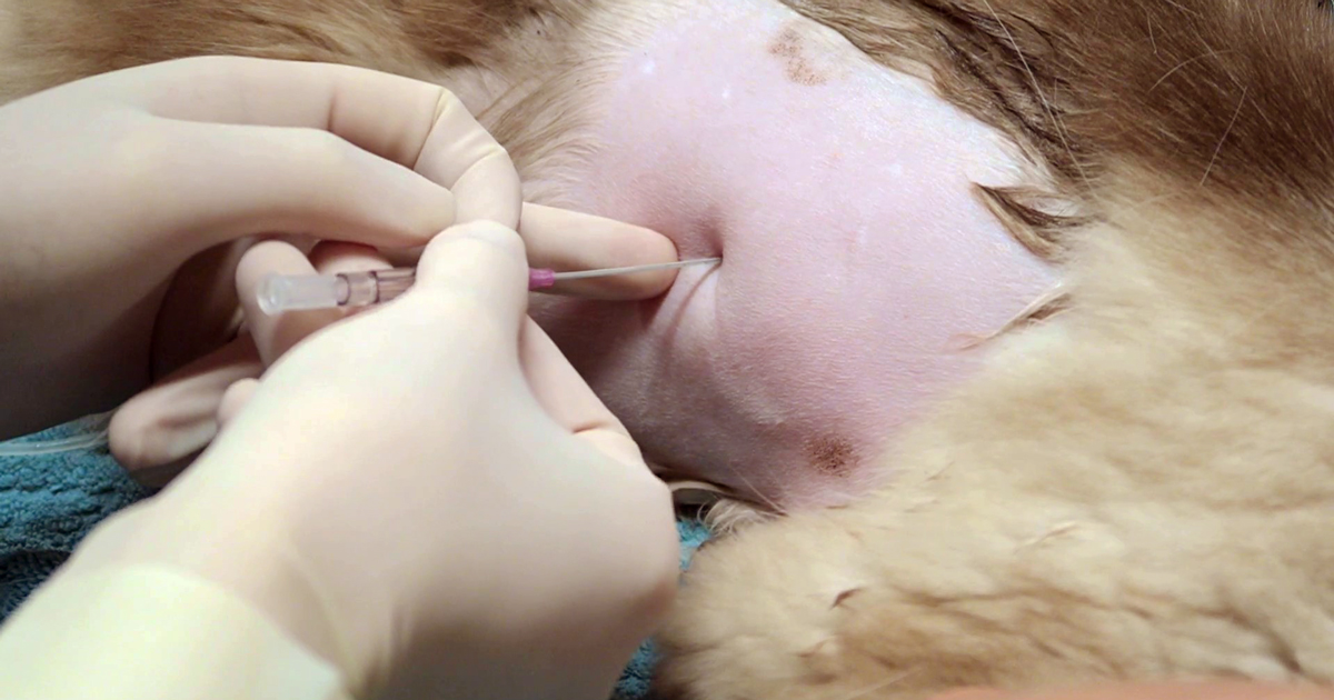
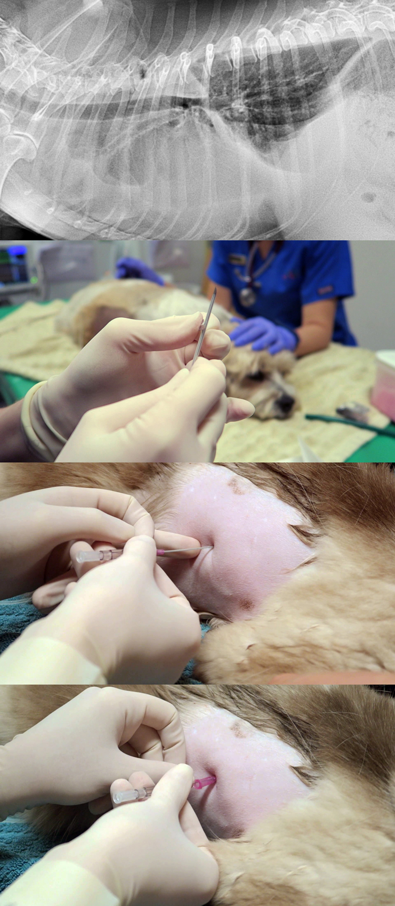
    Thoracocentesiscombined
    An approach to performing a thoracentesis.

    a. Options include local anaesthetic infiltration of the intended centesis site, and/or IM or IV opioid pain relief at standard doses.

    b. Opioid pain relief, such as butorphanol, is great for sedation that facilitates the process.

    c. Depending on the case, I often use opioid pain relief without local. This is sufficient in the vast majority of cases.

    d. If severely dyspnoeic, anaesthesia and intubation can help facilitate the process. It will reduce patient stress, enable manual ventilation and administration of 100% oxygen, and allow for larger volumes of air/fluid to be removed.

    2. Patient positioning

    Generally, sternal is easiest – otherwise, lateral recumbency or standing (if the animal will tolerate it).

    3. Site

    a. Locate the seventh to ninth intercostal spaces.

    b. To remove air, clip the dorsal two-thirds of the chest.

    c. To remove fluid, clip the ventral two-thirds of the chest.

    d. Clip a larger area than you expect.

    e. Prepare the area for an aseptic procedure.

    4. Connect everything

    a. The syringe to the three-tap and extension set should be ready prior to connecting the butterfly catheter.

    b. Often, a rush occurs to connect everything after the catheter is in place.

    5. Needle insertion

    a. Insert the needle on the cranial edge of the rib to avoid the nerves and blood vessels that run along the back of the rib.

    b. Ultrasound guided is best for fluid; you can lube the inside of a sterile glove and put the probe inside the glove to keep the area sterile.

    c. An IV catheter can also be used. I partially fenestrate a 20g IV catheter with two extra holes – once the catheter is advanced into the chest minimal risk exists of trauma to the lungs, and larger volumes of fluid and air can be removed.

    6. After insertion

    a. Once through the skin, connect to the extension set and apply gentle negative pressure. This can help determine how far you need to advance the needle into the chest.

    b. Sometimes a small syringe, such as 10ml, is better for smaller volumes as it creates less negative pressure. Pulse the negative pressure.

    7. Collect samples

    Make sure you collect the required samples from the first collection, as this is often the best sample and means you don’t forget.

    Overall, if you feel it is necessary to perform an emergency thoracentesis then do not delay. Most animals will tolerate the procedure well and have immediate dramatic improvements in respiratory rate, effort and oxygen saturations – all great outcomes for any dyspnoeic patient.

    Next week, we will look at what to do with the collected sample.

  • 5 things I would tell my vet school self, part 3

    5 things I would tell my vet school self, part 3

    So far in this five-part series I have stressed the importance of signalment and finding practical work while studying – both of which should be crucial in student learning.

    In this third part I decided to focus on the incredible support nursing and reception teams offer vets – both on a professional and personal level.

    Lifeline

    I did not appreciate how much I would come to rely on nurses and receptionists for support. For me, the support is both clinical and emotional. Not long after graduation, I remember I asked my head nurse at the time for advice on how to treat a hot spot, as I had never done so before.

    It was an extremely humbling experience – especially since it had never occurred to me until that point that I may need my nursing staff to offer clinical tips or perspective. Then again, I forget a lot of the nurses have more experience than me, having assisted vets years before I even graduated.

    Support

    Nurses are also there for you emotionally – they are the ones with you when you treat your patients; so, just like you, they share all the patients’ wins and also the losses. You will not be able to find someone else who can empathise with you more.

    Sometimes, when faced with particularly difficult consults, you will be surprised how often you offload your stress by talking to them, and it’s quite a relief to know you have someone to listen.

    I don’t think I could possibly explain to myself when starting out just how much I would rely on support teams.