Christmas can be a busy time for vet clinics, so here is a list of common intoxications and conditions to keep an eye out on during the festive period.
Chocolate
 Numerous online calculators can determine whether a toxic dose has been consumed and they are a great place to start.
- I always perform emesis in patients that have ingested chocolate, even hours after ingestion as often large amounts can reside in the stomach.
- Remember that cardiac arrhythmias can also occur in clinically normal looking patients, so perform an ECG.
- The toxic components can be reabsorbed through the bladder wall; therefore, urinary catheterisation is a part of management of this intoxication.
Onions
- Onions used in roasts and on BBQ’s can cause Heinz body formation, haemolytic anaemias and pigmenturia.
- This is not a common intoxication, but should be considered in anaemia patients and those with discoloured urine.
Raisins
- Commonly used in Christmas cakes and puddings. They can cause acute kidney failure, the exact mechanism of action is unknown, and there does not appear to be a dose-dependent relationship.
- It should always be a differential for azotemic patients this time of year.
- IV fluid induced diuresis for 48 hours is the safest way to manage raisin exposure.
Mistletoe
- The berries can be fatal, even if only a couple are ingested.
Ethylene glycol
- In colder climates, ethylene glycol can be a very common toxicity.
- This sweet liquid is very attractive to pets and can cause acute renal failure, with the first signs being acute onset ataxia.
Macadamia nuts
- Macadamia nuts are common in some parts of the world. They result in joint pain in the hocks and carpus leading to weakness and ataxia.
- Often confused with trauma and soft tissue injuries. Hyperextension of the hocks and sometimes flexion of the carpus are the clinical features.
Xylitol
- Xylitol is a sugar-free product used in lollies and baking.
- In dogs, it triggers endogenous insulin to be released and a subsequent hypoglycemia develops. It can also cause hepatic failure.
- As a general rule, I approach all intoxications as if they could be fatal as it is rare to know exactly how much of the toxic agent they have been exposed to. I consider if a patient I am treating for intoxication never develops clinical signs and wonder whether it was going to or not is the best outcome.
Strings
- Look under the tongue.
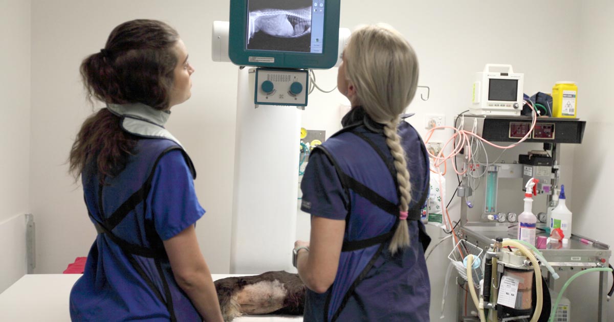
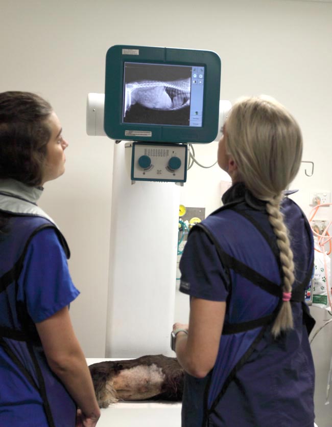
- Linear foreign bodies can be difficult to diagnose. Some features on abdominal radiographs to look out for include abnormal bunching of the small intestines, and “c” and “comma” shaped gas patterns.
Christmas meals
- Gastroenteritis is the most common presenting condition over the Christmas period, with dietary change and indiscretion often being the culprit.
- Bones can lead to obstructions from oral cavity to the intestines and can also cause constipation.
- Leftover meat trimmings, often fat laden, are a common cause of pancreatitis.
BBQ skewers
- In some parts of the world (Australia especially) BBQs are common around Christmas time.
- BBQ skewers can cause gastrointestinal tract perforation and septic peritonitis.
- Because they are not radiopaque they are often difficult to diagnose.