Tag: Sodium

  • Ionised hypocalcaemia, pt 3: acute treatment and management

    Ionised hypocalcaemia, pt 3: acute treatment and management

    Treatment of ionised hypocalcaemia (iHCa) is reserved for patients with supportive clinical signs, then divided into acute and chronic management.

    Since the most common cases of clinical hypocalcaemia in canine and feline patients are acute to peracute cases, this blog will focus on the acute treatment and management of hypocalcaemia.

    Clinical signs

    The severity of clinical signs of iHCa is proportional to the magnitude, as well as the rate of decline in ionised calcium (iCa) concentration.

    The normal reference range for iCa is 1.2mmol/L to 1.5mmol/L in dogs and 1.1mmol/L to 1.4mmol/L in cats. Serum iCa concentrations in younger dogs and cats are, on average, 0.025mmol/L to  0.1mmol/L higher than adults.

    Mild iHCa (0.9mmol/L to 1.1mmol/L) – as seen in critically ill dogs and cats with diabetic ketoacidosis, acute pancreatitis, protein-losing enteropathies, sepsis, trauma, tumour lysis syndrome or urethral obstructions – often has no observable clinical signs.

    Moderately (0.8mmol/L to 0.9mmol/L) to severely (lower than 0.8mmol/L) affected animals – in the case of eclampsia and those with parathyroid disease – often display severe signs.

    Early signs of iHCa are often non-specific, and include:

    • anorexia
    • rubbing of the face
    • agitation
    • restlessness
    • hypersensitivity
    • stiff and stilted gait

    As the serum iCa concentration further decreases, patients often progress to:

    • paresthesia
    • tachypnoea
    • generalised muscle fasciculations
    • cramping
    • tetany
    • seizures

    In cats, the gastrointestinal system can also be affected, presenting as anorexia and vomiting.

    Treatment

    The need for treatment of hypocalcaemia is dependent on the presence of clinical signs, rather than a specific cut-off of serum concentration of iCa itself.

    Moderate to severe iHCa should always be treated. Mild hypocalcaemia, on the other hand, may not be necessary, especially if it is well tolerated. It should be remembered the threshold for development of clinical signs is variable, and treatment may benefit critical cases with an iCa concentration of less than 1.0mmol/L.

    Treatment is divided into the acute treatment phase and chronic management.

    In the tetanic phase, IV calcium is required – 10% calcium gluconate (equivalent to 9.3mg/ml) administered at 0.5ml/kg to 1.5ml/kg dosing to effect. This should be administered slowly with concurrent ECG monitoring. Infusion of calcium needs to be stopped if bradycardia develops or if shortening of the QT interval occurs.

    Some suggest calcium gluconate (diluted 1:1 with 0.9% sodium chloride) of half or the full IV dose can be given SC and repeated every six to eight hours until the patient is stable enough to receive oral supplementation. However, be aware calcium salts SC can cause severe necrosis or skin mineralisation.

    Calcium chloride should never be given SC, as it is a severe perivascular irritant.

    Correcting iCa

    Irrespective of the chronicity of the treatment, the rule of thumb is correction of calcium should not exceed 1.1mmol/L.

    Correction of iCa to normal or hypercalcaemic concentration should always be avoided, as this will result in the desensitisation of the parathyroid response, predisposing renal mineralisation and formation of urinary calculi.

    Some of the more common calcium supplementation medications – both parenteral and oral formulas – are detailed in Table 1. Supplementation of magnesium may also benefit some patients, as it is a common concurrent finding in critically ill patients with iHCa.

    Table 1. Common calcium supplementation medications
    Drug Calcium Content Dose Comment
    Parenteral calcium
    Calcium gluconate
    (10% solution)
    9.3mg/ml
    i) slow IV dosing to effect (0.5ml/kg to 1.5ml/kg); acute crisis, 50mg/kg to 150mg/kg over 20 to 30 minutes
    ii) 5mg/kg/hr to 15mg/kg/hr IV or 1,000mg/kg/day to 1,500mg/kg/day (or 42mg/kg/hr to 63mg/kg/hr)
    Stop if bradycardia or shortened QT interval occurs.
    Infusion to maintain normal Ca level
    SC calcium salts can cause severe skin necrosis/mineralisation.
    Calcium chloride
    (10% solution)
    27.2mg/ml 5mg/kg/hr to 15mg/kg/hr IV Do not give SC as severe perivascular irritant
    Oral calcium
    Calcium carbonate
    (many sizes)
    40% tablet 5mg/kg/day to 15mg/kg/day
    Calcium lactate
    (325mg, 650mg)
    13% tablet 25mg/kg/day to 50mg/kg/day
    Calcium chloride
    (powder)
    27.2% 25mg/kg/day to 50mg/kg/day May cause gastric irritation
    Calcium gluconate (many sizes) 10% 25mg/kg/day to 50mg/kg/day

    Next time…

    The next blog will look at the pathophysiology behind iHCa among critically ill animals. It will also look at the controversy regarding treatment of non-clinical iHCa cases and the prognostic indications of iCa concentrations.

  • Hyponatraemia, pt 3: correcting a sodium concentration of 110mEq/L

    Hyponatraemia, pt 3: correcting a sodium concentration of 110mEq/L

    The amount of sodium required to increase serum sodium concentration to a desired value can be calculated from the following formula:

    Sodium deficit = 0.6 × bodyweight (kg) × (normal sodium [mEq/L] – patient sodium [mEq/L])

    Table 1. Sodium content of various fluids
    Fluid Type Sodium content (mEq/L)
    0.9% sodium chloride 154
    Normosol-R 140
    Hartmann’s solution
    130
    3% sodium chloride 513
    7.5% sodium chloride 1,300

    This sodium deficit is then replaced over “x” hours, at an average rate of 0.5mEq/L/hr.

    In hypovolaemic hyponatraemia patients – where the fluid deficits also need correcting – it is important to select a fluid where the sodium concentration is within 5mEq/L to 10mEq/L of the patient plasma sodium level.

    Table 1 shows the sodium content of various fluids. If none of the fluids listed in Table 1 are suitable – for example, the patient’s sodium level is 110 – you can make your own fluid by mixing 5% dextrose in water using the formula below:

    Volume of 5% dextrose in water to be added (ml) =

    ([current IV fluid Na+] – [desired IV fluid Na+]) × 1,000ml ÷ ([desired IV fluid Na+] – [supplemental IV fluid Na+])

    Hartmann’s example

    The most common cause of severe hyponatraemia is hypoadrenocorticism. Using an example of a severe hyponatraemia of 110mEq/L, I select Hartmann’s solution first, as it has the lowest sodium concentration. How low I dilute Hartmann’s depends on the patient’s volume status.

    If the patient requires fluid resuscitation because it is showing signs of poor perfusion – such as elevated heart rate, poor pulse quality, pale gums, prolonged capillary refill time, dull mentation, low core body temperature and elevated lactate – I aim for a sodium concentration the same as the patient. For this example, I would dilute the Hartmann’s to 110mEq/L, as then I can bolus therapy this without elevating the patient’s sodium concentration.

    So, aiming for 110mEq/L, the volume of 5% dextrose in water (D5W) required to dilute Hartmann’s is:

    = ([130 – 110] × 1,000) ÷ (110 – 0)

    = (20 × 1,000) ÷ 110

    = 181ml of D5W.

    This volume may not fit in the bag, so I remove 150ml from the Hartmann’s bag first and insert 850ml into the equation:

    = ([130 – 110] × 850) ÷ (110 – 0)

    = (20 × 850) ÷ 110

    = 154ml of D5W to be added to the bag for a total volume of 1,054ml with a sodium concentration of 110mEq/L.

    TIP:

    Electrolytes can be used on custom solutions to check the final sodium concentration. It will be a couple of mEq/L above or below, due to variations in each Hartmann’s bag.

    I bolus with this 110mEq/L of custom solution for correct perfusion, reassess the patient and sodium concentration – and make a solution between 5mEq/L and 10mEq/L higher – and administer at much slower rates with repeated monitoring.

    The treatment of hypervolaemic hyponatraemic patients will not be discussed here, as it revolves around treating the underlying medical condition.

    Conclusion

    Hyponatraemia is a common and potentially life-threatening change in our critical patients.

    It is crucial to establish whether this is an acute or chronic change, to avoid development of osmotic demyelination syndrome. If I have any doubt about the timeline, I treat as a chronic change and increase slowly.

  • Hyponatraemia, pt 2: causes

    Hyponatraemia, pt 2: causes

    The causes of hyponatraemia can be divided into three major categories, based on serum osmolality. This is further divided based on the patient’s volume status (Table 1).

    Most patients we see in clinic fall into the hypovolaemic category, except patients with diabetes mellitus.

    Table 1. Causes of hyponatraemia based on osmolality and volume status (from Guillaumin and DiBartola, 2017).
    Hypo-osmolar Hyperosmolar Normo-osmolar
    Hypovolaemic Normovolaemic Hypervolaemic
    Gastrointestinal fluid loss
    Third-space fluid losses
    Shock
    Hypoadrenocorticism (Addison’s disease)
    Renal insufficiency
    Excessive diuretic administration
    Salt-losing nephropathy
    Cerebral salt wasting syndrome
    Syndrome of inappropriate antidiuretic hormone secretion (SIADH)
    Hypotonic fluid administration
    Hypothyroidism
    Glucocorticoid insufficiency
    Psychogenic polydipsia
    Reset osmostat (SIADH type B)
    Congestive heart failure
    Acute or chronic renal failure
    Nephrotic syndrome
    Hepatic cirrhosis
    Accidental ingestion or injection of water (water intoxication)
    Hyperglycaemia
    Mannitol
    Severe azotaemia
    Hyperlipidaemia
    Hyperproteinaemia

    Common causes

    In dogs, the three most common causes of hyponatraemia are:

    • gastrointestinal (GI) fluid loss
    • third-space fluid loss
    • fluid shift from intracellular fluid to extracellular fluid (ECF) as a result of hyperglycaemia

    In cats, the three most common causes of hyponatraemia are:

    • urologic diseases
    • GI fluid loss
    • third-space fluid losses

    In most patients, more than one pathophysiologic factor is likely to be contributing to the hyponatraemia.

    Circulating volume

    Hypovolaemic patients – those with, for example, GI losses, hypoadrenocorticism, renal losses and haemorrhagic shock – have a reduced effective circulating volume. ECF contraction triggers antidiuretic hormone (ADH) secretion, which leads to increases in free water absorption and thirst, and results in dilution of the serum sodium concentration. Aldosterone secretion is reduced in hypoadrenocorticism, so an overall reduction in sodium reabsorption compounds the problem.

    Hypervolaemic patients are those with an increased fluid retention state, such as:

    • congestive heart failure (pulmonary oedema)
    • advanced hepatic failure (ascites, third-space fluid)
    • renal failure
    • free water ingestion

    Congestive heart failure patients have a reduced cardiac output and, therefore, a decreased effective circulating volume, despite the presence of the extra fluid status. Renin-angiotensin activation leads to release of ADH and aldosterone, resulting in sodium and free water reabsorption, and increased thirst. Both lead to an excess of free water retention.

    Advanced hepatic (cirrhosis) or renal failure (nephrotic syndrome) both result in hypoalbuminaemia, leading to fluid shifting into the interstitial space and third space, reducing effective circulating volumes. This leads to activation of ADH to increase free water reabsorption, to restore the circulating volume in the face of existing hypervolaemia and hyponatraemia.

    Diabetic patients

    Moderate to severe hyperglycaemic diabetic patients can be either hyperosmolar or normo-osmolar, depending on the serum blood glucose concentration. Hyponatraemia occurs when water shifts from the intracellular fluid to the ECF down the osmotic gradient, diluting the serum sodium content.

    Despite this osmotic shift, not all diabetic patients develop hyponatraemia. Glucosuria also causes also causes a renal osmotic shift, sometimes resulting in urine water loss in excess to sodium. This offsets the hyponatraemia – in some cases, hypernatraemia results.

    Treatment

    Treatment of hyponatraemia hinges on how quickly it developed and the volume status of the patient. The rule of thumb is to correct hyponatraemia slowly – not exceeding 0.5meq/L/hr – especially in chronic cases, or cases where the duration of hyponatraemia is unknown. Keeping to this rate is paramount until serum sodium concentration reaches 130meq/L.

    In acute patients with severe clinical signs, such as seizures, some clinicians may choose to use a higher rate of 1meq/L/hr to 2meq/L/hr until clinical signs resolved.

    It should be emphasised, once again, this rate should never be used in chronic patients, patients with an unknown duration of hyponatraemia, or where frequent serum sodium concentration cannot be monitored. The rapid correction of hyponatraemia can lead to osmotic demyelination syndrome (myelinolysis).

    Its effect will not be apparent until three or four days after therapy, and can result in neurological abnormalities such as:

    • weakness
    • ataxia
    • dysphagia
    • paresis
    • coma

    For that reason, frequent electrolyte measurements are required, starting hourly then once a suitable rate of increase has been established and less frequently thereafter.

    • Part 3 will look at how to correct patients with hyponatraemia.

    Reference

    Guillaumin J and DiBartola SP (2017). A quick reference on hyponatremia, Veterinary Clinics of North America: Small Animal Practice 47(2): 213-217.

  • Hyponatraemia, pt 1: clinical signs

    Hyponatraemia, pt 1: clinical signs

    Hyponatraemia is a relatively common electrolyte disturbance encountered in critically ill patients, and the most common sodium disturbance of small animals.

    In most cases, this is caused by an increased retention of free water, as opposed to the loss of sodium in excess of water.

    Low serum sodium concentration

    Hyponatraemia is defined as serum concentration lower than 140mEq/L in dogs and lower than 149mEq/L in cats.

    The serum sodium concentration measured is not the total body sodium content, but the amount of sodium relative to the volume of water in the body. For this reason, patients with hyponatraemia can actually have decreased, increased or normal total body sodium content.

    This series will look briefly at the modulators of the sodium and water balance, clinical signs associated with hyponatraemia, the most common causes in small animals, the pathophysiology behind these changes, and treatment and management.

    ECF volume

    hyponatraemia
    An example of hyponatraemia.

    Sodium is the main osmotically active particle in the extracellular fluid (ECF), so is the main determining factor of the ECF volume. Any disease process that alters the patient’s ECF volume will lead to hyponatraemia, such as:

    • dehydration
    • polyuria
    • polydipsia
    • vomiting
    • diarrhoea
    • cardiac diseases
    • pleural or peritoneal effusion

    The modulators of water and sodium balance are also different, so should be thought of as different processes.

    Water balance is modulated by thirst and antidiuretic hormone, and the effect of this is to maintain normal serum osmolality and serum sodium concentration.

    Modulators of sodium balance aim to maintain normal ECF volume. It adjusts this by altering the amount of renal sodium excretion; an expansion of ECF volume will lead to an increased sodium excretion, while a reduction in ECF volume will lead to increased sodium retention.

    Rate and magnitude

    The clinical signs of hyponatraemia are both dependent on the magnitude of the decrease and the rate at which it developed.

    In mild or chronic patients, no visible clinical signs can exist. In severe (lower than 125mEq/L) and acute cases, clinical signs exhibited are typically neurological, reflecting cerebral oedema. Possibilities include:

    • lethargy
    • anorexia
    • weakness
    • incoordination
    • disorientation
    • seizures
    • coma

    Patients with acute hyponatraemia – for example, water intoxication – are more likely to show clinical signs, compared to those with chronic hyponatraemia, because the brain takes time (at least 24 to 48 hours) to produce idiogenic osmoles, osmotically active molecules that help shift free water out of brain cells.

    Therefore, any acute hyponatraemia that develops within a 24 to 48-hour period tend to show clinical signs, whereas chronic cases are less likely.

    • Next week’s blog will look into the different causes of hyponatraemia and how they result in sodium loss.
  • Blood gas analysis, pt 5: metabolic acidosis and alkalosis

    Blood gas analysis, pt 5: metabolic acidosis and alkalosis

    Base excess (BE) and bicarbonate (HCO3-) represent the metabolic components of the acid base equation. In general, both components will change in the same direction.

    Decreased HCO3– and BE indicate either a primary metabolic acidosis or a metabolic compensation for a chronic respiratory alkalosis. Elevated HCO3– and BE indicate either a primary metabolic alkalosis or a metabolic compensation for a chronic respiratory acidosis.

    The exception to this rule arises when a patient hypoventilates or hyperventilates.

    Carbonic acid equation

    CO2 + H2O ↔ H2CO3 ↔ HCO3– + H+

    When a patient hypoventilates, CO2 will increase as a result of reduced expiration, so a shift to the right of the equilibrium will occur. The shift to the right will increase the bicarbonate levels proportional to the increase in CO2.

    The opposite occurs when a patient hyperventilates; the equilibrium shifts to the left, so a decrease in HCO3– is present.

    Since HCO3– is not independent to the patient’s respiratory status, it is an inaccurate way of measuring the metabolic component in patients with respiratory changes. For this reason, the BE value is the preferred.

    The BE represents the amount of acid, or base, needed to titrate 1L of the blood sample until the pH reaches exactly 7.4, with the assumption the blood sample is equilibrated to a partial pressure of CO2 of 40mmHg (the middle of the reference range) and the patient’s body temperature is normal.

    Possible causes

    The possible causes of the primary disease are:

    Metabolic acidosis

    • lactic acidosis – shock and poor perfusion
    • renal failure – reduced hydrogen ion (H+) excretion and increased loss of HCO3
    • diabetic ketoacidosis – ketone acids
    • gastrointestinal (GI) losses – loss of HCO3– through vomiting and diarrhoea

    Metabolic alkalosis

    • GI outflow obstructions – loss of H+ and chloride via vomiting
    • reduced chloride levels and resultant poor perfusion – body attempts to reabsorb water and sodium to increase intravascular volume, but inadvertently also reabsorbs HCO3– in the process, despite existing alkalosis
    • refeeding syndrome
    • severe hypokalaemia:
      • transcellular shift – potassium ions leave and H+ ions enter the cell
      • transcellular shift in cells of proximal tubules → intracellular acidosis → promotes ammonium production and excretion
      • H+ excretion in the proximal and distal tubules increases → further reabsorption of HCO3– and net acid excretion
    • renal insufficiency
    • diuretic therapy (contraction alkalosis – loss of bodily fluids that do not contain HCO3-; this causes the extracellular volume to contract around a fixed quantity of HCO3-, resulting in a rise in the concentration of HCO3– without an actual increase in HCO3– levels)

    Next step

    After ruling out the differential causes of either respiratory or metabolic acidosis/alkalosis, the next step is to determine whether a compensatory response is present and, if so, if this is adequate or whether a true mixed acid-base disorder exists.

  • Blood gas analysis, pt 1: why everyone needs to know about it

    Blood gas analysis, pt 1: why everyone needs to know about it

    For those of you who have received referral histories from emergency or specialists hospitals, blood gas analysis is probably no stranger to you. For those who have never heard of them before, fear not – you are in for a treat.

    In my emergency hospital, the blood gas analyser is arguably one of the most frequently used bench top lab machines, second only to centrifuge, and for good reasons…

    Acid-base disturbances are common in critically ill and emergency patients, and it can help determine the severity of their condition and sometimes provide the answer. Tracking changes in blood gas parameters can provide information about the patient’s response to your interventions.

    blood-gas-analyser_output
    Blood gas analysis can help assess the severity of a patient’s condition and help guide your diagnostic plan.

    The information gained from pulse oximetry is very limited in patients with severe respiratory compromise, and the only way to accurately assess their oxygenation and/or ventilation status is by looking at their blood gas status.

    So what does the blood gas analysis actually measure?

    Most blood gas panels assess the pH of the blood, partial pressure of oxygen (PO2) and partial pressure of carbon dioxide (PCO2). From these, the machine is able to derive the percentage of haemoglobin saturated with oxygen (SO2), bicarbonate (HCO3) concentration and base excess of the extracellular fluid (BEecf).

    In most machines, they are also able to measure other parameters, such as electrolytes (Na, K, Ca, Cl), glucose and lactate.

    While arterial blood gas samples are required for determining the ability of the body to oxygenate the haemoglobin, venous samples are suitable for determining the ventilation status, assessing acid base balance, electrolytes, glucose and lactate levels.

    So how can this help as a point-of-care test?

    As mentioned previously, blood gas analysis can help assess the severity of a patient’s condition and help guide your diagnostic plan. It can also provide a diagnosis (such as diabetic ketoacidosis, typical hypoadrenocorticism and high gastrointestinal obstructions).

    The changes in these parameters over time can be essential in managing critical patients in the emergency setting; it will help guide you in developing an appropriate IV fluid therapy regime and fluid choice, address the patient’s oxygenation and/or ventilation needs, correct any electrolyte and glucose abnormalities, and – although fallen out of favour – the administration of sodium-bicarbonate therapy.

    In upcoming blogs, I will teach you how to interpret the blood gas results. At the end of this, I hope everyone will incorporate blood gas analysis as their standard point-of-care test for the better assessment and management of patients.

    If given the choice between a biochemistry and a blood gas panel in a critical patient, I would hands down select blood gas every time.

  • Intoxication: decontamination advice

    Intoxication: decontamination advice

    Building on from last week’s blog on telephone advice, this is what I advise owners they can do at home if their pet has been exposed to a toxin.

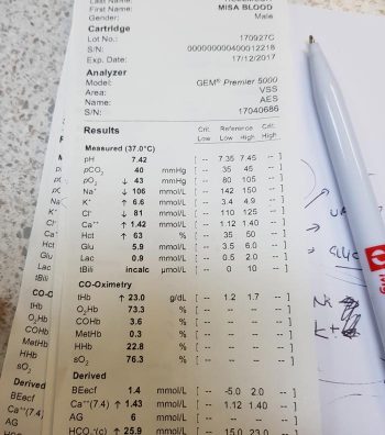
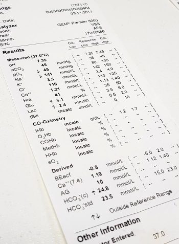
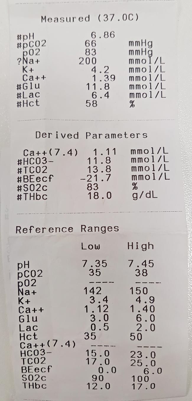
    The patient’s blood gas analysis and electrolyte panel. Note the sodium concentration.
    Figure 2. The patient’s blood gas analysis and electrolyte panel. Note the sodium concentration.

    The main exposure routes are ocular, dermal and gastrointestinal.

    Ocular

    Acids and alkalis cause the most severe effects, as they can cause ongoing damage for some time after initial contact.

    Eye irrigation

    Avoid contact lens solution as this can cause further irritation. Instead, I recommend:

    • tepid water, saline or distilled water
    • 20 to 30 minutes (ideally)
    • rinse from medial to lateral, to avoid contamination of the other eye

    Once the eye(s) have been flushed, recommend the animal be taken to the veterinary clinic for further assessment. Corneal ulceration can be difficult to see with the naked eye.

    Dermal

    Owners need to take precautions to protect themselves from contact with the toxin. The aim here is for owners to remove as much of the toxin off the skin of their pet without exposing themselves to it.

    The most common method is bathing or rinsing with a mild dish soap in warm water. If it is a dry power and it safe, vacuuming off the powders may be tried, unless risk of aerosolisation of the toxin is high.

    Gastrointestinal

    Oral exposure

    Ideally wearing gloves, instruct the owner to wipe the inside of the lips and over the gums using a damp dish cloth to try to remove any toxin remaining on the mucous membranes. Warn the pet may try to bite and, if it does, to stop immediately.

    Ingested toxins

    Inducing emesis depends on the type of toxin, but, either way, I do not recommend emesis induction to be performed at home. I have seen disastrous effects from salt slurries (Figures 1 and 2).

    Emesis induction is most safely performed in a clinical setting where the medications that can be administered are safer and more effective.

    Seizures

    Nothing can be done at home to stop a seizure. If a toxin is causing a pet to seizure then it is unlikely they will stop, so will require medications. The pet will need bringing into the clinic immediately.

    I suggest owners do not try to put their fingers in their pet’s mouth, as they are very unlikely to choke on their own tongue.

    Wrap them in a blanket to help prevent injury to the owners. Once in the car, keep the head slightly down – if they do vomit or have large amounts of foam then it is allowed to fall out of the mouth, not build at the back of the mouth and lead to aspiration.

  • Lipaemia – the bane of biochemistry

    Lipaemia – the bane of biochemistry

    Last week we covered haemolysed samples – this week we’re looking at lipaemic samples.

    Lipaemic samples are caused by an excess of lipoproteins in the blood, creating a milky/turbid appearance that interferes with multiple biochemical tests and can even cause haemolysis of red blood cells.

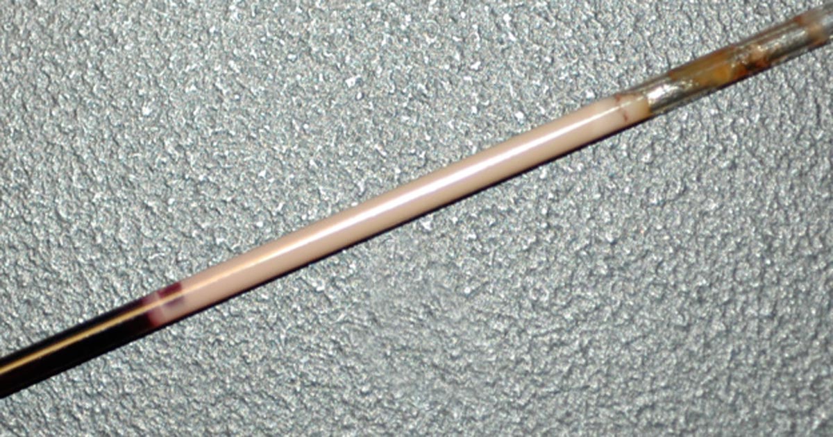
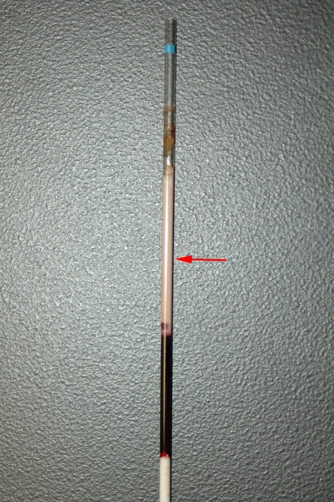
    lipaemic sample
    A severely lipaemic sample (red arrow). IMAGE: eClinPath.com (CC BY-NC-SA 4.0).

    Lipaemia can follow recent ingestion of a meal – especially one high in fat. Although not pathognomonic for any diseases, its presence can help increase the suspicion of certain diseases, including:

    • pancreatitis
    • diabetes mellitus
    • hypothyroidism
    • hyperadrenocorticism
    • primary hyperlipidaemia (in some specific breeds, such as the miniature schnauzer)

    It warrants further investigation in patients that have been ill and inappetent.

    Irksome interpretations

    Lipaemia can dramatically impact laboratory testing and is often troublesome in critically ill patients, making interpretation of biochemistry particularly difficult, if not impossible.

    Lipaemia can affect different analysers in different ways, most commonly causing:

    • Falsely increased calcium, phosphorus, bilirubin, glucose and total protein (via refractometer) and some liver parameters such as alkaline phosphatase, alanine aminotransferase, aspartate aminotransferase, haemoglobin concentration, and mean corpuscular haemoglobin concentration.
    • Falsely decreased sodium, potassium, chloride, albumin and bicarbonate.

    Tube tips

    Assessment of a centrifuged haematocrit tube before running a biochemistry panel can help reduce wasted biochemistry consumables.

    If the sample is lipaemic in the haematocrit tube then maybe try some of the following tips.

    • If blood tests are planned in advance, try fasting the patient beforehand for 12 to 24 hours.
    • Repeat sampling a couple of hours later may yield a less lipaemic sample.
    • Collecting and centrifuging a larger amount of blood (3ml to 5ml, for example) can sometimes yield enough clear sample between the lipid layer and red blood cells.
    • Refrigeration of the sample can help the separation.
    • Extract lipids using polar solvents, such as polyethylene glycol.
    • Centrifugation at higher than normal speeds (if possible) can also assist in clearing the layer.
  • PCV/total solids interpretation: serum colour

    PCV/total solids interpretation: serum colour

    When interpreting the often misinterpreted and underused PCV and total solids test, it is important to take note of the serum colour as this may give clues into the diagnosis.

    PCV tubes
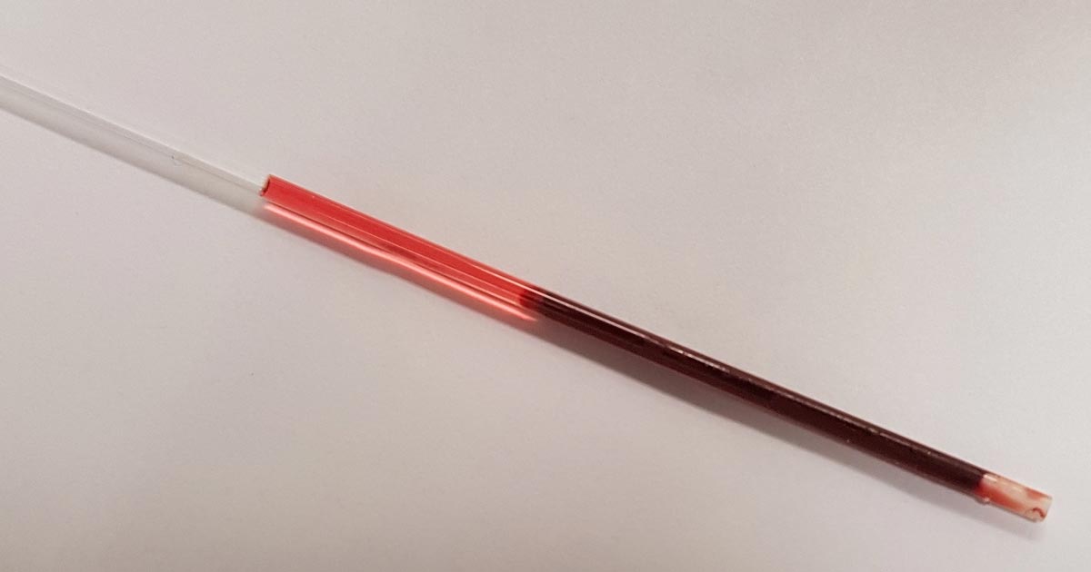
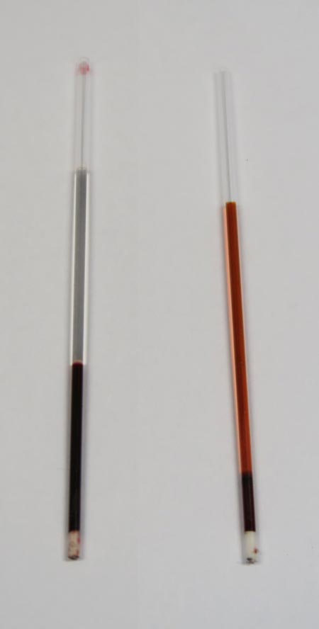
    Normal serum colour (left) compared to a patient with immune-mediated haemolytic anaemia. The serum is haemolysed and anaemia is present.

    The most common abnormalities seen in clinic are icteric, haemolysed and lipaemic serum.

    Clear serum can also be of importance – especially when you interpret it with blood counts and urine colour.

    Haemolysis

    The most common abnormality of serum colour changes is haemolysis. In my experience, the most common cause is suboptimal collection technique. To confirm this, simply collect another sample and repeat.

    If it is repeatable, and concurrent anaemia or pigmenturia is present, it warrants further investigation.

    Intravascular haemolysis can be caused by:

    • immune-mediated haemolytic anaemia
    • blood transfusion reactions
    • infectious diseases such as Mycoplasma haemofelis, Babesia canis, Ehrlichia canis, FeLV and others
    • Heinz bodies from the ingestion of heavy metal, onions or paracetamol
    • hypophosphataemia
    • macroangiopathic disease (neoplasia, for example)
    • envenomation – typically, snake bites

    Testing issues

    Haemolysis can also affect other laboratory testing. It can lead to an artefactual increase in glucose, phosphorus, bilirubin, total protein, fructosamine and triglycerides, and a decrease in sodium (pseudohyponatraemia), cholesterol, calcium, potassium and albumin.

    Extravascular haemolysis often does not cause haemolysed serum as it is generally slower and the body is able to clear the haemoglobin before it can lead to discolouration of the serum.

  • Maintenance fluids

    Maintenance fluids

    A while ago we discussed the components of a fluid therapy plan and talked about hydration deficits. This week I want to touch on maintenance fluids.

    Gerardo_IVF
    IV fluids

    Maintenance rates are typically calculated using the following formulae:

    ml/day = 80 × bodyweight (kg)0.75 (cats)
    ml/day = 132 × bodyweight (kg)0.75 (dogs)

    or
    ml/day = 30 × bodyweight (kg) + 70

    These formulae better estimate the needs of smaller and larger patients. The flat 3ml/kg/hr underestimates for small patients and overestimates for larger patients.

    This maintenance rate is in addition to rehydration rates.

    So what sort of fluid should you use for maintenance?

    True “maintenance” crystalloids:

    • used to replace ongoing fluid and electrolyte loss from normal metabolism, not to replace perfusion and hydration deficits or ongoing losses from diarrhoea, for example
    • sodium concentration less than plasma
    • potassium concentrations higher than plasma
    • glucose sometimes added to bring solute concentrations similar to extracellular fluid

    Do you have to use maintenance crystalloids or can you use replacement crystalloids?

    Replacement crystalloids are more frequently used for maintenance fluid therapy rather than maintenance crystalloids. This is because they are more readily available, we are more familiar with their use and effect, and patients are generally continued on these after perfusion and hydration deficits have been corrected.

    In reality, most of the time it doesn’t really matter if we are using replacement crystalloids for maintenance therapy as the patient can manage the excess sodium, but some patients – especially cats – may require potassium supplementation. The key point is regular assessment of the patient’s hydration status and electrolytes – for example, every 12 to 24 hours for patients on IV fluids and not eating.