Tag: Skin

  • Ionised hypocalcaemia, pt 3: acute treatment and management

    Ionised hypocalcaemia, pt 3: acute treatment and management

    Treatment of ionised hypocalcaemia (iHCa) is reserved for patients with supportive clinical signs, then divided into acute and chronic management.

    Since the most common cases of clinical hypocalcaemia in canine and feline patients are acute to peracute cases, this blog will focus on the acute treatment and management of hypocalcaemia.

    Clinical signs

    The severity of clinical signs of iHCa is proportional to the magnitude, as well as the rate of decline in ionised calcium (iCa) concentration.

    The normal reference range for iCa is 1.2mmol/L to 1.5mmol/L in dogs and 1.1mmol/L to 1.4mmol/L in cats. Serum iCa concentrations in younger dogs and cats are, on average, 0.025mmol/L to  0.1mmol/L higher than adults.

    Mild iHCa (0.9mmol/L to 1.1mmol/L) – as seen in critically ill dogs and cats with diabetic ketoacidosis, acute pancreatitis, protein-losing enteropathies, sepsis, trauma, tumour lysis syndrome or urethral obstructions – often has no observable clinical signs.

    Moderately (0.8mmol/L to 0.9mmol/L) to severely (lower than 0.8mmol/L) affected animals – in the case of eclampsia and those with parathyroid disease – often display severe signs.

    Early signs of iHCa are often non-specific, and include:

    • anorexia
    • rubbing of the face
    • agitation
    • restlessness
    • hypersensitivity
    • stiff and stilted gait

    As the serum iCa concentration further decreases, patients often progress to:

    • paresthesia
    • tachypnoea
    • generalised muscle fasciculations
    • cramping
    • tetany
    • seizures

    In cats, the gastrointestinal system can also be affected, presenting as anorexia and vomiting.

    Treatment

    The need for treatment of hypocalcaemia is dependent on the presence of clinical signs, rather than a specific cut-off of serum concentration of iCa itself.

    Moderate to severe iHCa should always be treated. Mild hypocalcaemia, on the other hand, may not be necessary, especially if it is well tolerated. It should be remembered the threshold for development of clinical signs is variable, and treatment may benefit critical cases with an iCa concentration of less than 1.0mmol/L.

    Treatment is divided into the acute treatment phase and chronic management.

    In the tetanic phase, IV calcium is required – 10% calcium gluconate (equivalent to 9.3mg/ml) administered at 0.5ml/kg to 1.5ml/kg dosing to effect. This should be administered slowly with concurrent ECG monitoring. Infusion of calcium needs to be stopped if bradycardia develops or if shortening of the QT interval occurs.

    Some suggest calcium gluconate (diluted 1:1 with 0.9% sodium chloride) of half or the full IV dose can be given SC and repeated every six to eight hours until the patient is stable enough to receive oral supplementation. However, be aware calcium salts SC can cause severe necrosis or skin mineralisation.

    Calcium chloride should never be given SC, as it is a severe perivascular irritant.

    Correcting iCa

    Irrespective of the chronicity of the treatment, the rule of thumb is correction of calcium should not exceed 1.1mmol/L.

    Correction of iCa to normal or hypercalcaemic concentration should always be avoided, as this will result in the desensitisation of the parathyroid response, predisposing renal mineralisation and formation of urinary calculi.

    Some of the more common calcium supplementation medications – both parenteral and oral formulas – are detailed in Table 1. Supplementation of magnesium may also benefit some patients, as it is a common concurrent finding in critically ill patients with iHCa.

    Table 1. Common calcium supplementation medications
    Drug Calcium Content Dose Comment
    Parenteral calcium
    Calcium gluconate
    (10% solution)
    9.3mg/ml
    i) slow IV dosing to effect (0.5ml/kg to 1.5ml/kg); acute crisis, 50mg/kg to 150mg/kg over 20 to 30 minutes
    ii) 5mg/kg/hr to 15mg/kg/hr IV or 1,000mg/kg/day to 1,500mg/kg/day (or 42mg/kg/hr to 63mg/kg/hr)
    Stop if bradycardia or shortened QT interval occurs.
    Infusion to maintain normal Ca level
    SC calcium salts can cause severe skin necrosis/mineralisation.
    Calcium chloride
    (10% solution)
    27.2mg/ml 5mg/kg/hr to 15mg/kg/hr IV Do not give SC as severe perivascular irritant
    Oral calcium
    Calcium carbonate
    (many sizes)
    40% tablet 5mg/kg/day to 15mg/kg/day
    Calcium lactate
    (325mg, 650mg)
    13% tablet 25mg/kg/day to 50mg/kg/day
    Calcium chloride
    (powder)
    27.2% 25mg/kg/day to 50mg/kg/day May cause gastric irritation
    Calcium gluconate (many sizes) 10% 25mg/kg/day to 50mg/kg/day

    Next time…

    The next blog will look at the pathophysiology behind iHCa among critically ill animals. It will also look at the controversy regarding treatment of non-clinical iHCa cases and the prognostic indications of iCa concentrations.

  • Intoxication: decontamination advice

    Intoxication: decontamination advice

    Building on from last week’s blog on telephone advice, this is what I advise owners they can do at home if their pet has been exposed to a toxin.

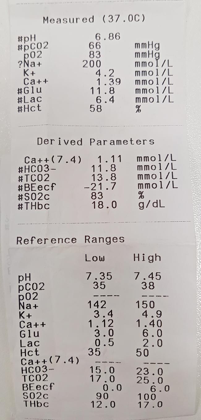
    The patient’s blood gas analysis and electrolyte panel. Note the sodium concentration.
    Figure 2. The patient’s blood gas analysis and electrolyte panel. Note the sodium concentration.

    The main exposure routes are ocular, dermal and gastrointestinal.

    Ocular

    Acids and alkalis cause the most severe effects, as they can cause ongoing damage for some time after initial contact.

    Eye irrigation

    Avoid contact lens solution as this can cause further irritation. Instead, I recommend:

    • tepid water, saline or distilled water
    • 20 to 30 minutes (ideally)
    • rinse from medial to lateral, to avoid contamination of the other eye

    Once the eye(s) have been flushed, recommend the animal be taken to the veterinary clinic for further assessment. Corneal ulceration can be difficult to see with the naked eye.

    Dermal

    Owners need to take precautions to protect themselves from contact with the toxin. The aim here is for owners to remove as much of the toxin off the skin of their pet without exposing themselves to it.

    The most common method is bathing or rinsing with a mild dish soap in warm water. If it is a dry power and it safe, vacuuming off the powders may be tried, unless risk of aerosolisation of the toxin is high.

    Gastrointestinal

    Oral exposure

    Ideally wearing gloves, instruct the owner to wipe the inside of the lips and over the gums using a damp dish cloth to try to remove any toxin remaining on the mucous membranes. Warn the pet may try to bite and, if it does, to stop immediately.

    Ingested toxins

    Inducing emesis depends on the type of toxin, but, either way, I do not recommend emesis induction to be performed at home. I have seen disastrous effects from salt slurries (Figures 1 and 2).

    Emesis induction is most safely performed in a clinical setting where the medications that can be administered are safer and more effective.

    Seizures

    Nothing can be done at home to stop a seizure. If a toxin is causing a pet to seizure then it is unlikely they will stop, so will require medications. The pet will need bringing into the clinic immediately.

    I suggest owners do not try to put their fingers in their pet’s mouth, as they are very unlikely to choke on their own tongue.

    Wrap them in a blanket to help prevent injury to the owners. Once in the car, keep the head slightly down – if they do vomit or have large amounts of foam then it is allowed to fall out of the mouth, not build at the back of the mouth and lead to aspiration.

  • Occupational hazards

    Occupational hazards

    Before I started vet school, I attended a workshop for aspiring vets where students shared anecdotes about the various occupational ailments they had experienced or witnessed over the years.

    Despite having to defer the start my veterinary degree due to a horse-related incident, I got through university largely unscathed by veterinary-related disease. I contracted a skin infection while on placement in Bolivia, but I don’t think that was anything zoonotic.

    However, some colleagues were not so lucky…

    Illnesses and injuries

    The various vet-student ailments that have affected friends include:

    • rotavirus caught while on a dairy EMS placement
    • an odd reaction to the BCG vaccine we received en masse in the first few weeks of vet school – after investigating a persistent cough, it transpired it was actually latent tuberculosis that would flare up periodically
    • a mumps epidemic – while not zoonotic, the disease spread like wildfire through those who attended “Vetski” one year (a number of other skiing-related injuries were suffered on the same trip, including two damaged knees)
    • the notorious cryptosporidiosis, which claimed at least one victim on every farm rotation group
    • ringworm – despite having been in close proximity to cattle heavily infected with ringworm, I have avoided it thus far (the same cannot be said for one friend, who had to claim she had thrush to convince the pharmacist to sell her the necessary antifungal cream)

    Appreciating dangers

    TB testing
    TB testing – one of the most dangerous veterinary tasks.

    Since qualifying, a good proportion of my work in practice has consisted of one of the most dangerous veterinary tasks – TB testing.

    While I remained relatively unharmed for the first few months, I did appreciate how easy it could be to get injured, with some dodgy crushes to contend with and, often, largely unhandled beasts.

    Having tested hundreds of cattle unscathed, my final test was quite eventful…

    During a previous test at this particular farm, the vet had considered sedating one of the cows because it was so wild – but this time I was prepared, with sedation at the ready in case it was needed and the crush chained so the cow couldn’t go flying out the front door.

    Crushed crush

    Although a great deal of jumping about took place, I managed to test the cow without needing to resort to xylazine. However, the bull, which could barely squeeze into the crush, decided to stick his head under the front door and bend it nearly in half as the farmer, his son and I watched in horror.

    Luckily, the bull seemed to think better of this plan and retreated before destroying the crush.

    After the farmer had bashed the door back into some resemblance of its original shape, one calf somehow jumped out of the side of the crush and ended in a neighbour’s field.

    First-hand experience

    Just as we thought we’d had enough entertainment for one day, one of the six-month-old sucklers managed to squash my hand between it and the crush.

    The world went green for a moment and I had to park myself on an upturned bucket.

    Having taken a bit of a breather, we got the next calf in and I tried to continue, but the world kept spinning and I didn’t want to take my glove off to look at the damage. Feeling highly embarrassed, I sat back down and telephoned the practice for backup while the farmer went off to fetch a cup of sugary tea.

    Not the only ones

    An x-ray thankfully found no breaks, but a lot of swelling and bruising.

    I joked with my doctor about occupational injuries, saying I didn’t suppose GPs would be likely to get into that kind of situation. However, he said he’d had a couple of knives pulled on him – one from someone demanding a prescription!

    So, maybe we’re not the only medical profession at such a high risk of injury, we’re just exposed to slightly different dangers.

  • Getting the most out of EMS placements

    Getting the most out of EMS placements

    A few weeks ago, I finished my last final year rotation, and I’m days away from finishing my last ever EMS placement – where has the time gone?!

    Everybody warned us final year would fly by, but this is ridiculous. Surely it can’t be almost a year since I sat in one of the small animal hospital meeting rooms, practically shaking with fear on my first day of emergency medicine – my first day of final year – and yet, here I am.

    EMSWith 12 weeks of preclinical and 26 weeks of clinical EMS under my belt, I’ve picked up a few handy tips along the way.
    On the whole, I’ve had some fantastic placements, seen some incredible things and travelled to a number of far-flung destinations, but (as ever), with hindsight, there are a few things I may have done differently.

    So, for those students in their earlier years, here are some things to bear in mind.

    Plan your placements

    This sounds obvious, and the majority of placements will need to be planned well in advance (often a year or more), purely due to practice availability. But, if you have the luxury of choice (if the practice has lots of free dates), really think about what you want to achieve before final year, and try to have at least a taste of small animal, farm and equine before rotations, if possible.

    I got to final year and realised I’d had very limited experience in farm practice, so felt a bit lost at the beginning of the farm rotation. However, having now done more farm EMS – which accidentally ended up all happening at the end of final year – I feel so much more confident.

    Also, think about how much EMS you want to do at which stages of the course.

    You have to spread it out and start somewhere, and while you need to give yourself some downtime in final year, I think you also get a lot more out of placements the further through the course you are.

    Listen to recommendations

    Talk to people in your year and in the years above.

    Some practices are really keen to teach, which results in a much more helpful experience for you and them alike.

    The quality of your EMS placements can make a real difference – don’t get stuck somewhere you’re only allowed to stand in the corner and watch.

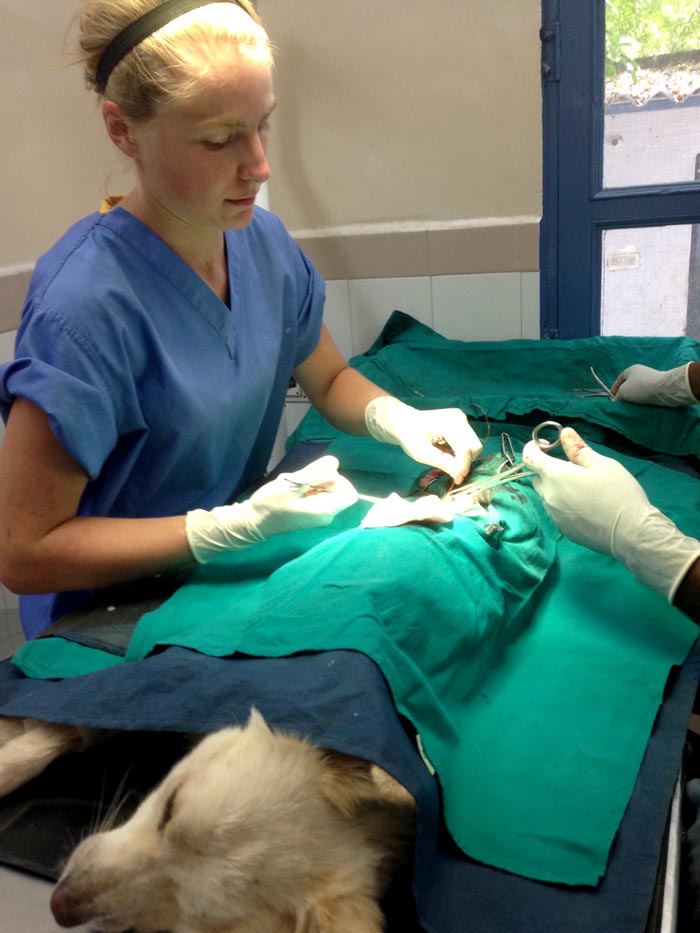
    Do a spay clinic

    spay
    Spay clinics allow you to gain hands-on experience that may be tricky to come by otherwise.

    The quality of the surgery might be different to what you would see in the UK, but these clinics allow you to get hands-on experience, which may be more tricky to come by at home.

    You will practise tissue handling, suturing and ligature placement – all transferable skills.

    Again, listen carefully for recommendations as some clinics are better than others.

    Be adaptable

    There’s more than one way to skin a cat – likewise, you’ll see many different approaches to the same technique, which could be something as simple as giving an IV injection.

    I was “told off” in the university hospital for giving a horse an IV the way I’d been shown on EMS. The following week – on the first opinion part of our rotation – I was told to do it a different way, again.

    Learn how your supervising vet would like you to do things to stay out of trouble, but in the end you will find your own preference.

    Take tips on board

    The vets you meet on EMS have been in practice a while – they’ve made their mistakes and got the T-shirt, so take stock of any handy tips they might give.

    Recently, one vet expressed surprise I was rectalling cows with my “strong” hand (I’m right handed), but didn’t really expand on why they were taught to use the left.

    Another, older vet, strongly advised me to switch to my weaker arm because “years of having your hand squeezed inside a cow will give you horrendous arthritis”, and you’d rather that happen to your left hand and keep your right hand working. Subsequently, I swapped and soon felt competent with my left hand. I do still think the right hand is better for horses though…

    Most importantly, while it can be difficult to tread the line between being too imposing and too shy, you do need to put yourself out there. Offer to do things to help you know you’re capable of, such as setting up a fluid bag.

    And, most importantly, enjoy yourself!

  • Needle aspirate subcutaneous masses

    Cytology of a mast cell tumor from a Labrador retriever at a magnification of 1,000x. By Joel Mills (Own work) [GFDL (http://www.gnu.org/copyleft/fdl.html), CC-BY-SA-3.0 (http://creativecommons.org/licenses/by-sa/3.0/) or CC-BY-SA-2.5-2.0-1.0 (http://creativecommons.org/licenses/by-sa/2.5-2.0-1.0)], via Wikimedia Commons.
    Cytology of a mast cell tumor from a Labrador retriever at a magnification of 1,000x. Image by Joel Mills [GFDL, CC-BY-SA-3.0 or CC-BY-SA-2.5-2.0-1.0], via Wikimedia Commons.
    Fine needle aspiration (FNA) is a valuable tool in subcutaneous skin masses.

    We have all had those lumps that, on palpation, you are sure are lipomas (being soft, freely mobile and slow growing). Indeed, the vast majority are just such benign problems – however, it is worth aspirating them to be sure.

    Felt like lipoma

    We recently had a case in a nine-year-old Labrador with a soft subcutaneous mass the owner had been aware of for a month. It felt just like a lipoma.

    On checking the lump again two weeks later, this had got noticeably bigger. FNAs of the mass revealed the presence of a mast cell tumour.

    Following excision with good 3cm margins, the Lab went on to make a full recovery.

  • RCVS VN council election manifesto: Wendy Nevins RVN

    RCVS VN council election manifesto: Wendy Nevins RVN

    WENDY NEVINS RVN

    Wendy Nevins RVN.

    Operations manager, The Webinar Vet

    T: 07809 702074

    E: wendy@wendynevins.com

    Wendy began her VN career in 1991 as a trainee at a small animal practice in Surrey. On qualifying, she moved to Yorkshire and started work in mixed practice, and after becoming head nurse, left to relocate to Germany with her forces husband.

    Back in the UK in 2000, Wendy moved to Devon and spent two years at a two-branch, small animal practice as a nurse, VN assessor and eventually, practice manager.

    Moving to the north-west in 2002, she worked as a nurse in a small animal practice before becoming the Veterinary Nursing Approved Centre coordinator at Myerscough College. After four years there, she returned to practice part-time, where she started working for Anthony Chadwick at his Skin Vet practice. During this time, Anthony launched The Webinar Vet, and, once established, sold up. Wendy stayed behind as operations manager and is often known as “Webinar Wendy”.

    In her role, Wendy organises CPD for vets, nurses and SQPs. She also deals with dermatology referral clients and spends at least one day a month working in practice.

    Why is she standing?

    Wendy says she is “extremely proud” to be a veterinary nurse and wants to take an “active part” in ensuring the profession moves forward.

    “I feel I have gained valuable experience throughout my different career roles, which will benefit the role requirements of an elected member and our cause as a whole,” she said. “I have a very good understanding of the varying levels of our profession and I am in a privileged position of having contact with members daily, ranging from students to the most experienced VNs.”

    On education, Wendy says she feels “passionately”, and has a “wide exposure”, thanks to her time at Myerscough and The Webinar Vet.

    “I believe I will also add value… establishing and reviewing schemes for post-qualification and CPD for VNs. This also includes recommending to the council amendments to the rules relating to the registration, conduct and discipline of veterinary nurses, if required. I am widely accessible to nurses due to my current role, so I can offer effective communication between VNs and the council. On future challenges and issues? You tell me. I aim to be an effective voice for the nurse in practice – an effective voice for you.”

    Hustings highlights

    Wendy tackled lack of VN engagement in the RCVS in her passionate video, citing poor voting turnout figures and calling the electorate to action. “I want to help increase engagement, hopefully by… carrying on communicating with nurses via social media and within my role at The Webinar Vet,” she said. “Voting in an election is important.

    Can you get other VNs to vote? Do you know nurses who don’t vote? If everyone who votes gets one more VN voting, we can increase that [2015 turnout] 11 per cent to 20 per cent.

    “It would be great if you voted for me to represent you on VN council, but more importantly though – please, please vote.”

  • Barking up the right tree – with Trusty Paws

    Barking up the right tree – with Trusty Paws

    Originally a charity set up by veterinary students for the homeless hounds of Glasgow in October 2014, Trusty Paws has become incredibly successful and has received a huge amount of public support.

    The Trusty Paws Clinic logo

    This success has allowed the charity to run monthly clinics at the Simon Community Scotland drop-in centre, providing free health checks, vaccinations, microchipping, and flea and worming treatment for the pets of the homeless.

    Essential supplies, such as food, coats and harnesses for the dogs, are also given out at these clinics for those in need.

    Branching out

    The success of the Glasgow clinics has led to a branch of the charity opening in London, with the first clinic taking place in November 2015.

    Run by fourth year RVC students, the clinics take place at the West London Day Centre in Marylebone, which also provides other services for the homeless.

    The expansion of the charity is excellent news for the pets of the homeless, for whom we can continue to provide the veterinary care they deserve.

    In at the deep end

    The Glasgow clinics are organised by the student committee, but health checks are conducted by other fourth year student volunteers. Last week I had the chance to get involved and, under the supervision of a volunteer vet, conducted my first full consultation that didn’t involve actors in a communication skills class.

    My patient, Bruno, wasn’t particularly well. The owner said he was not himself (he certainly looked depressed), had lost a significant amount of weight since his last visit and had a slow heart rate, in addition to some evident skin issues.

    The vet suspected Cushing’s disease, so we referred him to the local Pets’n’Vets branch that undertakes any secondary veterinary care Trusty Paws patients require. They have conducted blood tests, paid for by the charity, and have confirmed the diagnosis.

    Offering support

    While a little daunting to be thrown straight into a full consultation, I thoroughly enjoyed helping out at the clinic and would certainly encourage other students (whether in Glasgow or London) to do so in the future.

    The clients are extremely appreciative and evidently love their pets dearly, so it’s easy to see the benefits of such a charity to everyone involved.

    • If you can’t get directly involved, but wish to offer financial support, donations can be made via PayPal.
    • Alternatively, the charity has two Amazon wishlists (one for Glasgow, one for London), enabling supporters to purchase specific products that each clinic requires.
  • Quick cytology preps for assessing skin microbial flora

    Purple stain
    Does full immersion make your tape strips hard to examine? Image ©iStock.com/BanksPhotos

    One of the pleasures of having completed my dermatology certificate is that I get to peer down a microscope at cytology preps a fair amount.

    Now, if you’re anything like me, you’ll have become frustrated with Diff-Quik preps (particularly if you are using tape strips and all three solutions) as the strip becomes soggy and opaque, making it hard to examine.

    A handy tip I learned from a specialist dermatologist working in the States was to just use the blue/purple stain, pop a drop onto a slide and then stick your strip over the liquid and slide. Leave this a few minutes and examine as usual.

    This is quick and allows easy identification of cocci and yeast microorganisms under the microscope.

    However if it’s cells you’re interested in – in, say, suspected cases of pustular disease – I’d suggest you still go through the full staining process.