Tag: Respiratory

  • Blood gas analysis, pt 6: compensatory response

    Blood gas analysis, pt 6: compensatory response

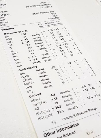
    Simple acid-base disorders are compensated by predictable compensatory changes. The primary disorder shifts the pH, while the compensatory mechanisms aim to normalise the pH and bring it back to neutral.

    This is achieved by attempting to normalise the bicarbonate (HCO3-) to partial pressure of CO2 (PCO2) ratio in a paralleled manner.

    For example, an increase in HCO3– (metabolic alkalosis) is compensated by an increase in PCO2 (respiratory acidosis). Similarly, a respiratory alkalosis (decrease in PCO2) is compensated by a metabolic acidosis (decrease in HCO3-).

    Ruling out secondary process

    However, before jumping to the conclusion an opposing change is the result of compensation, we must rule out the presence of a secondary process. This can only be determined by calculation (Table 1).

    Table 1. Calculating compensatory change
    Component Expected compensation
    Metabolic acidosis
    ↓HCO3 (↓BE)
    per 1mEq/L ↓ in HCO3 = ↓ PCO2 of 0.7mmHg
    Metabolic alkalosis
    ↑HCO3 (↑BE)
    per 1mEq/L ↑ in HCO3 = ↑ PCO2 of 0.7mmHg
    Respiratory acidosis (acute)
    ↑PCO2
    per 1mmHg ↑ PCO2 = ↑ 0.15mEq/L HCO3
    Respiratory acidosis (chronic)
    ↑PCO2
    per 1mmHg ↑ PCO2 = ↑ 0.35mEq/L HCO3
    Respiratory alkalosis (acute)
    ↓PCO2
    per 1mmHg ↓ PCO2 = ↓ 0.25mEq/L HCO3
    Respiratory alkalosis (chronic)
    ↓PCO2
    per 1mmHg ↓ PCO2 = ↓ 0.55mEq/L HCO3

    By comparing the reported to what the calculated compensatory change should be, you can determine whether the patient’s reported value is due to compensation or a separate disorder – for example, multiple primary acid-base disorders (a mixed acid-base disorder).

    An example of a mixed disturbance could be a hyperventilating (respiratory alkalosis) dog with renal failure (metabolic acidosis).

    The level of decrease in PCO2 change is in excess of the calculated compensation for the metabolic acidosis, therefore confirming a mixed acid-base disturbance. In fact, the most common causes of hyperventilation – pain, fear and excitement – often complicate blood gas analysis.

    Another example of a mixed disorder could be a patient with traumatic haemothorax experiencing both lactic acidosis (hypoperfusion) and hypoventilation (respiratory acidosis) due to pleural space disease.

    Waiting game

    Another thing to keep in mind is compensation takes time – respiratory processes take approximately 8 to 12 hours, while metabolic processes take one to three days.

    The lungs are able to alter PCO2 levels relatively quickly by adjusting the rate of ventilation. The kidneys, on the other hand, take a longer time to adjust the pH, as the change in rate of absorption and excretion of HCO3– takes much longer in comparison.

    Regardless of the rate, physiologic compensation for a primary acid-base disturbance is almost never able to return pH to neutral.

    Summary

    A simple acid-base disorder should be suspected when the patient’s reported values are similar to the calculated compensation value, and a mixed acid-base disorder when the values fall outside the calculated range.

    Another hint that a mixed acid-base disturbance is present is if the pH falls within the normal reference range, but the HCO3– or the PCO2 are not; or if the HCO3– and PCO2 are in opposite directions as opposed to being parallel.

    Remember, the body can never overcompensate nor return the pH to neutral.

  • Blood gas analysis, pt 5: metabolic acidosis and alkalosis

    Blood gas analysis, pt 5: metabolic acidosis and alkalosis

    Base excess (BE) and bicarbonate (HCO3-) represent the metabolic components of the acid base equation. In general, both components will change in the same direction.

    Decreased HCO3– and BE indicate either a primary metabolic acidosis or a metabolic compensation for a chronic respiratory alkalosis. Elevated HCO3– and BE indicate either a primary metabolic alkalosis or a metabolic compensation for a chronic respiratory acidosis.

    The exception to this rule arises when a patient hypoventilates or hyperventilates.

    Carbonic acid equation

    CO2 + H2O ↔ H2CO3 ↔ HCO3– + H+

    When a patient hypoventilates, CO2 will increase as a result of reduced expiration, so a shift to the right of the equilibrium will occur. The shift to the right will increase the bicarbonate levels proportional to the increase in CO2.

    The opposite occurs when a patient hyperventilates; the equilibrium shifts to the left, so a decrease in HCO3– is present.

    Since HCO3– is not independent to the patient’s respiratory status, it is an inaccurate way of measuring the metabolic component in patients with respiratory changes. For this reason, the BE value is the preferred.

    The BE represents the amount of acid, or base, needed to titrate 1L of the blood sample until the pH reaches exactly 7.4, with the assumption the blood sample is equilibrated to a partial pressure of CO2 of 40mmHg (the middle of the reference range) and the patient’s body temperature is normal.

    Possible causes

    The possible causes of the primary disease are:

    Metabolic acidosis

    • lactic acidosis – shock and poor perfusion
    • renal failure – reduced hydrogen ion (H+) excretion and increased loss of HCO3
    • diabetic ketoacidosis – ketone acids
    • gastrointestinal (GI) losses – loss of HCO3– through vomiting and diarrhoea

    Metabolic alkalosis

    • GI outflow obstructions – loss of H+ and chloride via vomiting
    • reduced chloride levels and resultant poor perfusion – body attempts to reabsorb water and sodium to increase intravascular volume, but inadvertently also reabsorbs HCO3– in the process, despite existing alkalosis
    • refeeding syndrome
    • severe hypokalaemia:
      • transcellular shift – potassium ions leave and H+ ions enter the cell
      • transcellular shift in cells of proximal tubules → intracellular acidosis → promotes ammonium production and excretion
      • H+ excretion in the proximal and distal tubules increases → further reabsorption of HCO3– and net acid excretion
    • renal insufficiency
    • diuretic therapy (contraction alkalosis – loss of bodily fluids that do not contain HCO3-; this causes the extracellular volume to contract around a fixed quantity of HCO3-, resulting in a rise in the concentration of HCO3– without an actual increase in HCO3– levels)

    Next step

    After ruling out the differential causes of either respiratory or metabolic acidosis/alkalosis, the next step is to determine whether a compensatory response is present and, if so, if this is adequate or whether a true mixed acid-base disorder exists.

  • Blood gas analysis, pt 4: respiratory acidosis and alkalosis

    Blood gas analysis, pt 4: respiratory acidosis and alkalosis

    Assessing the respiratory component is simple. A quick glance at the partial pressure of carbon dioxide (PCO2) level can tell you whether a respiratory acidosis or alkalosis is present.

    If the PCO2 level is elevated (respiratory acidosis) then either a primary respiratory acidosis is present, or it is the result of a compensatory response to a metabolic alkalosis.

    Similarly, if the PCO2 level is low (respiratory alkalosis) then it could either be a primary respiratory alkalosis, or compensation to metabolic acidosis has occurred.

    The respiratory component should always be assessed before the metabolic component, due to the ability to respond to pH shifts almost immediately. This, therefore, is a more accurate reflection of the patient’s clinical disease.

    Respiratory acidosis – increased CO2

    Respiratory acidosis occurs anytime the patient is hypoventilating and not eliminating CO2 appropriately.

    As hypoventilation can be associated with hypoxia, these patients are often critical and require immediate interventions.

    Causes of respiratory acidosis include:

    • drugs (depress respiratory centre, relax thoracic muscles)
    • neuromuscular disease (for example, tick paralysis, botulism and snake envenomation)
    • upper airway obstruction
    • pleural disease (for example, pneumothorax, pleural effusion and diaphragmatic hernia)
    • gas exchange disorders (for example, pulmonary thromboembolism, pneumonia and pulmonary oedema)

    Respiratory alkalosis – loss of CO2

    Respiratory alkalosis occurs when a patient is hyperventilating – excessive loss of CO2 causes the pH to increase.

    The health effect of this is usually minimal, since, in most cases, the effect is secondary and correction of the underlying cause usually resolves this problem. The exception is when respiratory alkalosis is a primary disorder. This is usually quite rare, but can occur with brain stem trauma where the respiratory centre is affected.

    Causes of respiratory alkalosis are:

    • hyperventilation (for example, fear, pain, stress, anxiety and hyperthermia)
    • neurological (for example, head trauma/neoplasia involving the respiratory centre)

    Anticipating changes

    Correctly identifying the primary disorder is essential for anticipating the changes the patient is likely to experience. This will help identify the underlying disease, and is essential for patient monitoring and disease management.

    In the next blog, we will discuss assessment of the metabolic component.

  • Blood gas analysis, pt 3: interpreting pH

    Blood gas analysis, pt 3: interpreting pH

    After taking note of the direction of the pH shift – acidaemia or alkalaemia – it is important to determine the primary and secondary causes.

    If an acidaemia is present (pH less than 7.35), an underlying respiratory or metabolic acidosis, or both, must exist. Similarly, if an alkalaemia is present (pH more than 7.45), an underlying respiratory or metabolic alkalosis, or both, must be present.

    This is usually very simple, with the exception of cases presenting with a normal pH (between 7.35 and 7.45, slightly higher for cats).

    Cases with normal pH

    uparrowFor cases with a normal pH, we need to determine which category it falls into:

    1. No acid-base disturbance
      • Both respiratory and metabolic components are within the normal reference range.
    2. Complete compensation for the acid-base disturbance
      • This requires specific calculations that will be discussed in a later blog.
      • This cannot be determined by glancing at the figures alone.
    3. Two opposing acid-base disturbances (a mixed disorder), which are cancelling the effect of each other out in terms of pH.
      • Both the respiratory and metabolic components will be outside of their reference range, going in the opposite direction to each other.

    Determining primary disorder

    Since these animals are within the normal pH range – particularly those with complete compensation – how can you tell which is the primary disease process?

    A golden rule of thumb is: even with maximal compensation, the pH will still usually move in the same direction as the primary problem.

    Therefore, if the pH lies towards the acidaemic side of the mid-point of the pH range (less than 7.4), the primary disease process is an acidosis. By the same token, if the pH lies towards the alkalaemic side of the midpoint of the pH range (more than 7.4), it will have a primary alkalosis disorder.

    The reason behind this is the body does not usually overcompensate for an acid-base disturbance.

    Secondary disorder

    downarrowOnce the primary disorder has been identified, we need to look at whether a secondary disorder is also present and, if so, whether this is the result of compensation or a true mixed process.

    To determine whether compensation occurred, you need to understand the timeline for when compensation usually occurs.

    With respiratory compensation, this typically starts immediately, but may take up to 8 to 12 hours to occur. This is because adjusting levels of CO2 is relatively easy, with the change of respiratory rate and patterns.

    On the other hand, metabolic compensations take approximately one to three days to occur, since renal excretion of hydrogen ions or retention of bicarbonate takes longer.

    Variation magnitude

    If the magnitude of the observed variation is compatible with compensation alone (this requires calculation), a compensatory mechanism is likely. A mixed process (mixed acid-base disorder) is present if the magnitude:

    • does not correspond to the clinical status of the patient
    • falls outside of the compensation time frame
    • is outside of the expected magnitude of compensation

    All other causes for why the acid base is moving in the opposite direction must be ruled out before determining a secondary process is present.

    Once the primary and, if present, secondary disorder are determined, the next step is to determine the cause of the respiratory and metabolic acidosis and alkalosis.

  • Blood gas analysis, pt 2: acid-base disturbances

    Blood gas analysis, pt 2: acid-base disturbances

    Acid-base disturbances are common in critical patients. These changes must be identified, as even minor deviations from the normal range can lead to significant abnormal body functions.

    Acidaemia and alkalaemia

    Acidaemia, which occurs when blood pH falls below 7.35, will lead to:

    • impedance of cardiac output
    • reduced cardiac contractility
    • a blunted response to catecholamine manifesting as hypotension
    • antagonism to insulin
    • a compensatory hyperkalaemia (extracellular movement of potassium in exchange for hydrogen ions [H+])

    Alkalaemia – blood pH above 7.45 – although less critical compared to acidosis, will result in:

    • muscle spasm
    • stuporous mentation
    • hypocalcaemia
    • hypokalaemia (intracellular movement of potassium in exchange for H+)

    As well as the aforementioned altered functions, H+ is essential for the normal function of enzymes and maintenance of normal cell structures. This is why the body maintains a very narrow pH range and uses multiple buffering mechanisms to achieve this.

    Buffering systems

    The two main buffering systems are the kidneys and lungs.

    Kidneys adjust the pH via the excretion of H+ and the uptake of bicarbonate (HCO3-), which is the primary extracellular buffer and has a linear relationship with pH.

    An increase in HCO3– concentration will result in a pH increase and vice versa. This mechanism can take hours or days from the time a shift in the pH is detected.

    The main respiratory buffer is CO2 – an acid. CO2 has an inverse relationship with pH, so an increase is equivalent to a lower pH level and vice versa. The effect of respiratory adjustments is immediate. This occurs by altering the respiratory rate to adjust CO2 levels.

    The first step towards interpretation of acid-base disturbances is identifying whether an alkalaemia or acidaemia is present. The next blog will discuss determining what is causing it – identifying the primary disorder and the compensatory mechanism employed to balance it out.

  • Blood gas analysis, pt 1: why everyone needs to know about it

    Blood gas analysis, pt 1: why everyone needs to know about it

    For those of you who have received referral histories from emergency or specialists hospitals, blood gas analysis is probably no stranger to you. For those who have never heard of them before, fear not – you are in for a treat.

    In my emergency hospital, the blood gas analyser is arguably one of the most frequently used bench top lab machines, second only to centrifuge, and for good reasons…

    Acid-base disturbances are common in critically ill and emergency patients, and it can help determine the severity of their condition and sometimes provide the answer. Tracking changes in blood gas parameters can provide information about the patient’s response to your interventions.

    blood-gas-analyser_output
    Blood gas analysis can help assess the severity of a patient’s condition and help guide your diagnostic plan.

    The information gained from pulse oximetry is very limited in patients with severe respiratory compromise, and the only way to accurately assess their oxygenation and/or ventilation status is by looking at their blood gas status.

    So what does the blood gas analysis actually measure?

    Most blood gas panels assess the pH of the blood, partial pressure of oxygen (PO2) and partial pressure of carbon dioxide (PCO2). From these, the machine is able to derive the percentage of haemoglobin saturated with oxygen (SO2), bicarbonate (HCO3) concentration and base excess of the extracellular fluid (BEecf).

    In most machines, they are also able to measure other parameters, such as electrolytes (Na, K, Ca, Cl), glucose and lactate.

    While arterial blood gas samples are required for determining the ability of the body to oxygenate the haemoglobin, venous samples are suitable for determining the ventilation status, assessing acid base balance, electrolytes, glucose and lactate levels.

    So how can this help as a point-of-care test?

    As mentioned previously, blood gas analysis can help assess the severity of a patient’s condition and help guide your diagnostic plan. It can also provide a diagnosis (such as diabetic ketoacidosis, typical hypoadrenocorticism and high gastrointestinal obstructions).

    The changes in these parameters over time can be essential in managing critical patients in the emergency setting; it will help guide you in developing an appropriate IV fluid therapy regime and fluid choice, address the patient’s oxygenation and/or ventilation needs, correct any electrolyte and glucose abnormalities, and – although fallen out of favour – the administration of sodium-bicarbonate therapy.

    In upcoming blogs, I will teach you how to interpret the blood gas results. At the end of this, I hope everyone will incorporate blood gas analysis as their standard point-of-care test for the better assessment and management of patients.

    If given the choice between a biochemistry and a blood gas panel in a critical patient, I would hands down select blood gas every time.

  • Focus on GDV, part 4: the recovery

    Focus on GDV, part 4: the recovery

    Postoperatively, gastric dilatation-volvulus (GDV) patients remain in our intensive care unit for at least two to three days.

    Monitoring includes standard general physical examination parameters, invasive arterial blood pressures, ECG, urine output via urinary catheter and pain scoring.

    I repeat PCV/total protein, lactate, blood gas and activated clotting times (ACT) immediately postoperatively and then every 8-12 hours, depending on abnormalities and patient progress.

    GDV recovery
    Patient recovering in the pet intensive care unit. As well as standard monitoring parameters, GDV patients have constant ECG, arterial blood pressure and urine output monitoring to enable the early detection and correction of abnormalities.

    I always repeat these blood tests postoperatively, as IV fluids given during the resuscitation and intraoperative period often cause derangements. I use the results to guide my fluid therapy, but also take it with a grain of salt.

    IV fluids

    I generally continue a balanced and buffered crystalloid. The rate depends on blood pressures, urine output and assessment of general physical examination parameters for perfusion and hydration, but I try to avoid fluid overload and reduce the IV fluids postoperatively as soon as possible.

    Coagulopathy

    Prolonged clotting times are frequently seen as a result of consumption in a dog with GDV. However, one should note it can also occur as the result of haemodilution.

    As the underlying disease process has been corrected, and haemostasis achieved during surgery, I usually monitor ACTs, but may not necessarily treat with blood products as prolonged ACTs do not always translate to clinical bleeding. Unless clinical evidence of bleeding exists, I generally hold off treatment and monitor.

    Hypoproteinaemia

    Low total protein is also common. This is generally due to haemodilution from fluid resuscitation. However, a low total protein does not mean oedema will develop, or that it requires management. I generally track the protein levels, use conservative fluid therapy and try to correct it by instituting enteral nutrition as soon as possible.

    Electrolyte imbalances

    Hypokalaemia is a common complication of fluid therapy. This can be rectified with potassium supplementation in the IV fluids.

    Hyperlactataemia

    If present post-surgery, this is usually corrected with a fluid bolus. However, I always assess for other things that may affect oxygen delivery to the tissues, such as poor cardiac output (arrthymias), hypoxaemia (respiratory disease) and anaemia (from surgical blood loss).

    Arrhythmias

    Ventricular arrhythmias are common post-surgery. Accelerated idioventricular rhythms are the most common cause, especially if a splenectomy was performed.

    arrhythmia
    Ventricular premature contractions are common postoperative arrhythmia.

    Before reaching for anti-arrythmia medications, first check and correct:

    • electrolyte abnormalities
    • hypoxaemia
    • pain control
    • hypovolaemia or hypotension

    If they are still present, despite correction of the above, consider treating the rhythm if:

    • multifocal beats (ventricular premature contractions of various sizes)
    • overall rate greater than 190 beats per minute
    • R-on-T phenomenon
    • low blood pressure during a run of ventricular premature contractions

    I start with a bolus 2mg/kg lidocaine IV and start a constant-rate infusion of 50ug/kg/min to 75ug/kg/min.

    Anaemia

    It is common to have a mild anaemia post-surgery, due to a combination of blood loss and haemodilution. In the absence of transfusion triggers – such as increased heart rate, increased respiratory rate or hyperlactataemia – it does not require treatment.

    Vomiting

    Anti-emetics are the first line of medication. Non-prokinetic anti-emetics, such as maropitant and ondansetron, can be used immediately; otherwise, after 12 hours, metoclopramide can also be used postoperatively. If the patient remains nauseous despite these medications, the placement of a nasogastric tube can ease nausea by removing static gastric fluid.

    Excessive pain relief may also contribute to the nauseous state.

    Pain relief

    I mostly rely on potent-pure opioid agonists, such as fentanyl constant-rate infusions and patches. This is generally sufficient for most patients. Ketamine is occasionally used.

    • Some drugs listed in this article are used under the cascade.
  • Focus on GDV, part 2: releasing the pressure

    Focus on GDV, part 2: releasing the pressure

    Last week we covered IV fluid resuscitation and pain relief. This week we will go into more detail about gastric decompression.

    stomach tube
    Passing the stomach tube inside the roll down into the oesophagus (click to zoom).

    Gastric decompression can be achieved in two ways:

    1. trocarisation
    2. stomach tube (orogastric tube) placement

    The decision on which method to use depends on many factors – personal preferences, past experiences and clinical protocols, to name a few.

    So, which one is best? A retrospective analysis of 116 gastric dilatation-volvulus (GDV) patients (Goodrich et al, 2013) found both methods of gastric decompression had low complication and high success rates, and either technique is acceptable.

    If one method fails to achieve gastric decompression, the other can be tried.

    How to decide

    Personally, I use either or sometimes both. Which one I choose first depends on the situation. My decision-making process goes something like this:

    Not clinically obvious or mild GDV

    These are often diagnosed based on supportive radiographic findings as history and presenting clinical signs making me suspicious of a GDV.

    I would always try to pass a stomach tube in these patients first, as the tube is passes easier when the gastric distention is milder. Although this procedure generally requires prior opioid analgesia administration to help reduce the stress, it can achieve rapid and lasting decompression of the stomach.

    I often leave the tube in throughout stabilisation, just prior to induction of anaesthesia for surgical correction of the torsion. The tube allows continual release of gastric gases that can accumulate again rapidly if the tube is removed prior to surgery.

    Obvious or severe GDV

    The abdomen in these animals is often distended and tympanic. I will perform trocarisation in these cases first, as passing a stomach tube in these patients is often unsuccessful. It allows rapid gastric decompression, which is particularly important in cases with evidence of respiratory compromise.

    After the trocar is no longer releasing gas, I will then pass a stomach tube. At this stage, it is often easier to pass the stomach tube once the gastric pressure has been reduced. Once again, I often leave the tube in during stabilisation.

    How to perform

    Stomach tube

    • The main risk is rupture of the oesophagus or gastric wall.
    • Pre-measure and mark the tube from the mouth to the level of the last rib.
    • Use a roll of adhesive bandaging material as the mouth gag. I prefer to use Elastoplast as it has an incompressible plastic core and the diameter is just large enough to fit our largest diameter stomach tube.
    • Unwrap approximately 30cm of Elastoplast before placing the roll of tape inside the mouth.
    • Wrap the tape snugly around the muzzle to prevent the dog from opening its mouth and dislodging the roll.
    • Lubricate the tube to reduce frictional trauma to the oesophagus.
    • Pass the stomach tube through the core of roll and into the mouth. You will feel a dead end at the level of the lower oesophageal sphincter, where the volvulus has torsed the oesophagus.
    • Apply gentle constant pressure and, most times, the tube will pass through into the stomach. Sometimes a puff of gas can be heard and felt from the aboral end of the tube when it enters the stomach. The tube can also be palpated when the stomach is decompressed.
    • The tube is taped to the muzzle to prevent dislodgement and the aboral end placed in a bucket to allow fluid to exit via gravity and siphon.
    • If it does not pass, reassess to see if trocarisation is required to relieve some pressure in the stomach

    As mentioned above, I generally leave the stomach tube in while continuing to stabilise the patient and prepare for surgery. Gas can rapidly accumulate in the stomach and cause rapid deterioration if the tube is not left in. The tube is removed just prior to induction of anaesthesia.

    tape
    Placing a roll in the mouth to prevent biting down on the stomach tube.

    Trocarisation

    • The main risk is hitting the spleen while trying trocarisation. To avoid this, identify the most tympanic site by palpation, or use the ultrasound to confirm the absence of the spleen.
    • A 3in, 14g catheter is usually sufficient.
    • Clip and surgically prep a 10cm by 10cm area where you intend to place the catheter.
    • Insert the catheter to the hub and remove the stylet.
    • Although local anaesthetic in the area is ideal, you will not have time to do this in most cases – especially the very unstable ones. Also, since I administer pure opioid agonist intravenously to most confirmed GDV cases on presentation, local anaesthetic is not required.
    • Remove the stylet and gas should come blowing out under pressure.
    • Once the gas flow starts to slow down, gently apply inward pressure or pressure on the dilated stomach, which helps ensure the stylet does not fall out of the stomach and as much of the gas is removed as possible.

    >>> Read Focus on GDV, part 3: surgery tips

  • Emesis: a thing of the past?

    Emesis: a thing of the past?

    Until I started researching this Tip of The Week, I did not know the medical profession has abandoned the routine use of emesis in oral poisoning.

    This is based on multiple medical literatures that have proven emesis induction does not influence the clinical severity of poisoning, the length of hospitalisation and the clinical outcome or mortality.

    Although the rationale for inducing emesis is obvious, it is not necessarily evidence based. It is also dependent on satisfying a few large assumptions, all of which are untrue:

    • Emesis is a very effective way of removing gastric contents.
    • No separation exists of poison from its vehicle while inside the acidic environment of the stomach.
    • Poison is not absorbed through the stomach wall.

    Ineffective method

    snail bait
    Snail bait ingestions: this patient ate 500g of snail bait containing metaldehyde.

    Emesis induction is an ineffective way of clearing stomach contents. A review of the effectiveness of induced emesis, with both human and canine participants, showed at 30 minutes post-ingestion of non-absorbable markers, the recovery rate averaged between 17.5% and 52.1%, but never exceeded 62%.

    In fasted puppies, this was even lower at 2% to 31%, despite inducing emesis immediately after marker administration. These have been confirmed by the presence of poisonous materials in the stomach of dead patients, despite effective emesis induction until clear fluid was brought up.

    The clinical outcome only improves if the systemic exposure of a toxicant is reduced by more than half. However, considering animals rarely practice restraint, the ingested amount is unlikely to be exactly the lethal dose and no more. Therefore, even reducing the ingested toxic dose by 62% is unlikely to make a clinical difference.

    Furthermore, most patients rarely present within 30 minutes of ingesting a toxicant, thus further reducing its efficiency.

    The absorption conundrum

    Some may argue the retrieval of metaldehyde or anticoagulant rodenticide granules from vomitus is indicative of reducing the toxicant dose. This could be true, but only if emesis was induced immediately after ingesting the poison.

    The poison itself is colourless and has a different absorption characteristic to the coloured vehicle (granule); therefore, the presence of granule only serves to confirm ingestion, but is of no indication whether the poison has already been absorbed.

    Contraindications

    Many well-recognised absolute contraindications also exist to inducing vomiting:

    • Ingestion of oils, which includes waxes that melt to oil in the internal body environment, as this poses a high risk of lipoid and bacterial pneumonia. This is of significant veterinary significance, as wax is routinely used in rodenticide baits.
    • Ingestion of hydrocarbons and other volatile substances, or caustic or corrosive substances.
    • When the mental status is altered – for example, hyperexcitable or depressed mental state.
    • Where the patient is at risk of seizures (seizures can be induced by emesis).
    • Increased intracranial pressure.
    • Risk of intracranial or cerebral haemorrhage – for example, thrombocytopenia or abnormal clotting parameters.

    Other less severe, yet important, reasons include:

    • delays administration of more effective treatment, such as activated charcoal, antidote or other treatments
    • risk of aspiration pneumonia
    • hypochloraemia in recurrent emetic patients
    • significant CNS and respiratory depression from apomorphine
    • rare, but reported, complications such as cerebral haemorrhage, oesophageal tear/ rupture, hiatal hernia, gastric rupture, pneumothorax and pneumomediastinum
    • legal implications – for example, if the product information clearly states emesis should not be induced

    A place for everything

    Emesis induction is not a benign procedure. It still has its place in certain circumstances, but its use in the routine management of oral poisonings may need to be reconsidered – especially if it means delaying administration of a more effective treatment, such as activated charcoal.

    So, after all this, how do I tackle this information? It is a bit hard to swallow. My clinical experience is emesis is generally safe, especially in canine patients using apomorphine. So, I still feel some merit exists in reducing the amount of toxicant in the stomach if you have a chance – and in some situations, you don’t know until you try.

    Emesis after ingestion of a toxic dose of chocolate can be incredibly rewarding, even six hours after ingestion, leading to patients not developing clinical signs at all.

    Overall, I am biased by my personal successes with emesis, so still feel a time and place exist for emesis induction. But I now stop and question my decision to induce emesis, whereas I did not hesitate before.

    • Some drugs listed are used under the cascade.
  • Isoflurane and oxygen: the dangers of 2 and 2

    Isoflurane and oxygen: the dangers of 2 and 2

    It is a common practice to place all patients on 2% isoflurane and 2l/min oxygen flow rate, but blanket isoflurane saturations and oxygen flow rates can be dangerous.

    2 and 2
    Take time to consider your anaesthetic approach.

    Without a doubt, the majority of patients seem to do just fine at these levels; but every patient is different, and simply placing all patients on 2% isoflurane and 2l/min oxygen may be introducing an easily avoidable risk into anaesthesias.

    Isoflurane

    Isoflurane can cause severe effects such as hypotension and respiratory depression, so 2% isoflurane may be too high – especially for patients that are critically ill or have been premedicated with sedatives/anxiolytics.

    In these patients, the isoflurane can be safely titrated down while monitoring the patient’s reflexes and vitals. Consider administering IV pain relief instead of turning up the isoflurane levels if the patients are too “light” and responding to pain. Examples include a low dose pure-opioid.

    It is important to titrate its use like any other anaesthetic agent, maintaining an appropriate level of anaesthesia while minimising potential side effects.

    Oxygen

    It is common to use a standard 2l/min oxygen flow for all anaesthetics regardless of the type circuit, but this will not meet the oxygen requirements for larger patients. It is best practice to work out the appropriate flow rate amount using an oxygen consumption chart.

    I cannot stress enough the importance of taking the time to consider your anaesthetic approach in patients, especially compromised ones such as those with renal/hepatic disease or circulatory deficits.

    Titrating anaesthetic agent levels can increase the stability of your patient under anaesthesia and significantly reduce the life-threatening complications.