Tag: Respiratory

  • Triage, pt 2: secondary survey

    Triage, pt 2: secondary survey

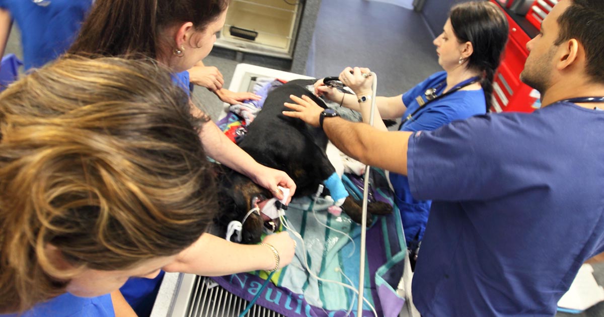
    Secondary survey refers to the detailed physical examination performed after the primary survey, and should only be performed once the patient has been adequately stabilised.

    It is always important to perform physical examinations systematically to avoid overlooking organ systems. This could be difficult in a stressful emergency situation, so one way to remind yourself is with the following acronym:

    A CRASH PLAN

    A – Airway

    • respiratory pattern
    • airway patency

    C – Cardiovascular

    • circulation
    • heart sounds
    • pulses
    • capillary refill time

    R – Respiratory

    • respiratory sounds
    • bruising
    • external wounds to chest

    A – Abdomen

    • palpation
    • bruising
    • external wounds
    • fast ultrasound:
      • abdomen:
        • free fluid (diaphragmatic-hepatic site, splenorenal site, cysto-colic site, hepatorenal site)
        • bladder integrity
      • thorax (do this at the same time as assessing the abdominal cavity):
        • ensure you do both left and right sides
        • chest tube site
        • pericardial site
        • wet/dry/third space

    S – Spine and tail

    • gait and posture
    • pain sensation
    • crepitus

    H – Head

    • mentation
    • cognitive function
    • cranial nerves
    • external wounds/bruising
    • eyes – including symmetry, third eyelids, eye position, haemorrhage and detailed ophthalmological examination
    • ears
    • nose
    CPR
    The secondary survey will help identify any concurrent problems not seen on the primary survey.

    P – Pelvis

    • wounds
    • bruising
    • pain
    • cepitus
    • perineum
    • external genitalia

    L – Limbs

    • deformities
    • fractures
    • pain
    • bruising
    • wounds
    • weight bearing vs non-weight bearing

    A – Arteries

    • all accessible superficial arteries – pulses and pressure

    N – Nerves

    • mentation
    • cranial nerves
    • conscious proprioception
    • postural reflexes
    • peripheral spinal reflexes
    • withdrawal reflexes
    • deep pain
    • cutaneous trunci reflex
    • anal tone

    Stable patient

    By following the primary and secondary triage processes consistently, you should be able to quickly determine the order of criticalness of patients, institute appropriate resuscitative measures and manage life-threatening injuries. Then, with your thorough physical examination, identify any other concurrent problems not seen on the primary survey.

    Overall, you have a stable patient, and are able to formulate an appropriate diagnostic and treatment plan.

  • Triage, pt 1: primary survey

    Triage, pt 1: primary survey

    The art of triage takes time to master – particularly in emergency hospitals, where critical patients arrive in quick succession to the crash area.

    Patients need to be examined quickly and effectively to ensure the most critical issues are identified and stabilised. To do this, I break triage into two categories – primary survey and secondary survey.

    In part one I will discuss the primary survey process. The second part will go into the details of the secondary survey.

    ABCDE approach

    Primary survey refers to the initial stabilisation stage, where the aim is to preserve life, manage life-threatening injuries and re-establish tissue perfusion with oxygenated blood.

    The approach to all deteriorating or critical patients is the same: ABCDEAirway, Breathing, Circulation, Disability and External.

    ABC – Airway, Breathing, Circulation

    Critically ill patients need to be examined quickly and effectively.
    Critically ill patients need to be examined quickly and effectively.

    Assess airway

    • Is it patent?
    • Does it require suctioning?
    • Does evidence exist of upper airway obstruction?
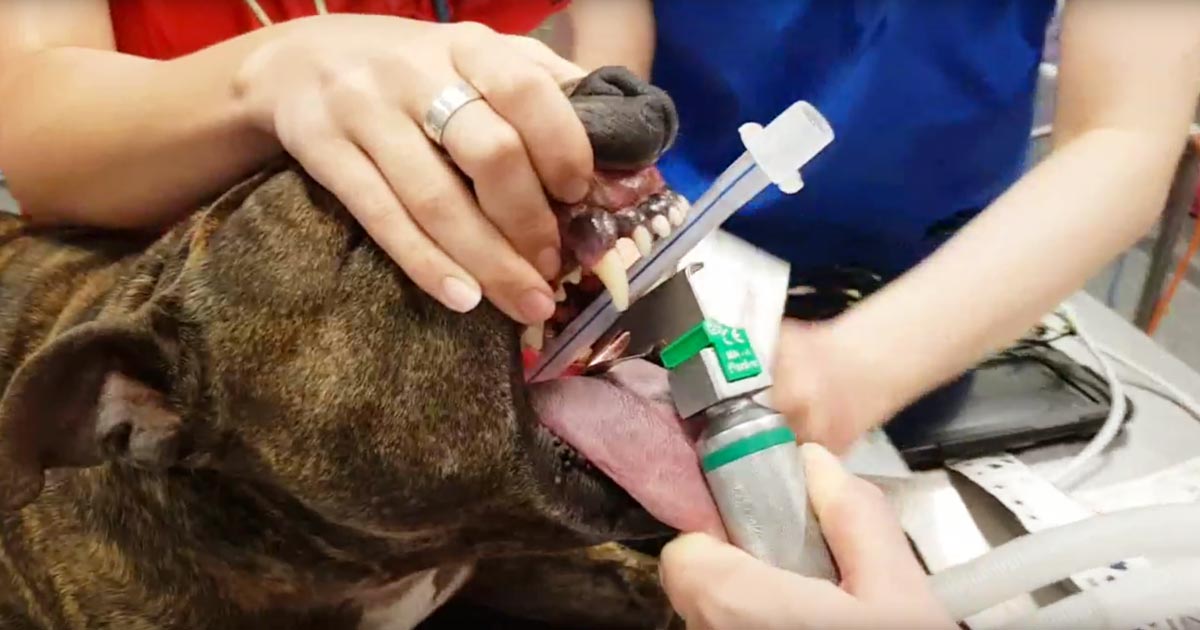
    • Intubate if you suspect the patient may need resuscitation.

    If you suspect cardiopulmonary arrest, begin basic life support immediately. This involves chest compressions, and intubation and ventilation in:

    • loss of consciousness
    • absence of spontaneous ventilation
    • absence of heart sounds on auscultation
    • absence of palpable pulses

    Assess breathing

    • Is the patient hyperventilating or hypoventilating?
    • How is the respiratory effort? Is it sustainable?
    • What is its oxygen saturation, or does the patient looks like it needs oxygen? If yes, provide supplemental oxygen therapy.
    • Does the patient need to be ventilated? Ventilation is required if any of these criteria are satisfied:
      • hypoxaemia (partial pressure of oxygen lower than 60mmHg or blood oxygen saturation level lower than 90%) and unresponsive to oxygen supplementation
      • hypercapnia or hypoventilation (partial pressure of carbon dioxiden greater than 60mmHg)
      • unsustainable respiratory effort?
    • Consider sedation. Reducing stress can make a dramatic difference in stabilising dyspnoeic patients. Butorphanol or low-dose acepromazine (try to rule out cardiac disease first) can be used in these situations.

    Assess cardiovascular system

    • Mucous membrane colour, capillary refill time, heart rate and rhythm, pulse pressure, and temperature.
    • If a heart murmur or arrhythmia is present, I try to rule out cardiogenic shock before resuscitation therapy.

    At the same time, draw enough blood to run baseline blood work and begin IV fluid resuscitation if signs of shock are present. Fluid boluses should be considered if any of these exist:

    • pale mucous membranes
    • slow capillary refill time
    • tachycardia
    • poor pulse pressures
    • hypothermia

    I generally start with 10ml/kg Hartmann’s Solution over five minutes and reassess.

    D – Disability assessment

    Any abnormalities in:

    • Mentation? Seizures?
    • Level of consciousness?
    • Altered pain sensation?

    E – External assessment

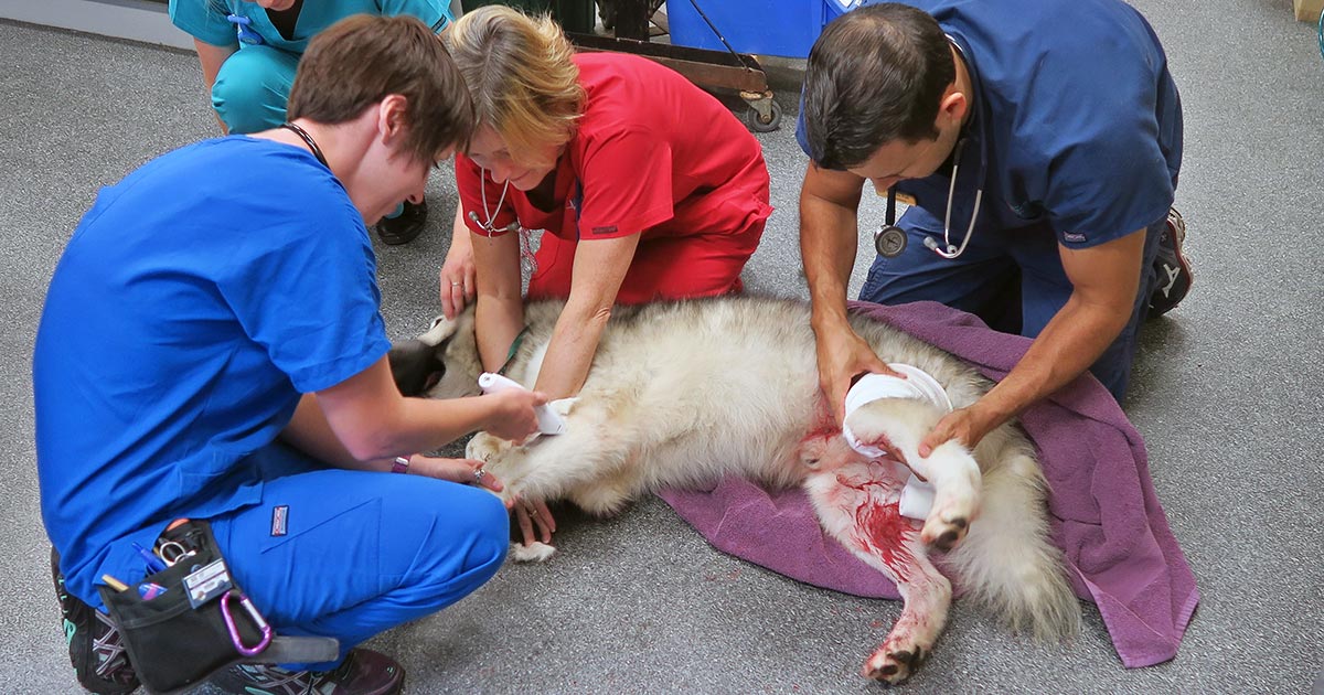
    • assessment for wounds or injuries
    • control any obvious bleeding, apply direct pressure – possibly tourniquets, but only for less than 30 minutes unless life-threatening bleeding
    • initial medications:
      • pain relief – generally opioids are safest with unstable patients
      • antiepileptics – diazepam
      • sedation for dyspnoeic patients – butorphanol or low-dose acepromazine IV

    Once you have finished your primary survey and performed the required intervention, you repeat your primary survey until the patient is appropriately stabilised.

  • Oh, CR*P! Using point-of-care C-reactive protein tests

    Oh, CR*P! Using point-of-care C-reactive protein tests

    Few companies now offer affordable point-of-care tests for canine C-reactive protein (CRP). As we did when we recently received our new box of CRP slides, you might soon be asking the question: what do we even do with this stuff?

    Here’s what we’ve learnt…

    CRP is one of the acute phase proteins produced by the liver in response to inflammation. Healthy patients have very low levels of CRP, but a systemic inflammatory condition will cause an increase in CRP within four to six hours. Conversely, increased levels will decrease rapidly on resolution of inflammation. This provides an almost real time measure of inflammation that is more responsive and reliable than the white blood cell response.

    In other words, CRP can indicate the presence of inflammation before the patient’s white blood cell count gives any clues, or before it becomes pyrexic – and, unlike the white blood cell count, stress and steroids do not affect CRP levels.

    Uses

    So, how do we use it?

    • I love it for early pickups of problems in those grey area cases: the dog seems okay on clinical examination, but something about it bothers me. A normal or mildly increased CRP test will make me sleep more easy, while a surprise high reading will prompt me to admit for full diagnostics, or at least get the patient in for a follow-up CRP the next day. Conversely, a localised problem – such as an abscess – combined with a normal CRP test might mean you can hold off on antibiotics and just recheck CRP in 24 hours.
    • It’s great for monitoring response to treatment. If my plan is working then I’d expect CRP to show a significant decrease by day two or three. If it’s not dipping by then, I need to reassess my treatment plan. Do I need to change antibiotics? Scan it again? Maybe we need to consider surgery? It can also be a good prognosticator. Research has shown failure of CRP to decrease significantly (around a 3× decrease) by around day three is generally bad news for patients with inflammatory conditions such as pancreatitis and immune-mediated haemolytic anaemia.
    • We are starting to play with it for post-surgical monitoring. Any surgery will cause inflammation with an increase in CRP levels, but in an uncomplicated postoperative period, you should expect levels to start decreasing by day three to five. A base line CRP 24 hours after surgery with a recheck on day three should pick up early signs of postoperative problems such as infection, and prompt investigation or intervention.
    • A potentially nifty use for it that we haven’t yet had the opportunity to use is in differentiating inflammatory lamenesses (arthritis, infection, injury) from a neurological causes – that is, is it arthritis or a nerve problem?

    Limitations

    • Remember, it’s very sensitive, so will increase with almost any inflammation. A mild upper respiratory infection or a bad gingivitis will likely induce some changes, so it’s important not to over-interpret (keep in mind that the magnitude of the increase in CRP does generally correspond with the severity of the inflammatory response). A pancreatitis case where the CRP fails to drop does not always mean death is looming – you may have just missed the concurrent skin disease. Always interpret CRP values in concert with your clinical examination.
    • Be aware that pregnancy and intense exercise can increase CRP values.
    • Not all serious conditions have an inflammatory component. CRP will be unchanged in most veterinary cases of heart disease; in common hormonal disease, such as adrenal disease and uncomplicated diabetes; urinary obstructions; many localised cancers; epilepsy and many others. Don’t presume that just because CRP is normal, everything is fine.
    • No similar test exists for cats.

    Sit up and say…

    My favourite way to explain how to use this test is by using its highly appropriate acronym – any unexpected increase should make you sit up and say: “Oh CR*P! What am I missing?”

  • Rat bait’s sneaky trick: bleeding into the dorsal tracheal membrane

    Rat bait’s sneaky trick: bleeding into the dorsal tracheal membrane

    Most of us are familiar with anticoagulant rodenticide toxicosis and the range of clinical signs it can present with, but there is one potentially fatal manifestation of coagulation pathology that is perhaps not as widely known…

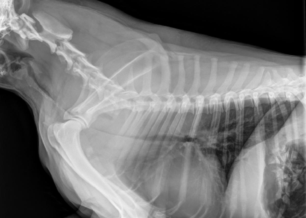
    Dogs with severe clotting problems will occasionally bleed into the dorsal tracheal membrane. This causes collapse of the thoracic trachea and can lead to severe respiratory distress.

    Presenting signs

    These cases can present with none of the other signs of bleeding normally associated with coagulopathies, so rat bait poisoning may not come to mind as a differential diagnosis if you are not aware of this syndrome.

    The typical case will present as an otherwise healthy dog that develops acute respiratory problems. Early signs can be as mild as a persistent cough, but it can quickly escalate into a life-threatening respiratory crisis.

    Severe cases will have an obvious stridor on both inspiration and expiration, cyanotic mucous membranes, and patients may be very distressed.

    It will look very much like:

    • a dog that is choking from a tracheal foreign body
    • an old dog with tracheal collapse
    • the end stages of laryngeal paralysis – except the stridor will come from much lower in the respiratory tract than it does in laryngeal paralysis

    So, what do you do?

    On initial presentation you would approach it as any respiratory distress case: oxygen, oxygen, oxygen, calm and stress-free handling, and light sedation (butorphanol, for example).

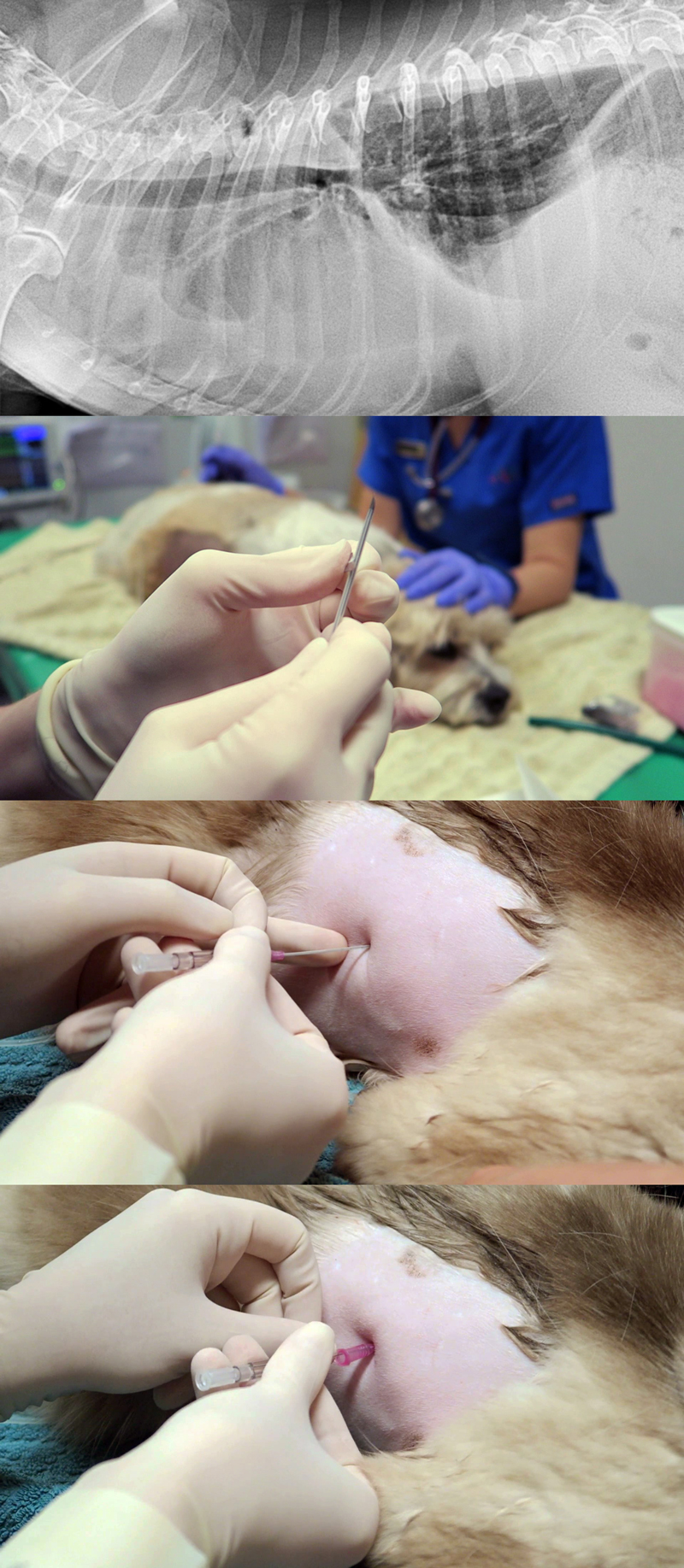
    bleeding_dorsal-tracheal-membrane

    Once it is safe to do so, you should take chest rads to look for what you’ll probably suspect is a tracheal foreign body, and you’ll get an image like the one above (although it may not be this severe). Then you’ll remember this article, have an “aha!” moment and run a clotting profile (but if it’s as bad as this case, you’ll obviously first save the animal’s life by passing an ET tube).

    Once a clotting problem is confirmed you’ll need to stop the bleeding with standard therapy for anticoagulant rodenticide toxicity: plasma and vitamin K.

    Severe cases

    In a severe case you may need to keep the dog intubated for several hours, until the clotting times have normalised, before cautiously attempting to extubate.

    If the patient is unable to stay well oxygenated without an ET tube (mucous membrane colour, pulse oximetry, blood gas), consider placing a long oxygen catheter past the narrowing – either via a tracheostomy or a nasal O2 catheter.

    If these cases are quickly recognised for what they are, and an open airway can be maintained, the prognosis should be good. These are potentially very satisfying cases with great potential for you to be a total hero.

  • Dystocia, pt 4: caesarean tips

    Dystocia, pt 4: caesarean tips

    Prolonged hypoxaemia, hypotension and hypoventilation are the top three causes of periparturient fetal mortality – for these reasons, all precautions must be taken to avoid it.

    As soon as authorisation has been obtained to proceed with a caesarean section, the patient should be stabilised immediately. This includes having perioperative blood work performed, and clinical hypoperfusion (common in patients that have gone through prolonged stage two labour) and hypotension corrected as soon as possible, usually with fluid boluses.

    While fluid deficits are being corrected, preoperative monitoring and surgical site preparation (clipping and the initial stages of surgical scrub) can be performed with the patient still conscious. This will significantly reduce the time the patient is anaesthetised, as isoflurane potentiates hypotension.

    Physiological changes

    A few physiological changes in periparturient patients must be considered before anaesthetising them.

    Higher oxygen demand

    Firstly, pregnant animals have a higher oxygen demand due to the developing fetuses. However, due to their large gravid uteruses, they have decreased functional residual capacity and total lung volume. This is further exacerbated when animals are placed in dorsal recumbency, with increased pressure on their diaphragms.

    For this reason, pregnant animals should always be preoxygenated prior to induction – with as much of the patient preparation completed – to reduce the risk of hypoxaemia. This is one of the main reasons the time from induction to delivery of the puppies should be as short as possible.

    Sensitivity to anaesthetic agents

    Secondly, pregnant animals have an increased sensitivity to anaesthetic agents. Blood volume and cardiac output also increase dramatically during pregnancy; therefore, if blood loss occurs and blood pressure is not maintained, significant hypotension can occur.

    Any medication that crosses the blood-brain barrier will equally cross the placental barrier; therefore, the effect of medications can be reduced by a few things. Firstly, the use of local anaesthetics (such as epidurals) can be employed to minimise inhalation anaesthetics, thus their hypotensive effects. Always use minimal drug dosages that achieve the desired effect. Short-acting, rapidly metabolised drugs and reversible drugs should be used whenever possible.

    Don’t premedicate

    Premedication of caesarean patients is strictly avoided at our hospital. Acepromazine can result in hypotension and has a long duration of action, while opioids can cause potent respiratory depressants in unborn fetuses as it crosses the placenta.

    Puppies and kittens born heavily narcotised or sedated will have bradycardia and may not take spontaneous breaths, further increasing the risk of mortality.

    Speedy delivery

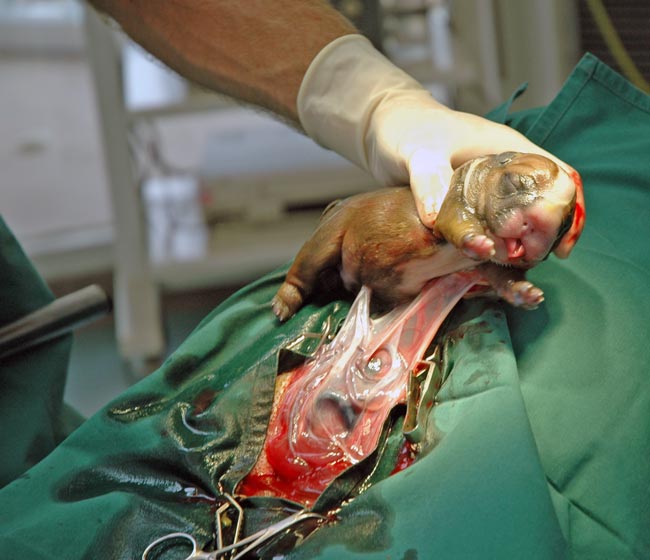
    IMAGE: Pilipipa / Adobe Stock
    Once the patient has been induced, the speed of delivering the fetuses is of paramount importance. Image © Pilipipa / Adobe Stock

    Once the patient has been induced, the speed of delivering the fetuses is of paramount importance. Inhalant anaeshetics causes maternal vasodilation and decreases uterine blood flow, as well as neonatal depression.

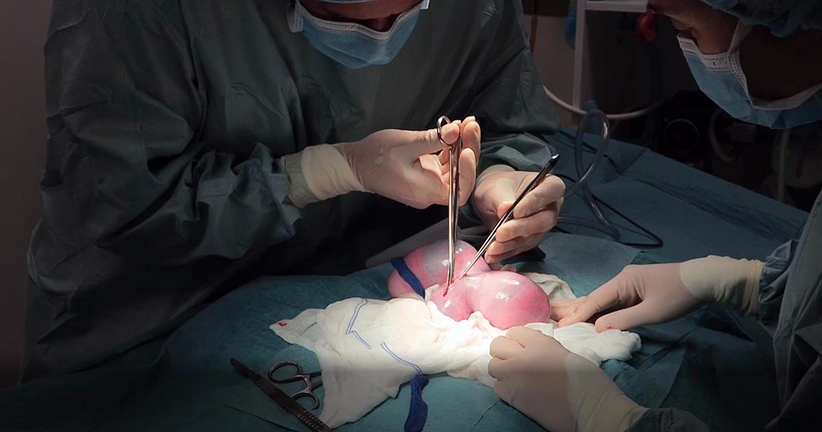
    Making a large abdominal incision is highly advised, despite the fact it may take longer to close, as it enables faster and more gentle manipulation of a large fetus-filled uterus.

    The traditional caesarean technique involves a single incision in the uterine body. Fetuses should be gently squeezed towards the incision. In patients with many fetuses, especially large-breed dogs, making a single uterine body incision may significantly delay delivery of the fetuses. Concern also exists with excessive traction and manipulation of uterine blood vessels when trying to manipulate the fetuses to the uterine body incision. In these cases, additional incisions in the uterine horns can be made.

    With this method, surgical time for closure will be longer and considered carefully in patients where future breeding is likely, as the risks of adhesions and uterine rupture in subsequent pregnancies increases compared to the single uterine body incision method.

    Closure of the uterine wall should always be in two layers – firstly, an appositional simple continuous pattern; followed by a second inverting (Cushing or Lembert) pattern.

    Post-fetal removal

    Once the fetuses have been removed, a few medications can be given safely intraoperatively.

    Firstly, opioids are safe at this time. Fast analgesia can be achieved when the opioid is given IV. Oxytocin can also be administered during this time, but beware uterine involution and contraction will be immediate; therefore, close attention needs to be paid to the uterine sutures to ensure they have not become loose.

    NSAIDs should be avoided in lactating queens and bitches, as most are excreted in the milk. Safety data has not been established in lactating animals, while previous animal studies have shown an adverse effect on the fetus.

    Tramadol, a synthetic opiate-like (μ receptor) agonist, has high analgesic effects. Tramadol and its active metabolite are known to enter maternal milk, albeit at very low levels. No animal reproduction studies exist to establish its safety in use in neonates, but it is an analgesic considered safe to use in young animals.

    Conclusion

    Caesarean section is the one emergency surgical procedure where speed is of essence.

    With prompt stablisation, pre-induction surgical preparation, fast delivery of fetuses and avoidance of certain medications, the chances of survival of the already distressed fetuses can dramatically increase.

  • Systemic antibiotics – a brief guide for new grads

    Systemic antibiotics – a brief guide for new grads

    A lot of information is available regarding different antibiotics and, for the newest generation of vets, the pressure to use them correctly and responsibly is greater than ever.

    One of main challenges when you start clinical practice is knowing the most appropriate antibiotic for common presenting conditions.

    Below is a rough guide for antibiotic selection according to body system. However, make sure you stick to the following rules:

    1. Limit antibiotic use to animals that actually require them – resist the urge to dispense them due to pressure from owners or when you feel there is nothing else to turn to.
    2. What is the likely type of bacteria you are aiming to target (such as anaerobes, Gram-positives and Gram-negatives)? Collect samples from lesions/discharge or effusions/blood and urine, and see if there is evidence of bacteria under the microscope.
    3. Use the most narrow spectrum antibiotic as possible.
    4. Perform a culture and sensitivity whenever possible – especially if a case does not respond to your first line antibiotic.
    5. Avoid using fluoroquinolones, third and fourth generation cephalosporins and amikacin without evidence of resistance from culture and sensitivity results.
    6. Use an appropriate dosage regime and make sure the owners have the capacity to administer them accordingly.

    Skin

    • Try topical chlorhexidine alone if surface pyoderma
    • Clindamycin
    • Cephalexin
    • Amoxicillin-clavulanic acid

    Upper respiratory tract

    • Doxycycline
    • Amoxicillin-clavulanic acid

    Lower respiratory tract

    • Amoxicillin-clavulanic acid
    • Ampicillin

    GI tract

    • Metronidazole (research questions the use of antibiotics for diarrhoea cases)
    • Tylosin (chronic diarrhoea)

    Urogenital tract

    • Remember that cystitis in cats is often stress-related rather than due to infection
    • Amoxicillin-clavulanic acid
    • Trimethoprim-sulpha (penetrates the prostate)

    Mastitis

    • Amoxicillin-clavulanic acid


  • Thoracentesis, part 1: indications, equipment and protocol

    Thoracentesis, part 1: indications, equipment and protocol

    Thoracentesis is a relatively straightforward and life-saving technique for seriously dyspnoeic animals with pleural space disease, and is a valuable diagnostic tool.

    Here are my tips for getting the most out of your approach to performing a thoracentesis.

    Indications

    • Therapeutic – relieve respiratory distress caused by pleural effusions and pneumothorax.
    • Diagnostics – cytological examination of pleural effusions will refine your differentials list and dictate subsequent management.

    Equipment required

    In addition to general equipment for clipping and prepping of the surgical site, the following tools are required to perform thoracentesis:

    • oxygen and mask
    • 20ml to 60ml syringe
    • 16G to 21G butterfly needle
    • three-way tap
    • extension set
    • ethylenediaminetetraacetic acid tubes (for cell counts)
    • sterile collection tubes (for culture and cytology)
    • fluid collection bowl (non-sterile collection)
    • +/- lidocaine 1mg/kg to 2mg/kg for centesis site

    Protocol

    1. Patient comfort

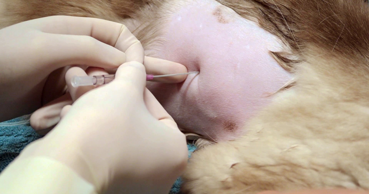
    Thoracocentesiscombined
    An approach to performing a thoracentesis.

    a. Options include local anaesthetic infiltration of the intended centesis site, and/or IM or IV opioid pain relief at standard doses.

    b. Opioid pain relief, such as butorphanol, is great for sedation that facilitates the process.

    c. Depending on the case, I often use opioid pain relief without local. This is sufficient in the vast majority of cases.

    d. If severely dyspnoeic, anaesthesia and intubation can help facilitate the process. It will reduce patient stress, enable manual ventilation and administration of 100% oxygen, and allow for larger volumes of air/fluid to be removed.

    2. Patient positioning

    Generally, sternal is easiest – otherwise, lateral recumbency or standing (if the animal will tolerate it).

    3. Site

    a. Locate the seventh to ninth intercostal spaces.

    b. To remove air, clip the dorsal two-thirds of the chest.

    c. To remove fluid, clip the ventral two-thirds of the chest.

    d. Clip a larger area than you expect.

    e. Prepare the area for an aseptic procedure.

    4. Connect everything

    a. The syringe to the three-tap and extension set should be ready prior to connecting the butterfly catheter.

    b. Often, a rush occurs to connect everything after the catheter is in place.

    5. Needle insertion

    a. Insert the needle on the cranial edge of the rib to avoid the nerves and blood vessels that run along the back of the rib.

    b. Ultrasound guided is best for fluid; you can lube the inside of a sterile glove and put the probe inside the glove to keep the area sterile.

    c. An IV catheter can also be used. I partially fenestrate a 20g IV catheter with two extra holes – once the catheter is advanced into the chest minimal risk exists of trauma to the lungs, and larger volumes of fluid and air can be removed.

    6. After insertion

    a. Once through the skin, connect to the extension set and apply gentle negative pressure. This can help determine how far you need to advance the needle into the chest.

    b. Sometimes a small syringe, such as 10ml, is better for smaller volumes as it creates less negative pressure. Pulse the negative pressure.

    7. Collect samples

    Make sure you collect the required samples from the first collection, as this is often the best sample and means you don’t forget.

    Overall, if you feel it is necessary to perform an emergency thoracentesis then do not delay. Most animals will tolerate the procedure well and have immediate dramatic improvements in respiratory rate, effort and oxygen saturations – all great outcomes for any dyspnoeic patient.

    Next week, we will look at what to do with the collected sample.

  • Oxyhaemoglobin dissociation curve, pt 2: pulse oximetry’s limitations

    Oxyhaemoglobin dissociation curve, pt 2: pulse oximetry’s limitations

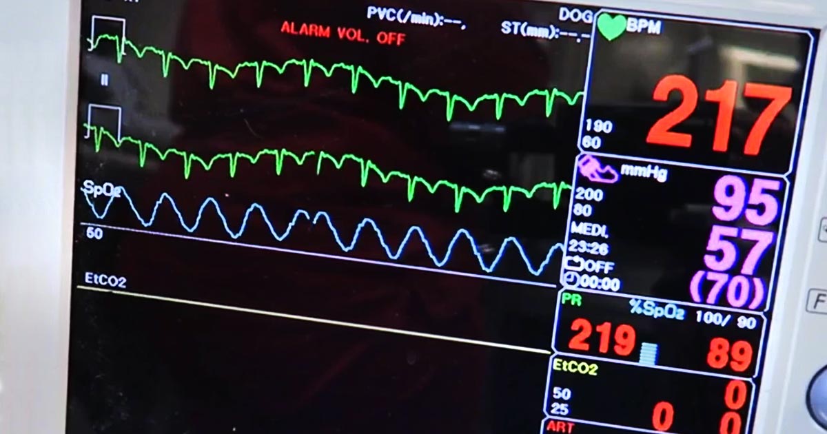
    Pulse oximetry is a useful, non-invasive method of measuring a patient’s oxygen saturation (SO2) and, under normal physiological circumstances, correlates well to the arterial oxygen saturation (SaO2).

    However, despite its ease of use and accessibility, it is not infallible. Circumstances exist that will undermine the accuracy of these readings – some with dire consequences if not recognised.

    Others causes are more technically associated, but also needs recognition.

    Unequal to task

    Pulse oximetry is incapable of assessing:

    • a patient’s haemoglobin levels
    • the haemoglobin’s functionality
    • the patient’s partial pressure of arterial carbon dioxide (PaCO2)

    The former is particularly apparent in anaemic patients, where peripheral capillary oxygen saturation (SpO2) readings could be greater than 95%, but animals still severely hypoxic. This is because the total numbers of haemoglobin is reduced; therefore, overall oxygen-carrying capacity is also decreased.

    Similarly, haemoglobin can be fully saturated with carboxyhaemoglobin or methaemoglobin strands, giving a misleadingly high SpO2 reading, yet patients are severely oxygen deprived.

    Finally, the ventilation status of the patient is not assessed by pulse oximetry. This is particularly important in animals with respiratory compromise, patients under heavy sedation and those under general anaesthetic or severe respiratory muscle paralysis from envenomation by a tick or snake. These patients can have near normal SpO2, but a dangerously high PaCO2.

    To overcome these problems, capnography or arterial blood gas analysis with cooximetry, and assessment of haemoglobin concentration is crucial.

    Accuracy issues

    The accuracy of pulse oximeter readings are also affected by several causes.

    Severe hypoxaemia (lower than 70% SpO2) is not accurately detected by pulse oximetry and requires partial pressure of arterial oxygen (PaO2) to confirm. Also, any cause of reduced peripheral perfusion can cause erroneously low readings, such as arrhythmias, hypotension, heart failure, hypothermia and severe vasoconstriction.

    Physical examination parameters that can indicate perfusion deficits are present include:

    • tachycardia
    • reduced pulse pressures
    • pale mucous membranes
    • prolonged capillary refill time
    • dull mentation/weakness
    • hypothermia

    It is not uncommon to stabilise a patient with hypovolaemic shock and find the SPO2 reading has normalised.

    Improving outcomes

    Although the accuracy of pulse oximetry readings are based on a large number of assumptions, it is still a valuable substitute for the measurement of PaO2 in clinically stable patients.

    Understanding the above concepts will allow you to derive a lot more information when used in the context of your patient’s oxyhaemoglobin dissociation curve and their clinical status.

    This will help improve patient outcomes, while early recognition of changes will allow prompt intervention and management of a patient’s disease.

  • Laryngeal paralysis

    Laryngeal paralysis

    This patient was brought to us for exercise intolerance, breathing difficulty and loud airway sounds.

    The patient has laryngeal paralysis. This is where the muscles controlling the arytenoids cartilages do not work and leads to failure of opening of the arytenoids during inspiration.

    Most commonly seen in middle-aged large breed dogs, it can occur acutely, but more often it is a chronic problem exacerbated by heat or stress. The cause is often unknown, but it can be caused by trauma or lesion to the cervical region or some kind of neuropathy, such as myasthenia gravis or tick paralysis. Diagnosis is based on visualisation of the arytenoid cartilages failing to abduct during inspiration under light anaesthesia.

    Treatment

    The management of the acute presentations include oxygen and sedation (butorphanol) to improve airway dynamics – with or without active cooling triggered by heat and with or without anti-inflammatories (dexamethasone) to reduce swelling secondary to airway turbulence.

    Patients in severe respiratory distress, anaesthesia and intubation may be required for a short period. Long-term management involves either surgery, such as laryngeal tieback, or conservative management strategies that involve weight loss, avoiding exercise and being kept in a cool environment.

  • Blood gas analysis, pt 7: evaluating oxygenation and ventilation

    Blood gas analysis, pt 7: evaluating oxygenation and ventilation

    In patients with respiratory compromise, it is important to look at the respiratory components of the blood gas to determine both oxygenation ability and adequacy of ventilation.

    To assess oxygenation, the partial pressure of arterial oxygen (PaO2) to fraction of inspired oxygen (FiO2) ratio, and alveolar-arterial gradient (A-a gradient) can be used. Conversely, the partial pressure of carbon dioxide (PCO2) is what dictates the adequacy of ventilation.

    Before going further, it should be noted only arterial samples can be used to evaluate oxygenation. Peripheral venous blood samples are rarely useful for oxygenation assessments, as the values are highly influenced by local factors and the degree of occlusion of the vessel from which the sample was collected from.

    Ventilation, conversely, can be assessed from either venous or arterial samples. The PCO2 values from the arterial samples are usually about 5mmHg to 10mmHg lower than the corresponding venous sample.

    Abnormalities

    Oxygenation and ventilation are not interchangeable terms. Adequate ventilation does not necessarily equate to adequate oxygenation, and vice versa.

    Oxygenation is determined by efficiency of oxygen absorption into the blood stream, after passing through the lungs, then delivered to the tissue. It is dependent on both ventilation (V) and perfusion (Q).

    For proper oxygenation to occur, matching ventilation and perfusion must occur at the level of the alveoli. When a V/Q mismatch occurs, blood is not properly oxygenated after passing through the lungs, referred to as venous admixture.

    Three forms of V/Q abnormalities exist:

    • Low V/Q can be caused by:
      • pneumonia
      • asthma
      • pulmonary oedema
      • inflammation
      • pulmonary thromboembolism
      • pulmonary neoplasia
    • Absent V/Q means blood has been diverted from parts of the lungs that are inadequately ventilated, such as those caused by atelectasis and alveolar collapse secondary to severe pleural effusion.
    • Diffusion impairment is caused by an increased distance between the gas exchange surfaces and pulmonary arterioles (rare in small animals). Pulmonary fibrosis and chronic obstructive pulmonary disease are among some of these causes where it can be severe enough to cause V/Q mismatch and, therefore, decreased PaO2.

    PaO2 to FiO2 ratio

    One way to determine the adequacy of oxygenation is by looking at the PaO2 to FiO2 ratio.

    FiO2 represents the percentage of the oxygen the patient is breathing. Room air is 21% oxygen, so the FiO2 is 0.21. PaO2 is normally about five times that of FiO2; therefore, the normal PaO2 of a patient with no pulmonary disease breathing room air should be about 100mmHg (5 × 21%).

    A normal PaO2 to FiO2 ratio is between 300 and 500. A number lower than 200 implies significant pulmonary disease or, possibly, acute respiratory distress syndrome. This ratio is particularly important in assessing patients requiring supplemental oxygen. An example is a congestive heart failure patient on bilateral nasal oxygen line (approximately FiO2 of 40%), with a PaO2 value of 80mmHg. The PaO2 appears to be adequate until the ratio is calculated (80/0.4 = 200). The change in FiO2 ratio is often more important than the PaO2 as a lone value.

    A-a gradient

    Once hypoxia has been established, the A-a gradient will help determine whether it is caused by ventilation failure or an underlying pulmonary disease. “A” stands for alveoli and “a” stands for arterial concentration of oxygen.

    A = [FiO2 × (Pb-PH20)] – (PaCO2/0.8) – Pb is the barometric pressure (760mmHg at sea level), and PH20 is saturated water vapour pressure, which is 50. The 0.8 is the respiratory quotient and affixed number.

    The simplified formula assumes the patients are breathing room air (FiO2 of 0.21) and at sea level, and the formula can be simplified to: A = 150 – (PaCO2/0.8)

    A normal A-a gradient should be less than 15. Abnormally high values indicate pulmonary parenchymal disease or an underlying heart disease, while a normal A-a gradient indicates the cause of hypoxia is likely secondary to ventilation failure.

    The ‘120 rule’

    Ventilation is particularly important to assess in animals with respiratory compromise, as it represents the entire mechanics of breathing.

    PCO2 is not only representative of the efficiency of ventilation, but also cellular metabolism and perfusion. Low PCO2, or hyperventilation, is rarely of any significance as aforementioned in the respiratory alkalosis section. High PCO2, however, is indicative of the lungs’ reduced ability to adequately shift air and can be caused by neurologic diseases, spinal cord injury, upper airway disease, trauma to the thoracic wall or muscles, and drugs that can cause respiratory depression.

    Ventilation must be assessed in light of oxygenation, as both are often affected by each other. An example of this would be a hypoxic animal (low PaO2) with a compensatory hyperventilation (low PCO2). The “120 rule” will help determine whether lung function is adequate.

    If the value you get, by adding the PaO2 and PaCO2, is greater or equal to 120, lung function is adequate. If the value is lower than or equal to 120, lung function is abnormal. This can only be calculated from patients breathing room air and at sea level.

    Ventilation adequacy

    Aside from looking at values on a blood gas, it is equally important to monitor the patient to determine whether the ventilation effort is sustainable.

    Animals with a significantly increased respiratory effort – despite normal blood gas values – are at risk of respiratory exhaustion and indicative of mechanical ventilation intervention.

    From these respiratory components of the blood gas value, clinicians should be able to determine the adequacy of ventilation, whether hypoxia is present and, if it is, whether the underlying cause is a result of ventilation failure or a possible underlying pulmonary or heart disease.

    Once oxygen supplementation has been implanted, the PaO2 to FiO2 ratio will help clinicians decide whether the response is adequate or mechanical ventilation is required.

    As with all laboratory measurements, it is extremely important to assess the patient itself. Non-sustainable respiratory efforts, in the face of normal blood gas parameters, is still an indication for mechanical ventilation.