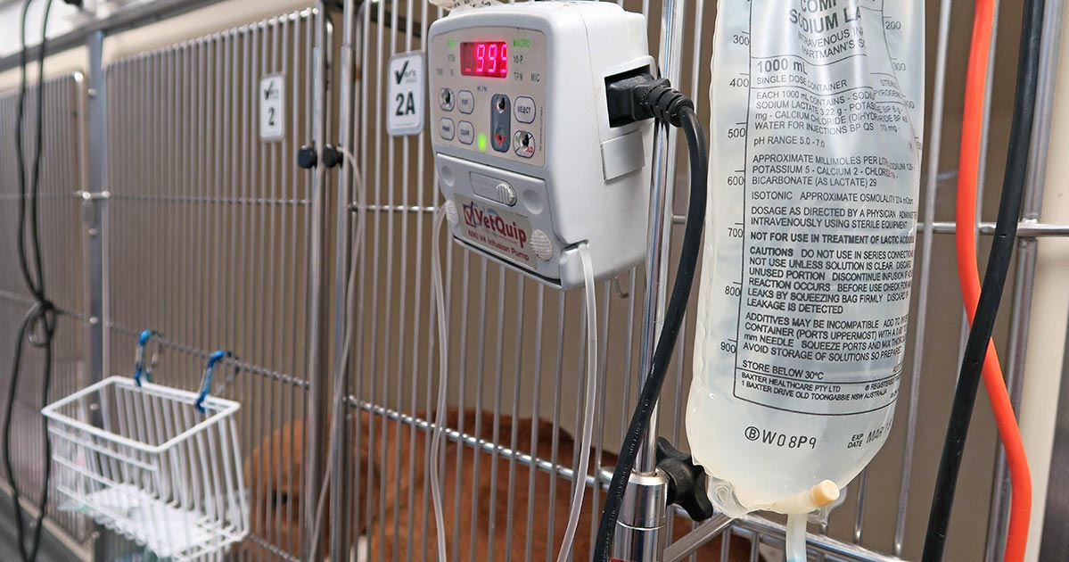
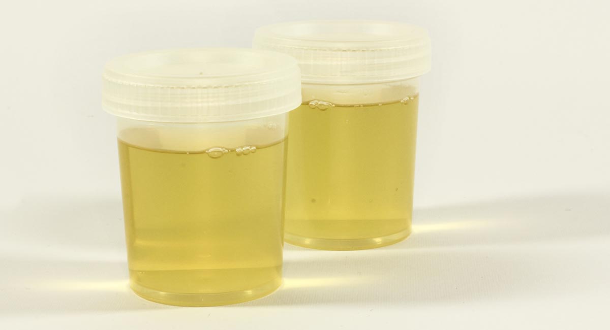
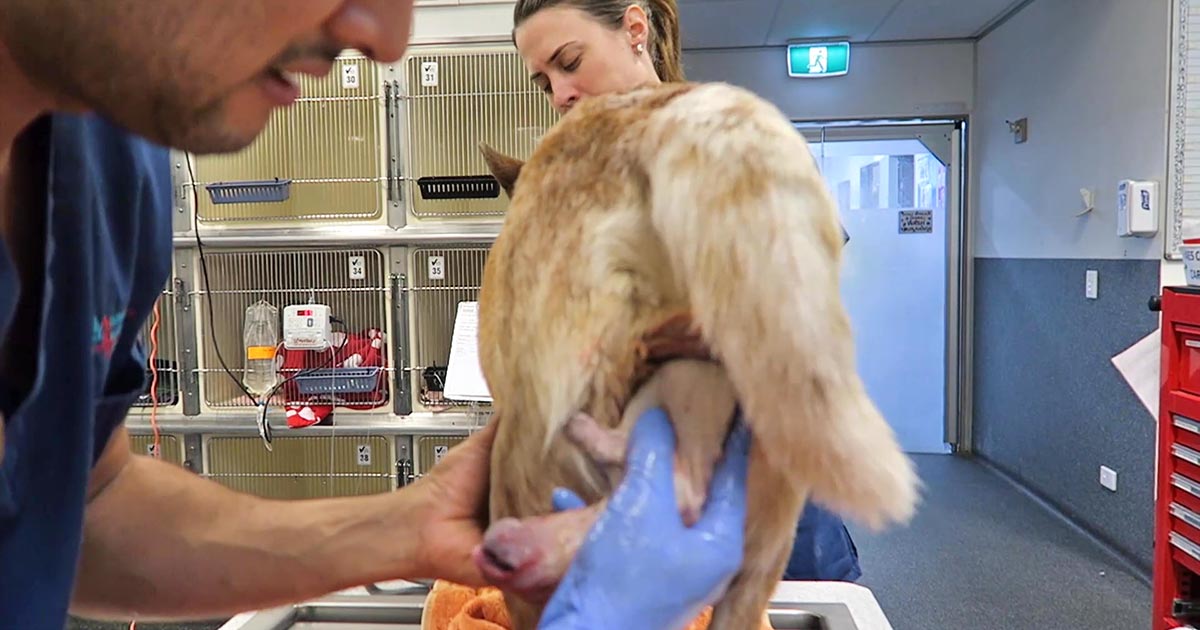
Urinalysis is an important diagnostic tool in veterinary practice. It is indicated for any patient that presents with polyuria or urinary tract signs, but also a necessary test to perform in conjunction with serum biochemistry.
Urine sediment exams on Christmas day.
Why do some clinicians fail to perform urinalyses even when they are indicated?
Reasons include:
clinicians not seeing the importance of obtaining a urine sample
the difficulty of obtaining a sample in some situations (the patient may not want to void)
no access to an ultrasound for a guided cystocentesis
patients not urinating upon bladder expression
Make it a priority
However, it is important clinicians make obtaining a urine sample a priority. Where possible, a sterile sample of urine using ultrasound-guided cystocentesis is recommended, especially when there is a possibility the urine may be sent to an external lab for culture and sensitivity.
For example, we’ve all been in situations were you start a patient on IV fluids only to find an azotemia on blood tests. Now you can’t determine whether it’s a pre-renal or renal cause, as you don’t have a pre-IV fluid urine sample.
Also it would be best to avoid the situation where you have run all other tests available on an ill patient only to find the answer lay in that urinalysis you did not collect earlier.
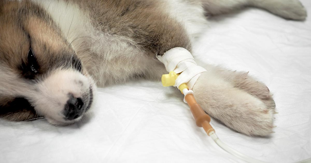
Treatment of ionised hypocalcaemia (iHCa) is reserved for patients with supportive clinical signs, then divided into acute and chronic management.
Since the most common cases of clinical hypocalcaemia in canine and feline patients are acute to peracute cases, this blog will focus on the acute treatment and management of hypocalcaemia.
Clinical signs
The severity of clinical signs of iHCa is proportional to the magnitude, as well as the rate of decline in ionised calcium (iCa) concentration.
The normal reference range for iCa is 1.2mmol/L to 1.5mmol/L in dogs and 1.1mmol/L to 1.4mmol/L in cats. Serum iCa concentrations in younger dogs and cats are, on average, 0.025mmol/L to 0.1mmol/L higher than adults.
Mild iHCa (0.9mmol/L to 1.1mmol/L) – as seen in critically ill dogs and cats with diabetic ketoacidosis, acute pancreatitis, protein-losing enteropathies, sepsis, trauma, tumour lysis syndrome or urethral obstructions – often has no observable clinical signs.
Moderately (0.8mmol/L to 0.9mmol/L) to severely (lower than 0.8mmol/L) affected animals – in the case of eclampsia and those with parathyroid disease – often display severe signs.
Early signs of iHCa are often non-specific, and include:
anorexia
rubbing of the face
agitation
restlessness
hypersensitivity
stiff and stilted gait
As the serum iCa concentration further decreases, patients often progress to:
paresthesia
tachypnoea
generalised muscle fasciculations
cramping
tetany
seizures
In cats, the gastrointestinal system can also be affected, presenting as anorexia and vomiting.
Treatment
The need for treatment of hypocalcaemia is dependent on the presence of clinical signs, rather than a specific cut-off of serum concentration of iCa itself.
Moderate to severe iHCa should always be treated. Mild hypocalcaemia, on the other hand, may not be necessary, especially if it is well tolerated. It should be remembered the threshold for development of clinical signs is variable, and treatment may benefit critical cases with an iCa concentration of less than 1.0mmol/L.
Treatment is divided into the acute treatment phase and chronic management.
In the tetanic phase, IV calcium is required – 10% calcium gluconate (equivalent to 9.3mg/ml) administered at 0.5ml/kg to 1.5ml/kg dosing to effect. This should be administered slowly with concurrent ECG monitoring. Infusion of calcium needs to be stopped if bradycardia develops or if shortening of the QT interval occurs.
Some suggest calcium gluconate (diluted 1:1 with 0.9% sodium chloride) of half or the full IV dose can be given SC and repeated every six to eight hours until the patient is stable enough to receive oral supplementation. However, be aware calcium salts SC can cause severe necrosis or skin mineralisation.
Calcium chloride should never be given SC, as it is a severe perivascular irritant.
Correcting iCa
Irrespective of the chronicity of the treatment, the rule of thumb is correction of calcium should not exceed 1.1mmol/L.
Correction of iCa to normal or hypercalcaemic concentration should always be avoided, as this will result in the desensitisation of the parathyroid response, predisposing renal mineralisation and formation of urinary calculi.
Some of the more common calcium supplementation medications – both parenteral and oral formulas – are detailed in Table 1. Supplementation of magnesium may also benefit some patients, as it is a common concurrent finding in critically ill patients with iHCa.
Table 1. Common calcium supplementation medications
Drug
Calcium Content
Dose
Comment
Parenteral calcium
Calcium gluconate
(10% solution)
9.3mg/ml
i) slow IV dosing to effect (0.5ml/kg to 1.5ml/kg); acute crisis, 50mg/kg to 150mg/kg over 20 to 30 minutes
ii) 5mg/kg/hr to 15mg/kg/hr IV or 1,000mg/kg/day to 1,500mg/kg/day (or 42mg/kg/hr to 63mg/kg/hr)
Stop if bradycardia or shortened QT interval occurs.
Infusion to maintain normal Ca level
SC calcium salts can cause severe skin necrosis/mineralisation.
Calcium chloride
(10% solution)
27.2mg/ml
5mg/kg/hr to 15mg/kg/hr IV
Do not give SC as severe perivascular irritant
Oral calcium
Calcium carbonate
(many sizes)
40% tablet
5mg/kg/day to 15mg/kg/day
Calcium lactate
(325mg, 650mg)
13% tablet
25mg/kg/day to 50mg/kg/day
Calcium chloride
(powder)
27.2%
25mg/kg/day to 50mg/kg/day
May cause gastric irritation
Calcium gluconate (many sizes)
10%
25mg/kg/day to 50mg/kg/day
Next time…
The next blog will look at the pathophysiology behind iHCa among critically ill animals. It will also look at the controversy regarding treatment of non-clinical iHCa cases and the prognostic indications of iCa concentrations.
As discussed in part one of this blog series, a myriad of disease processes can lead to ionised hypocalcaemia (iHCa).
Despite this, only hypocalcaemia caused by eclampsia and hypoparathyroidism (primary or iatrogenic – post-surgical parathyroidectomy) are severe enough to demand immediate parenteral calcium administration.
Hypoparathyroidism is quite rare, so this blog will not explore the detailed pathophysiology behind this syndrome. However, it is worthwhile mentioning – aside from primary hypoparathyroidism – no other disease state requires long-term calcium supplementation.
Eclampsia, on the other hand, is the most common cause of clinical hypocalcaemia in dogs and cats. Multiple factors can predispose animals to the development of this phenomenon, so understanding the pathophysiology behind this potentially fatal disease will not only help with future diagnosis and treatment, but also help prevent this issue.
Periparturient occurrence
Eclampsia – also known as puerperal tetany or periparturient hypocalcaemia – occurs in the periparturient period anywhere from the final few weeks of gestation to four weeks postpartum, with the latter being the more common time frame of manifestation.
The serum concentration of ionised calcium (iCa) is often less than 0.9mmol/L in bitches or less than 0.8mmol/L in queens. It presents as muscle fasciculation and tetany, but not usually in seizure since most patients maintain consciousness. Exceptions occur when these patients are left untreated – these patients may develop refractory seizures, cerebral oedema and death.
The increased muscle activity generates a lot of heat and uses a significant amount of glucose; therefore, hyperthermia and hypoglycaemia are common sequelae in patients with delayed presentations.
Reduced iCa
Eclampsia occurs as a result of reduced iCa in the extracellular compartment. In lactation-associated hypocalcaemia, it is the result of the body’s inability to maintain serum iCa through increased osteolytic activity and gastrointestinal calcium absorption, and reduced renal calcium excretion to compensate for the loss of calcium through milk production.
Other factors often predispose animals to developing eclampsia. These can include poor periparturient nutrition, excessive calcium supplementation and large litter size.
Excessive calcium supplementation in the prenatal period causes parathyroid gland atrophy, preventing parathyroid hormone release – resulting in reduced gastrointestinal calcium absorption and osteoclastic activity, and increased kidney calcium loss.
Clinical signs
Clinical signs can progress rapidly and become fatal if left untreated.
In the early phases, non-specific signs can present as:
facial pruritus
hyperaesthesia
panting
tremors
muscle fasciculations
paresis
ataxia
Within a few hours, these clinical signs rapidly progress to rigidity, and tonic and clonic spasms with opisthotonos. By this stage, animals will develop severe tachycardia, tachypnoea and hyperthermia. Without treatment, a high mortality rate exists.
Patients presenting with eclampsia require immediate medical intervention, as well as concurrent supportive therapy. The acute management of clinical iHCa is the same, regardless of the cause, and will be discussed in detail in part three.
Supportive therapies required to manage and prevent a patient relapsing in eclampsia often include active cooling and glucose supplementation. In cases that seizure, anti-seizure medications – such as diazepam and barbiturates – and mannitol for cerebral oedema may be required.
Prevention
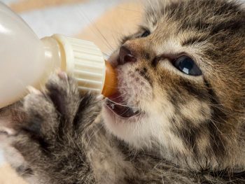
Even before getting to the stage where an animal requires treatment, all effort must be taken to prevent a dam from developing hypocalcaemia. This can be easily achieved by improving the calcium content of the food during the perinatal period, as well as reducing the milk demand by early weaning kittens or puppies. This is likely particularly helpful for those with a history of eclampsia or with large litters.
From the second half of gestation, it is recommended a commercial formulation of puppy/kitten food (1% to 1.8% calcium and 0.8% to 1.6% phosphorus) is to be fed to the dam without any additional minerals or vitamin supplementation.
Postpartum calcium is similar to the second half of gestation, requiring a diet containing at least 1.4% calcium with a 1:1 ratio with phosphorus (most balanced growth formula for puppies and kittens).
Less demand
Early supplementation of puppies and kittens with commercial milk formula will significantly reduce the lactation demand on the dam. Together with this, starting at aged three to four weeks, solids can be introduced at this time. These techniques will be particularly helpful to those with a history of previous eclampsia or those with large litter sizes.
Aside from the parenteral calcium supplementation required, other supportive therapy – such as active cooling, IV fluid therapy and glucose supplementation – may be required.
Long term, the dam’s nutritional content of calcium must be optimal from the second half of gestation. All additional calcium or other vitamins and mineral supplementations should not occur prior to parturition.
In the postpartum dam with a history of eclampsia or that is at risk, changing to a nutritionally balanced commercial food aim for growing puppies and kittens is ideal. Early weaning – or abrupt weaning if hypocalcaemia is severe – may be required in severe cases or those with a high risk of relapse/development.
Once the bitch presents to the clinic, a few basic diagnostic checks need completing to determine the status of the bitch/queen and the fetuses.
Physical examination
The first is a thorough physical examination, starting with the bitch or queen:
Demeanour, hydration status, vital signs, mucous membrane colour, capillary refill time and temperature are important.
Pregnancy anaemia is not uncommon; however, for patients with a haemorrhagic discharge, it is important to know their cardiovascular status.
A thorough abdominal palpation should be carried out to assess comfort level and palpation for the presence of fetuses. Palpating fetuses can be difficult and cannot confirm if no fetuses are present.
A digital vaginal examination should be performed. Feathering response – also known as the Ferguson reflex in human medicine – is the neuroendocrine reflex where the self-sustained cycle of uterine contractions is initiated by firm pressure on the dorsal aspect of the vestibulovaginal wall. If this is absent, the patient is unlikely to progress with the parturition unaided.
Palpation of fetuses in the canal can help decide whether surgical management is required. Obvious fetal malposition, malposture or malpresentation, or fetopelvic disparity, will be indications of caesarean. Abnormal pelvic diameter is also another reason to not proceed with medical management. To confirm these suspicions, abdominal radiography is required.
Radiographs will also help determine the number of fetuses to be expected, the signs of fetal death (presence of gas surrounding the fetus) and aforementioned fetomaternal abnormalities. I always repeat radiographs after the expected number of neonates is passed, to make sure I have not miscounted at the start.
Ultrasound
Panel 1. Heart rate ranges to help indicate stress of fetuses
Dogs:
normal – 180 to 220 beats per minute (bpm)
Stressed – 160bpm
Real concern – less than 160bpm
Cats:
normal – more than 220pbm
fetal stress – less than 180bpm
The second important diagnostic tool is ultrasound.
Fetal heart rates are good indicators of fetal stress. Some heart rate ranges that can help provide information about the status of the fetuses are detailed in Panel 1. These ranges vary between sources, but are good guidelines.
Ultrasounds can also help visualise the maturation status of the fetuses. At-term fetuses should have normal hepatic, renal and intestinal development. Intestinal peristalsis should be evident in at-term fetuses.
Other diagnostics
Other diagnostics may be indicated for patients, depending on the status of the bitch/queen:
If the patient is stable, but dystocia is present, a minimum database would include PCV/total protein, electrolytes, glucose, ionised calcium, lactate and acid-base balance.
Serum ionised calcium levels are important, as they influence the strength of contractions and how much supplementation is required.
Hypoglycaemia needs to be ruled out as a cause of dystocia, especially when large litters are involved.
If the patient is unstable or systemically unwell, include complete blood count, blood smears and biochemistry.
Physiological pregnancy anaemia can be present. The presence of regenerative response can help differentiate this from acute haemorrhage.
Abnormal leukocyte panel, especially with the presence of degenerative left shift, can indicate the presence of an infection – especially if toxic changes are present in the neutrophil.
Part three will briefly look at the medical management of dystocia and when surgical intervention is required.
The causes of hyponatraemia can be divided into three major categories, based on serum osmolality. This is further divided based on the patient’s volume status (Table 1).
Most patients we see in clinic fall into the hypovolaemic category, except patients with diabetes mellitus.
Table 1. Causes of hyponatraemia based on osmolality and volume status (from Guillaumin and DiBartola, 2017).
Hypo-osmolar
Hyperosmolar
Normo-osmolar
Hypovolaemic
Normovolaemic
Hypervolaemic
Gastrointestinal fluid loss
Third-space fluid losses
Shock
Hypoadrenocorticism (Addison’s disease)
Renal insufficiency
Excessive diuretic administration
Salt-losing nephropathy
Cerebral salt wasting syndrome
Syndrome of inappropriate antidiuretic hormone secretion (SIADH)
Hypotonic fluid administration
Hypothyroidism
Glucocorticoid insufficiency
Psychogenic polydipsia
Reset osmostat (SIADH type B)
Congestive heart failure
Acute or chronic renal failure
Nephrotic syndrome
Hepatic cirrhosis
Accidental ingestion or injection of water (water intoxication)
Hyperglycaemia
Mannitol
Severe azotaemia
Hyperlipidaemia
Hyperproteinaemia
Common causes
In dogs, the three most common causes of hyponatraemia are:
gastrointestinal (GI) fluid loss
third-space fluid loss
fluid shift from intracellular fluid to extracellular fluid (ECF) as a result of hyperglycaemia
In cats, the three most common causes of hyponatraemia are:
urologic diseases
GI fluid loss
third-space fluid losses
In most patients, more than one pathophysiologic factor is likely to be contributing to the hyponatraemia.
Circulating volume
Hypovolaemic patients – those with, for example, GI losses, hypoadrenocorticism, renal losses and haemorrhagic shock – have a reduced effective circulating volume. ECF contraction triggers antidiuretic hormone (ADH) secretion, which leads to increases in free water absorption and thirst, and results in dilution of the serum sodium concentration. Aldosterone secretion is reduced in hypoadrenocorticism, so an overall reduction in sodium reabsorption compounds the problem.
Hypervolaemic patients are those with an increased fluid retention state, such as:
Congestive heart failure patients have a reduced cardiac output and, therefore, a decreased effective circulating volume, despite the presence of the extra fluid status. Renin-angiotensin activation leads to release of ADH and aldosterone, resulting in sodium and free water reabsorption, and increased thirst. Both lead to an excess of free water retention.
Advanced hepatic (cirrhosis) or renal failure (nephrotic syndrome) both result in hypoalbuminaemia, leading to fluid shifting into the interstitial space and third space, reducing effective circulating volumes. This leads to activation of ADH to increase free water reabsorption, to restore the circulating volume in the face of existing hypervolaemia and hyponatraemia.
Diabetic patients
Moderate to severe hyperglycaemic diabetic patients can be either hyperosmolar or normo-osmolar, depending on the serum blood glucose concentration. Hyponatraemia occurs when water shifts from the intracellular fluid to the ECF down the osmotic gradient, diluting the serum sodium content.
Despite this osmotic shift, not all diabetic patients develop hyponatraemia. Glucosuria also causes also causes a renal osmotic shift, sometimes resulting in urine water loss in excess to sodium. This offsets the hyponatraemia – in some cases, hypernatraemia results.
Treatment
Treatment of hyponatraemia hinges on how quickly it developed and the volume status of the patient. The rule of thumb is to correct hyponatraemia slowly – not exceeding 0.5meq/L/hr – especially in chronic cases, or cases where the duration of hyponatraemia is unknown. Keeping to this rate is paramount until serum sodium concentration reaches 130meq/L.
In acute patients with severe clinical signs, such as seizures, some clinicians may choose to use a higher rate of 1meq/L/hr to 2meq/L/hr until clinical signs resolved.
It should be emphasised, once again, this rate should never be used in chronic patients, patients with an unknown duration of hyponatraemia, or where frequent serum sodium concentration cannot be monitored. The rapid correction of hyponatraemia can lead to osmotic demyelination syndrome (myelinolysis).
Its effect will not be apparent until three or four days after therapy, and can result in neurological abnormalities such as:
weakness
ataxia
dysphagia
paresis
coma
For that reason, frequent electrolyte measurements are required, starting hourly then once a suitable rate of increase has been established and less frequently thereafter.
Part 3 will look at how to correct patients with hyponatraemia.
Reference
Guillaumin J and DiBartola SP (2017). A quick reference on hyponatremia, Veterinary Clinics of North America: Small Animal Practice47(2): 213-217.
Hyponatraemia is a relatively common electrolyte disturbance encountered in critically ill patients, and the most common sodium disturbance of small animals.
In most cases, this is caused by an increased retention of free water, as opposed to the loss of sodium in excess of water.
Low serum sodium concentration
Hyponatraemia is defined as serum concentration lower than 140mEq/L in dogs and lower than 149mEq/L in cats.
The serum sodium concentration measured is not the total body sodium content, but the amount of sodium relative to the volume of water in the body. For this reason, patients with hyponatraemia can actually have decreased, increased or normal total body sodium content.
This series will look briefly at the modulators of the sodium and water balance, clinical signs associated with hyponatraemia, the most common causes in small animals, the pathophysiology behind these changes, and treatment and management.
ECF volume
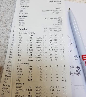
An example of hyponatraemia.
Sodium is the main osmotically active particle in the extracellular fluid (ECF), so is the main determining factor of the ECF volume. Any disease process that alters the patient’s ECF volume will lead to hyponatraemia, such as:
dehydration
polyuria
polydipsia
vomiting
diarrhoea
cardiac diseases
pleural or peritoneal effusion
The modulators of water and sodium balance are also different, so should be thought of as different processes.
Water balance is modulated by thirst and antidiuretic hormone, and the effect of this is to maintain normal serum osmolality and serum sodium concentration.
Modulators of sodium balance aim to maintain normal ECF volume. It adjusts this by altering the amount of renal sodium excretion; an expansion of ECF volume will lead to an increased sodium excretion, while a reduction in ECF volume will lead to increased sodium retention.
Rate and magnitude
The clinical signs of hyponatraemia are both dependent on the magnitude of the decrease and the rate at which it developed.
In mild or chronic patients, no visible clinical signs can exist. In severe (lower than 125mEq/L) and acute cases, clinical signs exhibited are typically neurological, reflecting cerebral oedema. Possibilities include:
lethargy
anorexia
weakness
incoordination
disorientation
seizures
coma
Patients with acute hyponatraemia – for example, water intoxication – are more likely to show clinical signs, compared to those with chronic hyponatraemia, because the brain takes time (at least 24 to 48 hours) to produce idiogenic osmoles, osmotically active molecules that help shift free water out of brain cells.
Therefore, any acute hyponatraemia that develops within a 24 to 48-hour period tend to show clinical signs, whereas chronic cases are less likely.
Next week’s blog will look into the different causes of hyponatraemia and how they result in sodium loss.
Simple acid-base disorders are compensated by predictable compensatory changes. The primary disorder shifts the pH, while the compensatory mechanisms aim to normalise the pH and bring it back to neutral.
This is achieved by attempting to normalise the bicarbonate (HCO3-) to partial pressure of CO2 (PCO2) ratio in a paralleled manner.
For example, an increase in HCO3– (metabolic alkalosis) is compensated by an increase in PCO2 (respiratory acidosis). Similarly, a respiratory alkalosis (decrease in PCO2) is compensated by a metabolic acidosis (decrease in HCO3-).
Ruling out secondary process
However, before jumping to the conclusion an opposing change is the result of compensation, we must rule out the presence of a secondary process. This can only be determined by calculation (Table 1).
Table 1. Calculating compensatory change
Component
Expected compensation
Metabolic acidosis
↓HCO3 (↓BE)
per 1mEq/L ↓ in HCO3 = ↓ PCO2 of 0.7mmHg
Metabolic alkalosis
↑HCO3 (↑BE)
per 1mEq/L ↑ in HCO3 = ↑ PCO2 of 0.7mmHg
Respiratory acidosis (acute)
↑PCO2
per 1mmHg ↑ PCO2 = ↑ 0.15mEq/L HCO3
Respiratory acidosis (chronic)
↑PCO2
per 1mmHg ↑ PCO2 = ↑ 0.35mEq/L HCO3
Respiratory alkalosis (acute)
↓PCO2
per 1mmHg ↓ PCO2 = ↓ 0.25mEq/L HCO3
Respiratory alkalosis (chronic)
↓PCO2
per 1mmHg ↓ PCO2 = ↓ 0.55mEq/L HCO3
By comparing the reported to what the calculated compensatory change should be, you can determine whether the patient’s reported value is due to compensation or a separate disorder – for example, multiple primary acid-base disorders (a mixed acid-base disorder).
An example of a mixed disturbance could be a hyperventilating (respiratory alkalosis) dog with renal failure (metabolic acidosis).
The level of decrease in PCO2 change is in excess of the calculated compensation for the metabolic acidosis, therefore confirming a mixed acid-base disturbance. In fact, the most common causes of hyperventilation – pain, fear and excitement – often complicate blood gas analysis.
Another example of a mixed disorder could be a patient with traumatic haemothorax experiencing both lactic acidosis (hypoperfusion) and hypoventilation (respiratory acidosis) due to pleural space disease.
Waiting game
Another thing to keep in mind is compensation takes time – respiratory processes take approximately 8 to 12 hours, while metabolic processes take one to three days.
The lungs are able to alter PCO2 levels relatively quickly by adjusting the rate of ventilation. The kidneys, on the other hand, take a longer time to adjust the pH, as the change in rate of absorption and excretion of HCO3– takes much longer in comparison.
Regardless of the rate, physiologic compensation for a primary acid-base disturbance is almost never able to return pH to neutral.
Summary
A simple acid-base disorder should be suspected when the patient’s reported values are similar to the calculated compensation value, and a mixed acid-base disorder when the values fall outside the calculated range.
Another hint that a mixed acid-base disturbance is present is if the pH falls within the normal reference range, but the HCO3– or the PCO2 are not; or if the HCO3– and PCO2 are in opposite directions as opposed to being parallel.
Remember, the body can never overcompensate nor return the pH to neutral.
Base excess (BE) and bicarbonate (HCO3-) represent the metabolic components of the acid base equation.In general, both components will change in the same direction.
Decreased HCO3– and BE indicate either a primary metabolic acidosis or a metabolic compensation for a chronic respiratory alkalosis. Elevated HCO3– and BE indicate either a primary metabolic alkalosis or a metabolic compensation for a chronic respiratory acidosis.
The exception to this rule arises when a patient hypoventilates or hyperventilates.
Carbonic acid equation
CO2 + H2O ↔ H2CO3 ↔ HCO3– + H+
When a patient hypoventilates, CO2 will increase as a result of reduced expiration, so a shift to the right of the equilibrium will occur. The shift to the right will increase the bicarbonate levels proportional to the increase in CO2.
The opposite occurs when a patient hyperventilates; the equilibrium shifts to the left, so a decrease in HCO3– is present.
Since HCO3– is not independent to the patient’s respiratory status, it is an inaccurate way of measuring the metabolic component in patients with respiratory changes. For this reason, the BE value is the preferred.
The BE represents the amount of acid, or base, needed to titrate 1L of the blood sample until the pH reaches exactly 7.4, with the assumption the blood sample is equilibrated to a partial pressure of CO2 of 40mmHg (the middle of the reference range) and the patient’s body temperature is normal.
Possible causes
The possible causes of the primary disease are:
Metabolic acidosis
lactic acidosis – shock and poor perfusion
renal failure – reduced hydrogen ion (H+) excretion and increased loss of HCO3–
diabetic ketoacidosis – ketone acids
gastrointestinal (GI) losses – loss of HCO3– through vomiting and diarrhoea
Metabolic alkalosis
GI outflow obstructions – loss of H+ and chloride via vomiting
reduced chloride levels and resultant poor perfusion – body attempts to reabsorb water and sodium to increase intravascular volume, but inadvertently also reabsorbs HCO3– in the process, despite existing alkalosis
refeeding syndrome
severe hypokalaemia:
transcellular shift – potassium ions leave and H+ ions enter the cell
transcellular shift in cells of proximal tubules → intracellular acidosis → promotes ammonium production and excretion
H+ excretion in the proximal and distal tubules increases → further reabsorption of HCO3– and net acid excretion
renal insufficiency
diuretic therapy (contraction alkalosis – loss of bodily fluids that do not contain HCO3-; this causes the extracellular volume to contract around a fixed quantity of HCO3-, resulting in a rise in the concentration of HCO3– without an actual increase in HCO3– levels)
Next step
After ruling out the differential causes of either respiratory or metabolic acidosis/alkalosis, the next step is to determine whether a compensatory response is present and, if so, if this is adequate or whether a true mixed acid-base disorder exists.
After taking note of the direction of the pH shift – acidaemia or alkalaemia – it is important to determine the primary and secondary causes.
If an acidaemia is present (pH less than 7.35), an underlying respiratory or metabolic acidosis, or both, must exist. Similarly, if an alkalaemia is present (pH more than 7.45), an underlying respiratory or metabolic alkalosis, or both, must be present.
This is usually very simple, with the exception of cases presenting with a normal pH (between 7.35 and 7.45, slightly higher for cats).
Cases with normal pH
For cases with a normal pH, we need to determine which category it falls into:
No acid-base disturbance
Both respiratory and metabolic components are within the normal reference range.
Complete compensation for the acid-base disturbance
This requires specific calculations that will be discussed in a later blog.
This cannot be determined by glancing at the figures alone.
Two opposing acid-base disturbances (a mixed disorder), which are cancelling the effect of each other out in terms of pH.
Both the respiratory and metabolic components will be outside of their reference range, going in the opposite direction to each other.
Determining primary disorder
Since these animals are within the normal pH range – particularly those with complete compensation – how can you tell which is the primary disease process?
A golden rule of thumb is: even with maximal compensation, the pH will still usually move in the same direction as the primary problem.
Therefore, if the pH lies towards the acidaemic side of the mid-point of the pH range (less than 7.4), the primary disease process is an acidosis. By the same token, if the pH lies towards the alkalaemic side of the midpoint of the pH range (more than 7.4), it will have a primary alkalosis disorder.
The reason behind this is the body does not usually overcompensate for an acid-base disturbance.
Secondary disorder
Once the primary disorder has been identified, we need to look at whether a secondary disorder is also present and, if so, whether this is the result of compensation or a true mixed process.
To determine whether compensation occurred, you need to understand the timeline for when compensation usually occurs.
With respiratory compensation, this typically starts immediately, but may take up to 8 to 12 hours to occur. This is because adjusting levels of CO2 is relatively easy, with the change of respiratory rate and patterns.
On the other hand, metabolic compensations take approximately one to three days to occur, since renal excretion of hydrogen ions or retention of bicarbonate takes longer.
Variation magnitude
If the magnitude of the observed variation is compatible with compensation alone (this requires calculation), a compensatory mechanism is likely. A mixed process (mixed acid-base disorder) is present if the magnitude:
does not correspond to the clinical status of the patient
falls outside of the compensation time frame
is outside of the expected magnitude of compensation
All other causes for why the acid base is moving in the opposite direction must be ruled out before determining a secondary process is present.
Once the primary and, if present, secondary disorder are determined, the next step is to determine the cause of the respiratory and metabolic acidosis and alkalosis.
This month, we will look at the final part of a fluid therapy plan – accounting for ongoing losses. This can be challenging, but some general rules can be helpful.
Regular assessment is essential to track patients’ responses.
When considering ongoing losses, try to not forget about patients with pre-existing polyuric diseases; chronic renal failure is a prime example. Patients with dehydrated chronic renal failure are unlikely to suddenly regain concentrating ability. Polyuria should be considered as an ongoing loss.
Other conditions that may result in additional urinary fluid losses include post-obstructive diuresis, diabetes mellitus, hyperadrenocorticism and hyperthyroidsim.
How much to add?
This is the tricky part. I often add an additional half to one maintenance and frequently reassess clinical parameters, or if a urinary catheter is placed matching ins and outs.
Gastrointestinal tract losses can be collected and weighed; 1g of vomitus or diarrhoea can be roughly equivalent to 1ml of water.
Fluid removed from drains placed in cavities or wounds should also be measured and accounted for.
Remember the key point is regular assessment of the patient’s hydration status, from repeat clinical exams, to track their response. Don’t forget regular retesting of electrolytes – for example, every 12 to 24 hours for patients on IV fluids and not eating.


 For cases with a normal pH, we need to determine which category it falls into:
 Once the primary disorder has been identified, we need to look at whether a secondary disorder is also present and, if so, whether this is the result of compensation or a true mixed process.