Tag: records

  • Abdominal ultrasound, pt 1: equipment controls

    Abdominal ultrasound, pt 1: equipment controls

    Abdominal ultrasound is an invaluable diagnostic tool that can give us far more information about abdominal organs than radiographs. It can also be a very daunting procedure – especially for clinicians unfamiliar with how the machine operates.

    One of the biggest frustrations with using the ultrasound machine stems from not knowing what each button is for, or how to use them to adjust the image quality appropriately, to obtain the information required.

    Over the next few posts, I will offer you hints and tips on how to get started. Once you understand what all those buttons and dials are for, you should be able to operate just about any ultrasound machine with confidence.

    I will also explain how to perform an abdominal ultrasound in a systematic manner, as well as how to get the most information out of it.

    Buttons and their uses

    Ultrasound control panel.
    Figure 1. An ultrasound machine control panel.

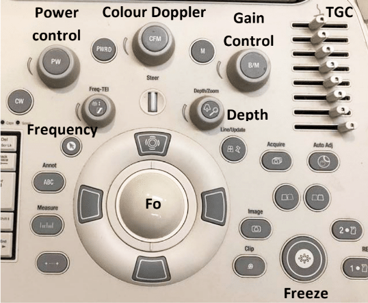
    Figure 1 is an example of a typical ultrasound machine control panel. The following explains the different buttons and dials present, and their uses.

    Power control

    The power or output control affects the strength of the emitted sound pulse. Keep this to as low as possible for the required depth.

    Gain control

    The gain control regulates amplification of all returning echoes non-selectively – that is, it makes the entire image brighter.

    Time gain compensation

    The time gain compensation (TGC) selectively regulates amplification of echoes returning from various depths.

    The objective of TGC is to provide a uniform image from top to bottom. Returning near-field echoes are already strong and may actually need to be suppressed. Far-field echoes are weaker and, therefore, may need to be amplified more according to the depth from which they return.

    Depth

    The depth allows you to choose how deeply into the tissues the image will display. Use an appropriate depth to view the structure of interest – that is, adjust the depth so the structure fills three-quarters of the field of view.

    Focus

    The focus (Fo) provides the best image at a certain depth and should be adjusted so it is at – or just behind – the area of interest. The focal point is the yellow arrow on the right side of the screen, which can be moved up and down with the Fo tracking ball.

    Freeze

    The freeze-frame control creates a frozen on-screen image for closer inspection, annotation, measurements using electronic calipers and producing permanent documentation with digital, film or paper images.

    Clip

    The clip button records a short video clip.

    Colour Doppler

    The colour Doppler assesses fluid flow. It is useful for differentiating between a vessel and structure of equal echogenicity, such as an adrenal gland or lymph node, and for determining the vascularity of a structure, such as a torsed spleen, mass lesion or abscess.

    Flow towards the transducer = red, while flow away from the transducer = blue.

  • Under the microscope: lessons from pathology rotation

    Under the microscope: lessons from pathology rotation

    The past couple of weeks on rotation have largely consisted of looking down a microscope or performing postmortems – and despite clinical and anatomic pathology being very different, a running theme seemed to exist across both.

    microscope
    Image ©iStock.com/The-Tor

    On the clinical pathology week, we pored over slide after slide of blood smears and cytology samples, trying to formulate differentials from minimal or no history about the case.

    We muddled through a number of biochemistry profiles and attempted interpretation, often with little or nothing to go on from the submitting clinician.

    Historical significance

    In anatomic pathology, we were often supplied with the history, but the clinicians would not reveal the full extent until after we had formed differential lists – even then the “full” history we were given before proceeding with postmortem examination would often be scarce.

    Although we would often get there in the end, or at least in the right ballpark, I think it’s safe to say the importance of a relevant history has been drummed into us for evermore.

    Going forward as new graduates – far sooner than many of us would like – I don’t think we’d dare send a sample to the university lab without filling the forms out meticulously and providing a relevant history. Making an accurate interpretation that fit the clinical picture was so much easier when a few, seemingly minor points were highlighted in the history.

    The write stuff

    Another lesson creeping into every rotation over the year is the sheer amount of paperwork involved in veterinary medicine – be it clinical notes, postmortem reports, lab submission forms, case reports, anaesthetic records… the list goes on.

    Keeping accurate records can be the difference between being sued and being able to prove your clinical judgement at the time. Most relevant to this rotation was accurate completion of lab submission forms, so samples can be correctly identified and results sent to the right place.

    forms
    Accurate completion of lab submission forms is essential. Image © gamjai / Fotolia.

    It sounds obvious, but the clinical pathology staff assure us the frequency of receiving samples with important information omitted is much higher than you’d think.

    Out of practice

    Having explored the different aspects of clinical and anatomic pathology, while dragging a lot of material from the depths of third-year knowledge, I can appreciate how quickly some skills and understanding can be lost when you don’t practise or use them regularly.

    Feeling considerably rusty at the beginning of the rotation, I feel a lot more comfortable now, but can see how vets can lose their ability or confidence to make a cytological diagnosis when in practice, especially when things get busy.

    Being able to send samples to the experts is a major advantage, but I think, as new grads, we should at least have a quick look down the microscope and make a provisional call to be confirmed by the lab, rather than just sending samples straight off.

  • The consequences of making a mistake

    The consequences of making a mistake

    beef-farm-crop-jordan
    Beef farms in Scotland can be quite picturesque.

    Fertility work makes up a large proportion of cattle veterinary work in the UK and, after spending a month on the farm rotation at university, I can appreciate the importance of getting it right – and how hard it can be.

    Experienced large animal vets make it look easy – they scan the uterus and ovaries, and decide what drug to give to aid getting the cow in calf, all in a minute or two.

    In the meantime, I’m still fumbling about trying to palpate what, I think, may be the uterus or ovaries while the cow squeezes and gradually cuts off the circulation in my arm until I can barely feel my fingers.

    Meanwhile, the vet has zipped through several cows already.

    When things go wrong

    cow-injection-lg
    What happens if you misdiagnose a pregnant cow as negative and administer prostaglandin, or give a cow steroids without realising she’s in calf?

    On one fertility visit this week, we discussed when things go wrong. For example, what if:

    • you misdiagnose a pregnant cow as negative and administer prostaglandin (PGF)?
    • a vet gives a cow steroids for any number of reasons without realising she’s in calf?

    The outcomes of both of these scenarios are almost inevitably abortion, which can have a number of repercussions on the farm – and, potentially, the vet.

    Negligence or misconduct?

    Many new graduates are terrified of being called up for “fitness to practice” for making a mistake such as those aforementioned. However, during a Veterinary Defence Society (VDS) workshop at the SPVS Lancaster weekend earlier this year, it was emphasised a difference exists between negligence and misconduct in the eyes of the RCVS.

    What I took away from this session was, in simple terms, negligence involves making a mistake – such as missing a diagnosis, giving the wrong treatment accidentally, eliciting side effects from something due to missing something in the case history – whereas misconduct is actively doing something you know you shouldn’t – such as trying to cover up a mistake, lying or misleading a client.

    Mistakenly giving PGF to a pregnant cow would be classed as negligence and, on the whole, the VDS would have your back; the farmer may receive some compensation for his losses, but the incident wouldn’t tarnish your career. In the same situation, misconduct would be denying you had administered any treatment.

    In short, you won’t get struck off for easily made, one-off mistakes, but you may if you lie about them.

    In the news

    Our conversation about accidentally aborting cows led us to discuss the case of Honey Rose, the optometrist convicted of gross negligence manslaughter. News reports vary, but, from what I can gather, she failed to diagnose papilloedema (swollen optic discs) in an eight-year-old boy, which would have been an indication of the hydrocephalus he died of five months later.

    It is reported she claimed to have been unable to examine his eyes properly because he had photophobia – a claim considered false by the judge. However, she had also failed to look at retinal photos taken by a colleague, on which the papilloedema was evident.

    Going by the RCVS’ rules, missing the diagnosis or not looking at the retinal photos would be negligent. However, denying she’d been able to carry out a thorough examination, despite written records suggesting otherwise, would be misconduct.

    Rose was found guilty of gross negligence manslaughter and was handed a two-year prison sentence, suspended for two years, and a 24-month supervision order, and was ordered to complete 200 hours of unpaid work.

    In your defence

    VDS logoAs vets, we have the VDS to help us in these situations – and, while it would be truly awful to make a mistake resulting in an animal’s death, we would not be at risk of imprisonment.

    Doctors and dentists also have defence societies that will fight their corner, but optometrists? To my knowledge, an equivalent does not exist – and this was the first case of an optometrist convicted of manslaughter in the UK, which added to the complexity of the legal battle.

    The death of a little boy is devastating, but, as medical professionals know, death is a risk with many procedures, no matter how small the risk may be. It’s frightening to think jail could be a consequence for those in the medical profession.

    So, while I gradually lost the sensation in my right arm as I tried to reach an ovary of the 10th cow in a row, I mulled this over and realised I was extremely thankful for the safety net the VDS provides and would not take working with animals for granted.

    After all, if I’d decided to be a doctor instead, I could be rummaging around in another human’s back end rather than a cow’s!

    • For further details of the Veterinary Defence Society and its services, visit www.thevds.co.uk