Tag: Peritonitis

  • How to do an autotransfusion

    How to do an autotransfusion

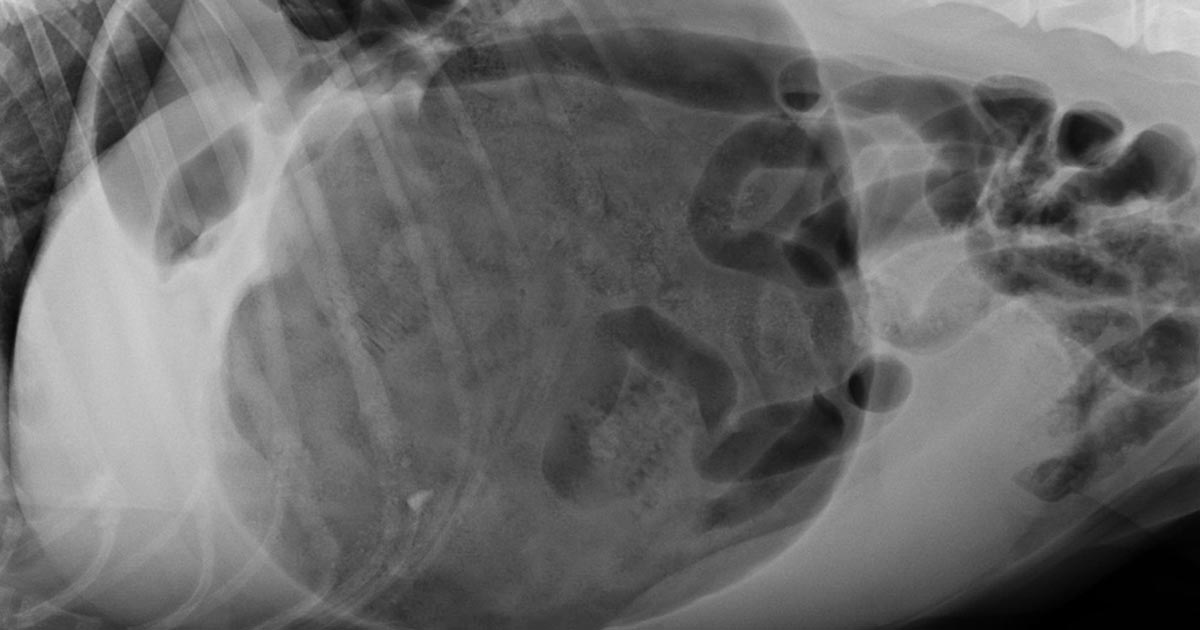
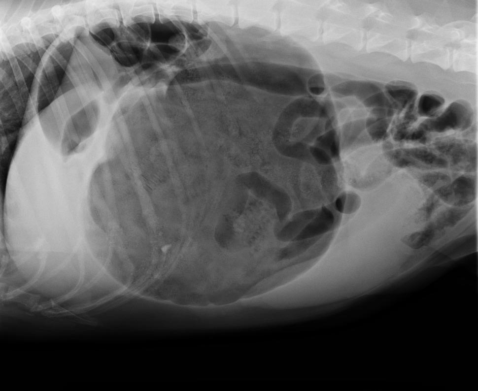
    You’re stuck in the clinic in the middle of the night with a dog that is dying – it’s bleeding into its abdomen and needs blood, but the bag in the fridge is expired.

    You’ve heard it’s possible to collect the blood out of the abdomen and safely give it to the patient, but you’re not sure how and don’t have time to google. How do you actually do it?

    It’s easier than you think. Depending on what equipment you have available in the clinic, here are two practical ways of administering an autotransfusion.

    Collection

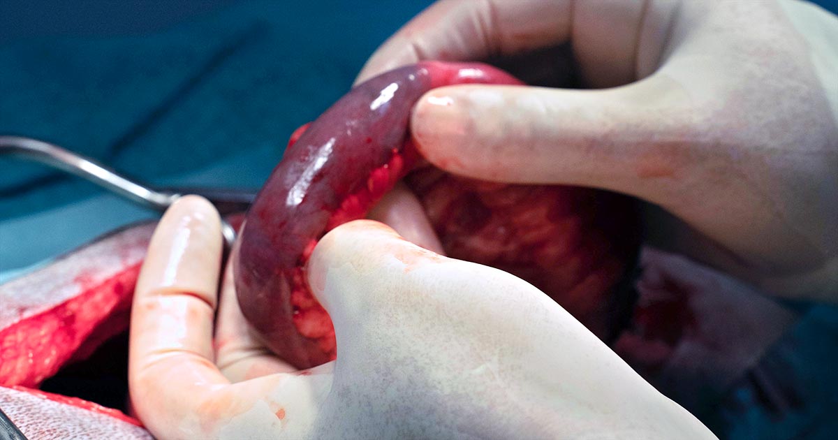
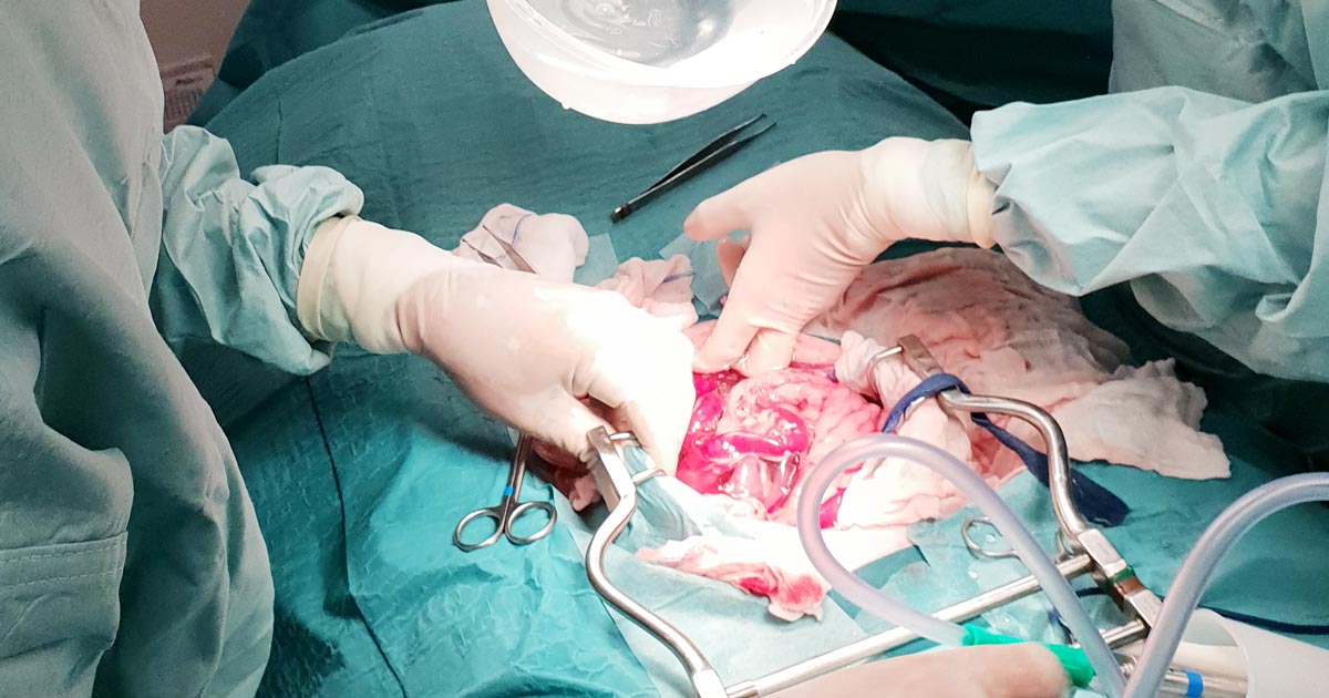
    Blood can be collected from the abdomen or thorax during exploratory surgery, simply by sucking it up with a 60ml syringe.

    Time is usually of the essence in these cases, so a handy tip is to use 60ml catheter tip syringes to aspirate the blood, then get someone to transfer the blood into a standard Luer-tipped syringe by shoving the smaller tip into the catheter tip. Like this:

    Autotransfusion

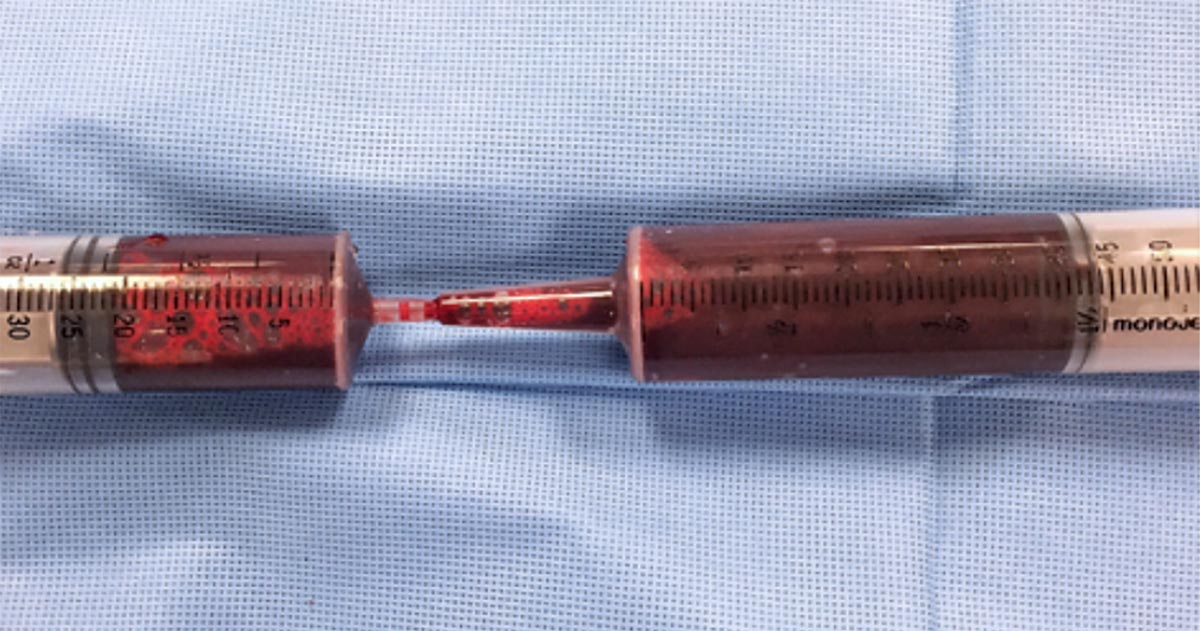
    Besides the faster collection time, the bigger aperture of the catheter tip syringe will also reduce negative pressure during collection and, therefore, the chances of haemolysis.

    If you are not going into surgery, you can collect the blood percutaneously – using a butterfly needle or catheter – as you would drain any body cavity fluid.

    You shouldn’t need to add any anticoagulants to your syringes, as fibrinolysis has already occurred – meaning the blood shouldn’t clot in your syringe.

    Administration

    Whichever method you use, it’s crucial the blood is administered through a blood filter.

    Method one

    The most straightforward way is to simply inject the blood back into the patient using the Luer-tipped syringe through a filter. For this technique, you will need a filter that can connect to a syringe.

    Method two

    If you only have a blood filter as part of a blood giving set, you could use a two-way stopcock valve to get your collected blood into a sterile bag – like an empty IV fluids bag, or a dedicated blood collection bag – and then run the blood through your blood giving set with the built-in filter, as you would for a normal transfusion.

    This is a bit more fiddly, so adding a few little blood filters to your emergency equipment could be well worth your time.

    When you shouldn’t do it

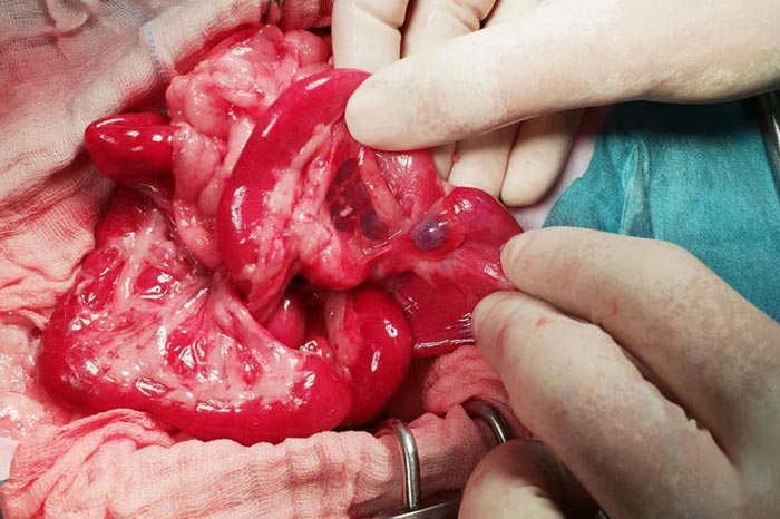
    Autotransfusions are most useful in cases of bleeding due to clotting deficiencies where the clotting problem is already being addressed, but a need for red blood cells still exists; and for cases of traumatic or postoperative bleeding.

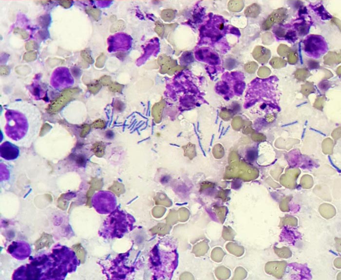
    It is contraindicated if contamination with urine, faeces, bile or bacteria – in case of septic peritonitis – is likely; or in cases of neoplasia in the body cavity where an autotransfusion has the potential to seed neoplastic cells to the rest of the body.

  • Ionised hypocalcaemia, pt 4: controversies and prognostic indications

    Ionised hypocalcaemia, pt 4: controversies and prognostic indications

    Ionised hypocalcaemia (iHCa) is a well-known electrolyte abnormality in critical human patients, which is also beginning to be recognised in our critical feline and canine patients.

    The exact mechanism for the development of iHCa is still unknown – making prevention difficult, if at all possible. Controversy also exists as to whether treating iHCa is of any benefit, especially in non-clinical cases.

    Despite these issues, serum concentration is proving to be an accurate prognostic indicator for the morbidity and mortality rates of some of the more critical patients.

    Research

    Over the past 30 years, significant resources have been put into trying to demystify the pathophysiological causes of iHCa in critically ill people; however, the exact mechanisms are still to be determined.

    Some proposed mechanisms include:

    • abnormal parathyroid hormone secretion or function
    • abnormal vitamin D synthesis or function
    • hypomagnesaemia
    • calcium chelation
    • alkalaemia
    • calcium sequestration in tissue or cells
    • an increase in calcitonin precursors (procalcitonin)

    In a canine study where endotoxaemia was induced, it was found hypovitaminosis D was associated with iHCa (Holowaychuk et al, 2012).

    Veterinary studies

    The true incidence of iHCa in critically ill canine and feline patients is yet to come to a consensus, due to the limited veterinary studies.

    In one retrospective study, 90% of 55 cats with septic peritonitis was reported to have iHCa (Kellett-Gregory et al, 2010), while only 24% of septic dogs (n=58) was reported to have iHCa (Luschini et al, 2010).

    Regardless of the true incidence, the commonness of this change questions whether a need exists to treat iHCa, especially cases in the mild or non-clinical categories.

    No consensus

    At this stage, no consensus exists to either support or prohibit the treatment of hypocalcaemia in critically ill patients.

    Well-designed prospective studies are scarce in human literature and non-existent in the veterinary field; no evidence-based guidelines are available for treatment.

    Based on logic, arguments for the administration of calcium to critically ill patients include:

    • iHCa during hospitalisation is a negative predictor for morbidity and mortality of patients.
    • Hypocalcaemia can cause decreased myocardial contractility.
    • In hypotensive patients dependent on vasopressors or inotropic agents, the supplementation of calcium may be beneficial.

    Arguments against calcium supplementation include:

    • Calcium accumulation within cells predisposes to hypoxia and ischaemia-reperfusion injury.
    • Increased mortality in experimental models of sepsis when calcium is supplemented, on top of the lack of evidence to support this act.

    Prognostic use

    Serum calcium concentrations – or, rather, the trend of it in hospital – appears to be of valuable prognostic indicators.

    Kellett-Gregory et al (2010) found although no direct associations existed between the presence or severity of iHCa at the time of patient admission, a positive correlation existed between the lowest iCa post-hospitalisation, and the length of hospitalisation and duration of intensive care stay.

    Of the cats that had iHCa, those that failed to return to a normal ionised calcium (iCa) during hospitalisation had a significantly lower rate of survival to discharge. Interestingly, iHCa was not associated with the status of hypotension, coagulopathy or arrhythmias, so cannot be used to predict the occurrence of these.

    These findings were echoed by Luschini et al (2010), where low mean ionised calcium and lowest documented ionised calcium concentrations were found to be associated with a poor outcome. The severity and duration of iHCa appears to be important in determining prognosis in these patients.

    Conclusion

    Controversy exists regarding whether treatment of mild iHCa in critically ill patients is recommended; however, one thing we now know is serum iCa concentration is a reliable predictor of mortality and morbidity in canine and feline patients.

    References

  • To resect, or not to resect…

    To resect, or not to resect…

    To resect or leave in a piece of intestine that is concerning you is a common source of stress when performing exploratory laparotomies.

    In many cases, this is straightforward; in some, it can be difficult to decide.

    The risk is if you leave in a piece of intestine and it devitalises, then leakage of intestinal contents into the abdomen occurs, leading to septic peritonitis – the situation we all dread.

    Criteria

    In this post, I cover some criteria I use to assess intestinal viability.

    These are all subjective criteria that require familiarity with what is normal, as well as good judgement, to be reliable; however, they are the only realistic criteria available for general use.

    Arterial pulsations in the mesenteric arteries that are feeding the affected areas

    This is the most important criteria, and it can be the most reliable criterion for assessing intestinal viability.

    It goes without saying – if there is no blood supply, there is no oxygen being delivered – resulting in tissue death.

    Intestinal tissue color

    It is important to remember the colour can change quite dramatically in a short period of time.

    Once an obstruction has been removed, perfusion to that area will be restored and the colour can change rapidly. Tissues that are dark in colour can turn dark red then bright red with time.

    Often, I give the section of questionable intestine time after I have removed an obstruction to see what colour it changes to. If the tissue is black or grey and does not improve, I would consider resection.

    Wall texture

    This can be a difficult thing to appreciate, but it can be helpful to compare a healthy segment of intestine to the affected segment of intestine.

    When you squeeze and roll a piece of healthy intestine between your thumb and index finger, it should feel thick and springy, whereas intestine that has lost viability can feel “thin”.

    Peristalsis

    The presence of peristalsis can be helpful and is a good sign. The absence of peristalsis is not an automatic indicator of poor intestinal viability, as it can take some time for motility to return in severe cases.

    Partial thickness incision

    Finally, a small partial thickness incision can help if you are really stuck.

    If there is effective blood supply to the tissues then the blood should be normal and bright red in colour, as it is oxygenated; poor blood supply will mean the blood will remain dark and blue tinged, indicating poor oxygenation.

    Concerning signs

    The criteria that makes me very concerned – and would make me lean towards resection – include:

    • black, grey discoloured intestine
    • absent arterial pulsations
    • absence of peristalsis
    • thin wall thickness
    • dark, deoxygenated blood from a partial thickness incision

    If I have concerns and it will not result in a significant loss of small intestinal length, I would remove the intestine rather than leave it in.

    If you are removing large segments, I would wait longer to see whether time and blood supply will help improve the health of the intestinal tract enough to feel comfortable leaving more of it in.

  • Leave no stone unturned

    Leave no stone unturned

    Although I have lots of clinical experience and have developed my diagnostic skills to a high level, at times I will be thrown a curveball, so I should never get complacent.

    Figure 2. The lymph node resection.
    Figure 1. The black lymph node.

    The more unusual cases are the ones I tend to enjoy most, as they really get my brain working and force me to look at every possibility – something I champion in my practice.

    An unusual case

    One particularly unusual case was of septic peritonitis – an inflammation of the peritoneal lining of the abdomen caused by a bacterial infection.

    Most commonly, this is due to perforation of the gastrointestinal tract, such as a foreign body, or ulceration or from some kind of abscess often secondary to neoplasia.

    Surprising findings

    Detection of bacteria found after performing a fine needle aspirate.
    Figure 2. Detection of bacteria found after performing a fine needle aspirate.

    We were unable to determine the cause of the bacterial infection in this patient via diagnostic imagery prior to exploratory laparotomy, but suspected it was due to some kind of gastrointestinal perforation due to dilated loops of hypomotile intestine.

    To our surprise, the only thing we found was this black lymph node at the ileocolic junction (Figure 1).

    We performed a fine needle aspirate and found bacteria under the microscope (Figure 2).

    The lymph node was resected, no perforation was present, the abdomen was copiously lavaged and a Jackson-Pratt drain was placed.

    This case highlighted perfectly the importance of leaving no stone unturned.

  • Christmas dangers

    Christmas dangers

    Christmas can be a busy time for vet clinics, so here is a list of common intoxications and conditions to keep an eye out on during the festive period.

    Chocolate

    • Michael Pettigrew / fotoliaNumerous online calculators can determine whether a toxic dose has been consumed and they are a great place to start.
    • I always perform emesis in patients that have ingested chocolate, even hours after ingestion as often large amounts can reside in the stomach.
    • Remember that cardiac arrhythmias can also occur in clinically normal looking patients, so perform an ECG.
    • The toxic components can be reabsorbed through the bladder wall; therefore, urinary catheterisation is a part of management of this intoxication.

    Onions

    • Onions used in roasts and on BBQ’s can cause Heinz body formation, haemolytic anaemias and pigmenturia.
    • This is not a common intoxication, but should be considered in anaemia patients and those with discoloured urine.

    Raisins

    • Commonly used in Christmas cakes and puddings. They can cause acute kidney failure, the exact mechanism of action is unknown, and there does not appear to be a dose-dependent relationship.
    • It should always be a differential for azotemic patients this time of year.
    • IV fluid induced diuresis for 48 hours is the safest way to manage raisin exposure.

    Mistletoe

    • The berries can be fatal, even if only a couple are ingested.

    Ethylene glycol

    • In colder climates, ethylene glycol can be a very common toxicity.
    • This sweet liquid is very attractive to pets and can cause acute renal failure, with the first signs being acute onset ataxia.

    Macadamia nuts

    • Macadamia nuts are common in some parts of the world. They result in joint pain in the hocks and carpus leading to weakness and ataxia.
    • Often confused with trauma and soft tissue injuries. Hyperextension of the hocks and sometimes flexion of the carpus are the clinical features.

    Xylitol

    • Xylitol is a sugar-free product used in lollies and baking.
    • In dogs, it triggers endogenous insulin to be released and a subsequent hypoglycemia develops. It can also cause hepatic failure.
    • As a general rule, I approach all intoxications as if they could be fatal as it is rare to know exactly how much of the toxic agent they have been exposed to. I consider if a patient I am treating for intoxication never develops clinical signs and wonder whether it was going to or not is the best outcome.

    Strings

    • Look under the tongue.
    • Linear foreign bodies can be difficult to diagnose. Some features on abdominal radiographs to look out for include abnormal bunching of the small intestines, and “c” and “comma” shaped gas patterns.

    Christmas meals

    • Gastroenteritis is the most common presenting condition over the Christmas period, with dietary change and indiscretion often being the culprit.
    • Bones can lead to obstructions from oral cavity to the intestines and can also cause constipation.
    • Leftover meat trimmings, often fat laden, are a common cause of pancreatitis.

    BBQ skewers

    • In some parts of the world (Australia especially) BBQs are common around Christmas time.
    • BBQ skewers can cause gastrointestinal tract perforation and septic peritonitis.
    • Because they are not radiopaque they are often difficult to diagnose.
  • All hands on deck: GDV diagnosis

    All hands on deck: GDV diagnosis

    Gastric dilatation-volvulus (GDV) is a true veterinary emergency and while it can be daunting to be presented with a sick dog with suspected GDV, the most important thing to remember is this patient will likely succumb to this condition without your intervention.

    First, a little pathophysiology: GDV is a broad term that can refer to gastric dilation on its own, gastric dilation with volvulus, and even chronic gastric volvulus. These conditions usually present in large or giant breeds and we still know little about the underlying causes.

    GDV
    Gastric dilatation-volvulus (click to zoom).

    Once dilation and volvulus occurs, perfusion to the stomach and other abdominal organs is compromised. Along with general shock – which can be fatal in its own right – decreased stomach wall perfusion can result in stomach wall necrosis, rupture and peritonitis.

    Clinical signs

    Quite often, a GDV case starts with a telephone call from a panicking owner. He or she usually reports an acute onset of retching, regurgitation or vomiting in their large or giant breed dog after feeding.

    Other common signs include:

    • hypersalivation
    • agitation
    • palpable abdominal distension

    When presented, many of these cases will be obvious and the animal already in some degree of shock. You need to institute fluid resuscitation and gastric decompression immediately to restore perfusion as soon as possible.

    Confirmation

    To confirm the patient truly has GDV, as some patients may present with simple gastric dilation from over-engorgement, you need to perform an abdominal radiograph.

    Always keep an eye out for the large, deep-chested dog that presents with vomiting or retching, but doesn’t appear bloated. Don’t be fooled into ruling out GDV in these patients based on physical examination alone – often, no visible or palpable gastric distension exists as the ribs cover the stomach. That is where the abdominal radiographs play an especially important role.

    It is common practice at our hospital to perform abdominal radiographs as soon as possible, so as to not miss a hidden or subtle GDV in these large breed dogs.

    Which view is best?

    The classic approach is to lie the patient in right-lateral recumbency, in this view, you would see the classic “Smurf’s hat”, “boxing glove”, “Popeye’s arm”, “double bubble”, etc. This is compartmentalisation of the stomach, indicating not only gastric dilatation, but volvulus as well.

    You should also look for evidence of pneumoperitoneum, as it may suggest gastric wall rupture.

    At this stage, it is also important to collect blood for biochemistry, haematology, electrolytes and, if available to you, blood gas analysis. ECG readings should also be taken to determine if the patient has any life-threatening arrhythmias, such as ventricular tachycardia.

    Next month, we will talk about stabilising and treating these patients.

  • Linear foreign bodies, part 3: should YOU take it to surgery?

    Linear foreign bodies, part 3: should YOU take it to surgery?

    In the previous post we covered what to look out for on ultrasound when assessing for a linear foreign body. Now we discuss the things you should consider before deciding to take the patient to surgery.

    Read the following statements and answer the questions – either yes or no…

    • Linear foreign body surgeries can be technically difficult and can take a prolonged period of time to perform. The longer the surgery, the higher the rate of complications. Are you (or do you have access to) an experienced surgeon who has performed a linear foreign body surgery before, and do you have an additional pair of hands for surgery to help reduce surgery time?
    • Were bacteria visualised or is their presence likely based on the comparison of the abdominal fluid glucose and lactate to peripheral blood (see previous post)? If yes, then septic peritonitis is present. Approximately 40% of dogs with linear foreign bodies will have septic peritonitis. This means you need to be prepared to perform a resection and anastomosis; sometimes two. Have you performed a resection and anastomosis before?
    • Often a combination of gastrotomy, enterotomy, and resection and anastomosis are required. Are you prepared to perform a combination of these surgeries?
    • Linear foreign body surgeries can often require a significant number of surgical instruments and consumables. Do you have Balfour retractors, forceps, clamps, additional kit for closure, supply of lap sponges, as well as substantial amounts of lavage and access to suction?
    • Linear foreign body patients are often critically ill due to septic peritonitis. They can present in shock and have biochemical derangements such as hypoalbuminaemia, which indicates the need for perioperative critical care. Do you have experience stabilising, performing anaesthesia and postoperatively managing a critically ill patient?
    • Hypotension can be caused by hypovolaemia; however, if the hypotension has not responded to reasonable volumes of a balanced isotonic crystalloid fluid – for example, 30ml/kg to 40ml/kg – then the hypotension could be caused by vasodilation from septic shock. This means vasopressor agents will be required in addition to crystalloids. Do you have access and experience with vasopressor therapy?
    Image: Gerardo Poli.

    Conclusion

    This list of questions is not meant to be conclusive or definitive, but merely a list of considerations before taking a linear foreign body patient to surgery.

    If you answered no to many of these questions, then consider referral to a facility that is prepared and equipped for the challenges that often accompany linear foreign body patients. However, if referral is not an option, consider the list above as a way to be as prepared as possible for tackling those situations.