Tag: Pathology

  • Don’t rush: a systematic approach to x-rays

    Don’t rush: a systematic approach to x-rays

    One of my responsibilities in our emergency hospital is the training and mentoring of vets new to the field of emergency and critical care.

    First look
    FIRST LOOK: The patient was bitten by another dog and presented with multiple puncture wounds and difficulty breathing. Radiographs were taken to assess for thoracic injuries (click to zoom).

    A common area I have found where clinicians request more training is radiographic interpretation.

    When I review radiographs and find pathology that was missed, it Is more often due to a lack of systematic approach to reviewing the radiograph than the clinician’s lack of experience or knowledge.

    There is, of course, no one set way you should go about interpreting a radiograph – but whatever the method, the entire radiograph should be assessed, not just the area of interest.

    Radiograph tips

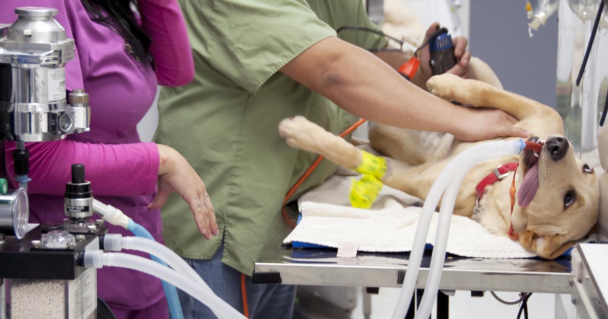
    1. Try not to struggle with your patient. If possible, appropriately sedate your patient (or anaesthetise if safe to do so). This reduces stress for everyone involved and improves your chance of getting a good radiograph. For musculoskeletal radiographs, you often need to manipulate painful joints and limbs to get diagnostic images.
    2. Take appropriate views. For example, I aim to get three plane projections for thorax and abdominal radiographs – i.e. left and right laterals and VD (or DV). Three views are critical for the assessment of both lung fields, and also to help interpret abdominal gas patterns more effectively.
    3. Collimate, rotate, crop, label and adjust the image appropriately. Displaying radiographs in a standardised format is important for proper assessment. Reviewing anatomy in the same way each time helps develop an understanding of what is normal, and makes identifying abnormalities easier.
    4. This is my top rule: At first, IGNORE the area you are interested in. This means, if you are interested in looking at the GI tract in a vomiting dog, try not to focus – albeit initially – on the stomach and intestines on your radiograph.
    5. Start at the periphery. This means things like the spine, subcutaneous tissue, etc – you would be surprised how often lesions are missed in these areas.
    6. reveal
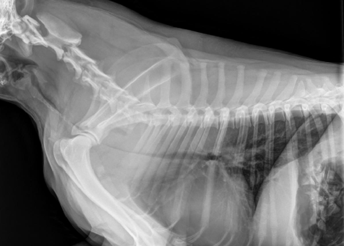
      SECOND LOOK: This fracture was missed on initial review of the radiographs (click to reveal).

      Now take a look at the cavity space (the pleural and abdominal space, for example). You should not be able to see the pleural space, and you should see no evidence of air or fluid in the abdominal space. When it comes to musculoskeletal radiographs, don’t forget to consider joint cavities and soft tissue structures such as ligaments and tendons.

    7. Lastly, make sure you assess every organ (again leaving your organ of interest until last). Things often overlooked include the prostate, kidneys and mediastinal region. At the end of this, I always ask myself, have I missed an organ?

    Tunnel vision

    Another thing I like to do sometimes is take a step back and assess the radiograph again. I find this gives a better global view of the projection, as opposed to staring at it closely.

    This is because when we focus on our area of interest, we start developing the habit of tunnel vision, introducing the potential for missing lesions.

  • Rat bait’s sneaky trick: bleeding into the dorsal tracheal membrane

    Rat bait’s sneaky trick: bleeding into the dorsal tracheal membrane

    Most of us are familiar with anticoagulant rodenticide toxicosis and the range of clinical signs it can present with, but there is one potentially fatal manifestation of coagulation pathology that is perhaps not as widely known…

    Dogs with severe clotting problems will occasionally bleed into the dorsal tracheal membrane. This causes collapse of the thoracic trachea and can lead to severe respiratory distress.

    Presenting signs

    These cases can present with none of the other signs of bleeding normally associated with coagulopathies, so rat bait poisoning may not come to mind as a differential diagnosis if you are not aware of this syndrome.

    The typical case will present as an otherwise healthy dog that develops acute respiratory problems. Early signs can be as mild as a persistent cough, but it can quickly escalate into a life-threatening respiratory crisis.

    Severe cases will have an obvious stridor on both inspiration and expiration, cyanotic mucous membranes, and patients may be very distressed.

    It will look very much like:

    • a dog that is choking from a tracheal foreign body
    • an old dog with tracheal collapse
    • the end stages of laryngeal paralysis – except the stridor will come from much lower in the respiratory tract than it does in laryngeal paralysis

    So, what do you do?

    On initial presentation you would approach it as any respiratory distress case: oxygen, oxygen, oxygen, calm and stress-free handling, and light sedation (butorphanol, for example).

    bleeding_dorsal-tracheal-membrane

    Once it is safe to do so, you should take chest rads to look for what you’ll probably suspect is a tracheal foreign body, and you’ll get an image like the one above (although it may not be this severe). Then you’ll remember this article, have an “aha!” moment and run a clotting profile (but if it’s as bad as this case, you’ll obviously first save the animal’s life by passing an ET tube).

    Once a clotting problem is confirmed you’ll need to stop the bleeding with standard therapy for anticoagulant rodenticide toxicity: plasma and vitamin K.

    Severe cases

    In a severe case you may need to keep the dog intubated for several hours, until the clotting times have normalised, before cautiously attempting to extubate.

    If the patient is unable to stay well oxygenated without an ET tube (mucous membrane colour, pulse oximetry, blood gas), consider placing a long oxygen catheter past the narrowing – either via a tracheostomy or a nasal O2 catheter.

    If these cases are quickly recognised for what they are, and an open airway can be maintained, the prognosis should be good. These are potentially very satisfying cases with great potential for you to be a total hero.

  • Seizures, part 2: the differentials

    Seizures, part 2: the differentials

    In part one of this series we discussed the important questions to ask when taking a history from owners of dogs and cats that are having seizures. In this part, we look at the differential diagnoses for these cases.

    There are many ways to classify the different causes of seizures, but the simplest is as follows:

    • Structural – where intracranial pathology is causing the seizures.
    • Reactive – where an extracranial issue is causing a seizure response in a normal brain.
    • Idiopathic – a diagnosis of exclusion where we are unable to identify a reason for the disturbances in brain activity.

    Structural

    Intracranial differential diagnoses include:

    • inflammatory processes (meningoencephalitis), such as steroid responsive meningitis-arteritis
    • viral diseases (for example, distemper)
    • metabolic storage diseases
    • neoplasia
    • vascular accidents involving clots or bleeds
    • hydrocephalus
    • trauma

    Reactive

    Extracranial differentials include:

    • hepatic encephalopathy due to hepatic failure or a portosystemic shunt
    • various toxicities, such as lead, chocolate, caffeine, ethylene glycol, parasiticides and slug/snail bait
    • metabolic issues, such as hypoglycaemia, hypocalcaemia and thiamine deficiency

    Idiopathic

    If diagnostic investigations (including advanced imagery, such as MRI) are unable to identify an underlying cause of recurrent seizures, this is referred to as idiopathic epilepsy.

    To break down this list of differentials into a more relevant and concise list is to consider the most common differentials according to signalment.

    In dogs less than a year old:

    • portosystemic shunts
    • inflammatory conditions of the brain
    • distemper
    • hydrocephalus or storage disease
    • toxicity

    In dogs one to five years old:

    • idiopathic epilepsy
    • inflammatory
    • toxicity
    • cerebral neoplasia

    In dogs of five years or older:

    • cerebral neoplasia
    • inflammatory
    • toxicity
    • idiopathic epilepsy
    • metabolic disease
    • vascular issues

    In cats:

    • toxoplasmosis
    • FIP, FeLV and FIV
    • audiogenic reflex seizures (older cats)
    • neoplasia
    • trauma
    • toxins
  • Public health: the less recognised role of vets

    Public health: the less recognised role of vets

    Bovine spongiform encephalopathy, commonly known as “mad cow disease”, hit the news again after an isolated incident was reported in Aberdeenshire, Scotland.

    In the 1990s, this disease resulted in the mass culling of hundreds of thousands of livestock, devastating the farming community and causing ripples throughout the British economy.

    Soaring meat prices, a ban on UK beef exports and sizeable public concern arising from the last outbreak led to extensive media coverage on a topic many people not involved in the medical field rarely entertain.

    Not all puppies and kittens

    Although you might not think it, food safety and public health are two of the most vital roles vets perform for society.

    I feel there’s a common misconception all vets are bound to the same path; when speaking to my uni friends about future careers, they never seem to assume I’ll end up anywhere other than small animal practice – and this may be true for the majority or veterinary graduates, at least those from the UK. Mixed practices are dwindling and veterinary food safety workers are predominantly sourced from overseas.

    That aside, veterinary involvement in the food industry is vital in keeping the public safe. Meat having the stamp of approval by professionals trained in parasites and pathology gives the British public – as well as importing countries – confidence in our farmers and serves to strengthen our economy, and build up domestic farmers and their businesses.

    Only the beginning

    Within the meat industry, the job of a vet is not over once the animal is slaughtered – they are just as involved in death as in life. Zoonotic diseases and parasites can be spread between people and animals, and vets ensure these never reach your supermarket shelves.

    At food crisis moments – such as the events that occurred over two decades ago – vets have two main responsibilities:

    • A duty of care to those animals who must be put down with a delicate combination of haste and humanity.
    • To work with farmers during the financial struggle they will most likely suffer following the loss of a large proportion of their livelihood.

    For the many, not the few

    Medical bills make up a substantial amount of costs when running a farm, and any vet going into the industry will quickly realise treating a herd animal is a far cry from treating a pet. They need to master the art of putting the needs of the herd over any individual.

    Of course, what might be the greatest responsibility of any farm vet is to aid farmers in preventing further outbreaks. To pool the resources of all field areas – researchers, clinicians, food technicians and farm workers – to improve productivity and ensure the continued safety of the public.

  • Under the microscope: lessons from pathology rotation

    Under the microscope: lessons from pathology rotation

    The past couple of weeks on rotation have largely consisted of looking down a microscope or performing postmortems – and despite clinical and anatomic pathology being very different, a running theme seemed to exist across both.

    microscope
    Image ©iStock.com/The-Tor

    On the clinical pathology week, we pored over slide after slide of blood smears and cytology samples, trying to formulate differentials from minimal or no history about the case.

    We muddled through a number of biochemistry profiles and attempted interpretation, often with little or nothing to go on from the submitting clinician.

    Historical significance

    In anatomic pathology, we were often supplied with the history, but the clinicians would not reveal the full extent until after we had formed differential lists – even then the “full” history we were given before proceeding with postmortem examination would often be scarce.

    Although we would often get there in the end, or at least in the right ballpark, I think it’s safe to say the importance of a relevant history has been drummed into us for evermore.

    Going forward as new graduates – far sooner than many of us would like – I don’t think we’d dare send a sample to the university lab without filling the forms out meticulously and providing a relevant history. Making an accurate interpretation that fit the clinical picture was so much easier when a few, seemingly minor points were highlighted in the history.

    The write stuff

    Another lesson creeping into every rotation over the year is the sheer amount of paperwork involved in veterinary medicine – be it clinical notes, postmortem reports, lab submission forms, case reports, anaesthetic records… the list goes on.

    Keeping accurate records can be the difference between being sued and being able to prove your clinical judgement at the time. Most relevant to this rotation was accurate completion of lab submission forms, so samples can be correctly identified and results sent to the right place.

    forms
    Accurate completion of lab submission forms is essential. Image © gamjai / Fotolia.

    It sounds obvious, but the clinical pathology staff assure us the frequency of receiving samples with important information omitted is much higher than you’d think.

    Out of practice

    Having explored the different aspects of clinical and anatomic pathology, while dragging a lot of material from the depths of third-year knowledge, I can appreciate how quickly some skills and understanding can be lost when you don’t practise or use them regularly.

    Feeling considerably rusty at the beginning of the rotation, I feel a lot more comfortable now, but can see how vets can lose their ability or confidence to make a cytological diagnosis when in practice, especially when things get busy.

    Being able to send samples to the experts is a major advantage, but I think, as new grads, we should at least have a quick look down the microscope and make a provisional call to be confirmed by the lab, rather than just sending samples straight off.

  • RCVS council election manifesto: Cheryl Scudamore

    RCVS council election manifesto: Cheryl Scudamore

    CHERYL SCUDAMORE

    Cheryl Scudamore.

    BVSc, PhD, FHEA, FRCPath, FRCVS

    Mary Lyon Centre, MRC Harwell, Harwell Campus, Didcot OX11 0RD.

    T 01235 841017

    M 07714 484522

    E c.scudamore@har.mrc.ac.uk

    PROPOSERS: Nicky Paull, Susan Rhind

    I graduated from the University of Liverpool in 1988 and completed a PhD at the Rowett Research Institute in Aberdeen. I continued my research interests and pathology training at the University of Edinburgh’s Royal (Dick) School of Veterinary Studies, becoming a senior lecturer in veterinary pathology.

    Pursuing an interest in human health research, I moved to the pharmaceutical industry in 2001 to work as a pathologist in the safety assessment of new medicines and managing a large technical team. While in industry, I maintained strong academic links, with particular interests in transferring technical skills between industry and academia, and encouraging vets to explore alternative career options.

    In recognition of these interests, I was awarded an Medical Research Council (MRC) skills gap grant, moving back to the RVC in 2009 and on to my current role as pathologist at MRC Harwell in 2012. I am an RCVS recognised specialist in veterinary pathology and hold visiting chairs at RVC and the University of Surrey.

    I have experience of serving on and chairing professional committees. I was part of the stakeholder group for the Vet Futures project and chair the Royal College of Pathologists’ specialty advisory committee for veterinary pathology.

    Manifesto

    The profession is undergoing modernisation and facing challenges that require new ideas to resolve. Key to development and innovation in any profession is the cultivation of diversity. Diversity includes the individuals within the profession, working practices and career pathways.

    Increasing diversity means, firstly, looking at our student intake, working with schools and aiming to attract a better balance of applicants that reflect our society today and will make resilient vets.

    A veterinary education provides a great starting point for a variety of potential career pathways. The Vet Futures project has shown the RCVS and the BVA recognise different career options are important for individuals and the professions. We need to build on this project to ensure graduates are prepared for different roles, practice and non-practice career paths are equally valued, career paths are signposted and people are supported throughout development in their chosen career.

    Mental health is another pressing issue for our profession and ensuring a satisfying career choice, with manageable levels of stress, are essentials for good mental health. We need to develop career pathways within the profession that offer progression and working practices that accept a good work-life balance is essential, not just desirable. We need to find ways to support vets through their early years and as they develop greater responsibilities later in their career.

    Diversity in the profession means having a range of resilient veterinary graduates able proactively to engage with a range of different career options and to adapt to changes in the external environment. Having worked in the university, research institutes and pharmaceutical industry as a lecturer, mentor, careers advisor, researcher and manager, I believe I have a range of skills and experience to contribute to the debate in these crucial areas.