QUICK TIP: Need to know if there is an oesophageal foreign body but can’t be certain on radiographs?
We have all been in the scenario where we are unsure whether there is an oesophageal foreign body on the radiographs we have just taken.
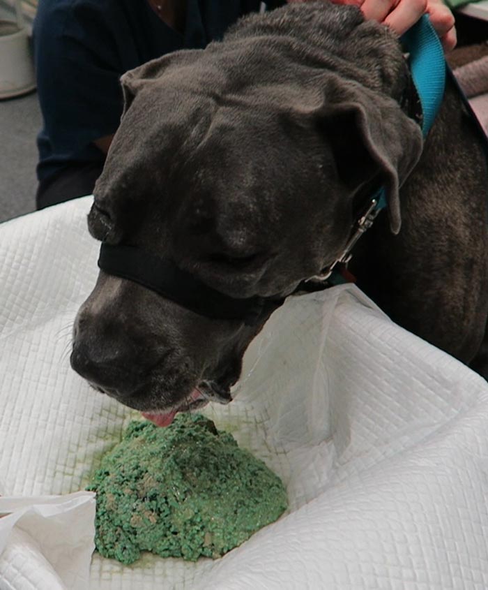
You might think of using a contrast medium to help, and the first that always comes to mind is barium. However, my personal first choice is a iodinated contrast medium – urografin, for example.
Advantages
Why? Their use is typically limited to myelograms or intravenous contrast studies but they can be given orally as well, for the assessment of oesophageal foreign bodies.
Lateral feline chest radiograph after contrast.
The advantage of using this over barium is that if this dye is accidentally aspirated it does not cause pneumonia like barium can.
How to
Using iodinated contrast medium is simple:
Given orally non-diluted, Dogs: 5-10ml, Cats: 5ml – you can give more if necessary
Immediately repeat the radiographs
If there is anything in the oesophagus, it will be highlighted
Nutrition is a key factor in a patients recovery; in fact, numerous studies show getting patients to eat as soon as possible or providing nutritional support early has several benefits:
Patients start to eat on their own earlier.
They are less nauseous once they start.
Reduced mortality.
Improved wound healing.
All of these contribute to overall improved outcomes for the patient.
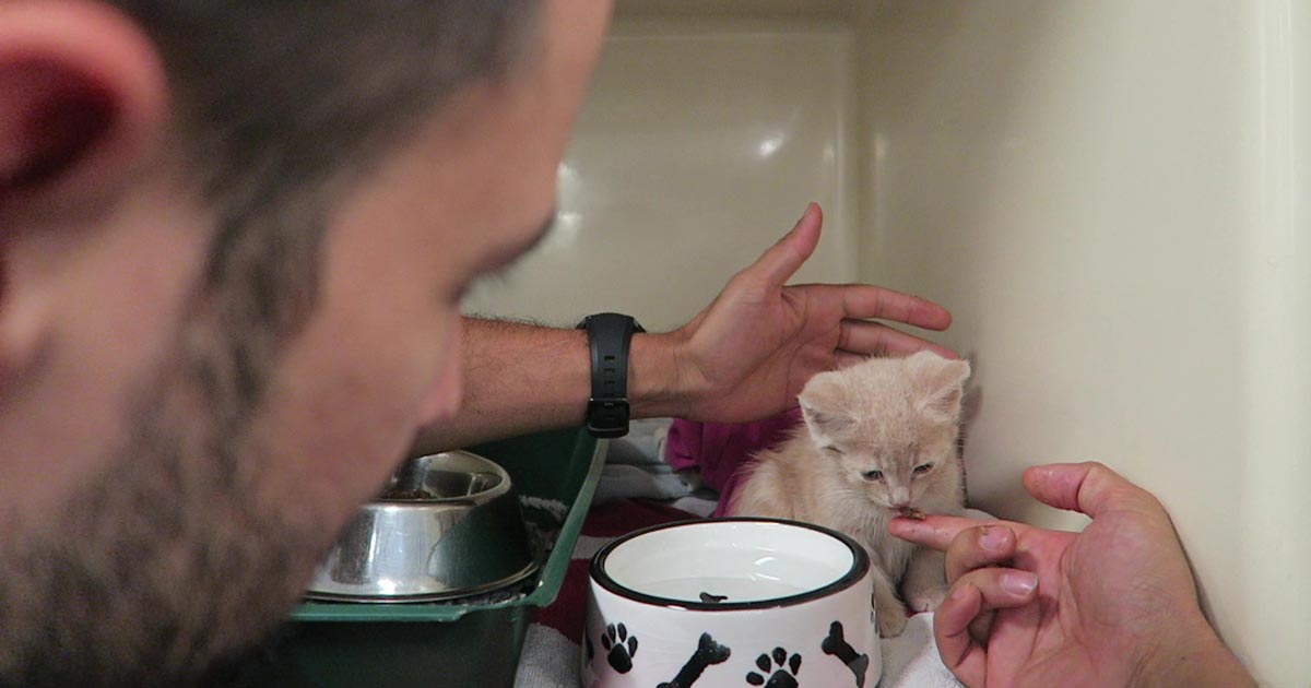
Encouraging patients
At Animal Emergency Service we treat the sickest of the sick so we work hard towards encouraging patients – just like the kitten pictured above – to eat as soon as possible. So, after they have recovered from their anaesthesia we make sure there are no contraindications, we address their nausea and pain, then offer food.
It is best for the patient and feeding to use as much of the gastrointestinal tract as possible, meaning it’s better if they eat on their own, otherwise the next best thing is an oesophageal tube, followed by a tube into the stomach, such as a nasogastric tube.
Focused approach
So, in combination with the management of pain, nausea and the underlying illness, we first encourage them to eat on their own. We begin with offering an assortment of different foods, warmed up to increase aromas, or ask owners to try to feed their pet.
If they are critically ill we will take a more focused approach with feeding tubes, as they are unlikely to eat on their own by themselves for several days.
We feel a proactive approach to early nutrition helps get our patients home to their families earlier.
Passing the stomach tube inside the roll down into the oesophagus (click to zoom).
Gastric decompression can be achieved in two ways:
trocarisation
stomach tube (orogastric tube) placement
The decision on which method to use depends on many factors – personal preferences, past experiences and clinical protocols, to name a few.
So, which one is best? A retrospective analysis of 116 gastric dilatation-volvulus (GDV) patients (Goodrich et al, 2013) found both methods of gastric decompression had low complication and high success rates, and either technique is acceptable.
If one method fails to achieve gastric decompression, the other can be tried.
How to decide
Personally, I use either or sometimes both. Which one I choose first depends on the situation. My decision-making process goes something like this:
Not clinically obvious or mild GDV
These are often diagnosed based on supportive radiographic findings as history and presenting clinical signs making me suspicious of a GDV.
I would always try to pass a stomach tube in these patients first, as the tube is passes easier when the gastric distention is milder. Although this procedure generally requires prior opioid analgesia administration to help reduce the stress, it can achieve rapid and lasting decompression of the stomach.
I often leave the tube in throughout stabilisation, just prior to induction of anaesthesia for surgical correction of the torsion. The tube allows continual release of gastric gases that can accumulate again rapidly if the tube is removed prior to surgery.
Obvious or severe GDV
The abdomen in these animals is often distended and tympanic. I will perform trocarisation in these cases first, as passing a stomach tube in these patients is often unsuccessful. It allows rapid gastric decompression, which is particularly important in cases with evidence of respiratory compromise.
After the trocar is no longer releasing gas, I will then pass a stomach tube. At this stage, it is often easier to pass the stomach tube once the gastric pressure has been reduced. Once again, I often leave the tube in during stabilisation.
How to perform
Stomach tube
The main risk is rupture of the oesophagus or gastric wall.
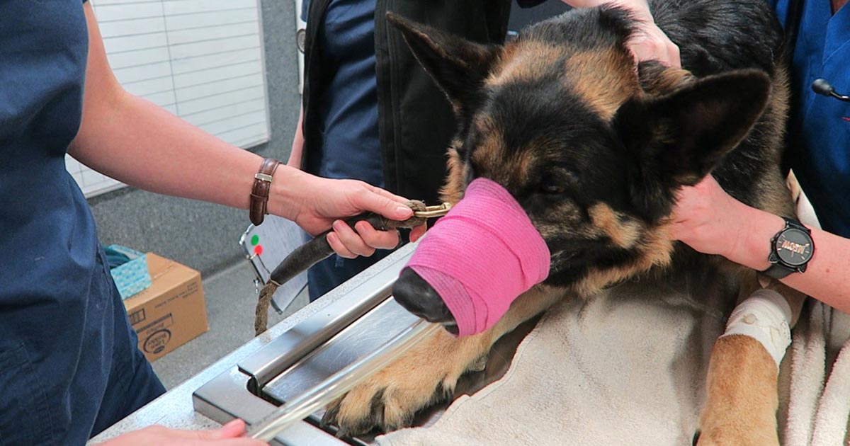
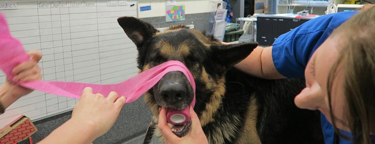
Pre-measure and mark the tube from the mouth to the level of the last rib.
Use a roll of adhesive bandaging material as the mouth gag. I prefer to use Elastoplast as it has an incompressible plastic core and the diameter is just large enough to fit our largest diameter stomach tube.
Unwrap approximately 30cm of Elastoplast before placing the roll of tape inside the mouth.
Wrap the tape snugly around the muzzle to prevent the dog from opening its mouth and dislodging the roll.
Lubricate the tube to reduce frictional trauma to the oesophagus.
Pass the stomach tube through the core of roll and into the mouth. You will feel a dead end at the level of the lower oesophageal sphincter, where the volvulus has torsed the oesophagus.
Apply gentle constant pressure and, most times, the tube will pass through into the stomach. Sometimes a puff of gas can be heard and felt from the aboral end of the tube when it enters the stomach. The tube can also be palpated when the stomach is decompressed.
The tube is taped to the muzzle to prevent dislodgement and the aboral end placed in a bucket to allow fluid to exit via gravity and siphon.
If it does not pass, reassess to see if trocarisation is required to relieve some pressure in the stomach
As mentioned above, I generally leave the stomach tube in while continuing to stabilise the patient and prepare for surgery. Gas can rapidly accumulate in the stomach and cause rapid deterioration if the tube is not left in. The tube is removed just prior to induction of anaesthesia.
Placing a roll in the mouth to prevent biting down on the stomach tube.
Trocarisation
The main risk is hitting the spleen while trying trocarisation. To avoid this, identify the most tympanic site by palpation, or use the ultrasound to confirm the absence of the spleen.
A 3in, 14g catheter is usually sufficient.
Clip and surgically prep a 10cm by 10cm area where you intend to place the catheter.
Insert the catheter to the hub and remove the stylet.
Although local anaesthetic in the area is ideal, you will not have time to do this in most cases – especially the very unstable ones. Also, since I administer pure opioid agonist intravenously to most confirmed GDV cases on presentation, local anaesthetic is not required.
Remove the stylet and gas should come blowing out under pressure.
Once the gas flow starts to slow down, gently apply inward pressure or pressure on the dilated stomach, which helps ensure the stylet does not fall out of the stomach and as much of the gas is removed as possible.
Until I started researching this Tip of The Week, I did not know the medical profession has abandoned the routine use of emesis in oral poisoning.
This is based on multiple medical literatures that have proven emesis induction does not influence the clinical severity of poisoning, the length of hospitalisation and the clinical outcome or mortality.
Although the rationale for inducing emesis is obvious, it is not necessarily evidence based. It is also dependent on satisfying a few large assumptions, all of which are untrue:
Emesis is a very effective way of removing gastric contents.
No separation exists of poison from its vehicle while inside the acidic environment of the stomach.
Poison is not absorbed through the stomach wall.
Ineffective method
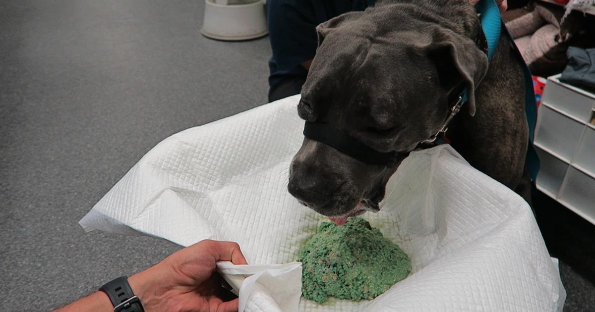
Snail bait ingestions: this patient ate 500g of snail bait containing metaldehyde.
Emesis induction is an ineffective way of clearing stomach contents. A review of the effectiveness of induced emesis, with both human and canine participants, showed at 30 minutes post-ingestion of non-absorbable markers, the recovery rate averaged between 17.5% and 52.1%, but never exceeded 62%.
In fasted puppies, this was even lower at 2% to 31%, despite inducing emesis immediately after marker administration. These have been confirmed by the presence of poisonous materials in the stomach of dead patients, despite effective emesis induction until clear fluid was brought up.
The clinical outcome only improves if the systemic exposure of a toxicant is reduced by more than half. However, considering animals rarely practice restraint, the ingested amount is unlikely to be exactly the lethal dose and no more. Therefore, even reducing the ingested toxic dose by 62% is unlikely to make a clinical difference.
Furthermore, most patients rarely present within 30 minutes of ingesting a toxicant, thus further reducing its efficiency.
The absorption conundrum
Some may argue the retrieval of metaldehyde or anticoagulant rodenticide granules from vomitus is indicative of reducing the toxicant dose. This could be true, but only if emesis was induced immediately after ingesting the poison.
The poison itself is colourless and has a different absorption characteristic to the coloured vehicle (granule); therefore, the presence of granule only serves to confirm ingestion, but is of no indication whether the poison has already been absorbed.
Contraindications
Many well-recognised absolute contraindications also exist to inducing vomiting:
Ingestion of oils, which includes waxes that melt to oil in the internal body environment, as this poses a high risk of lipoid and bacterial pneumonia. This is of significant veterinary significance, as wax is routinely used in rodenticide baits.
Ingestion of hydrocarbons and other volatile substances, or caustic or corrosive substances.
When the mental status is altered – for example, hyperexcitable or depressed mental state.
Where the patient is at risk of seizures (seizures can be induced by emesis).
Increased intracranial pressure.
Risk of intracranial or cerebral haemorrhage – for example, thrombocytopenia or abnormal clotting parameters.
Other less severe, yet important, reasons include:
delays administration of more effective treatment, such as activated charcoal, antidote or other treatments
risk of aspiration pneumonia
hypochloraemia in recurrent emetic patients
significant CNS and respiratory depression from apomorphine
rare, but reported, complications such as cerebral haemorrhage, oesophageal tear/ rupture, hiatal hernia, gastric rupture, pneumothorax and pneumomediastinum
legal implications – for example, if the product information clearly states emesis should not be induced
A place for everything
Emesis induction is not a benign procedure. It still has its place in certain circumstances, but its use in the routine management of oral poisonings may need to be reconsidered – especially if it means delaying administration of a more effective treatment, such as activated charcoal.
So, after all this, how do I tackle this information? It is a bit hard to swallow. My clinical experience is emesis is generally safe, especially in canine patients using apomorphine. So, I still feel some merit exists in reducing the amount of toxicant in the stomach if you have a chance – and in some situations, you don’t know until you try.
Emesis after ingestion of a toxic dose of chocolate can be incredibly rewarding, even six hours after ingestion, leading to patients not developing clinical signs at all.
Overall, I am biased by my personal successes with emesis, so still feel a time and place exist for emesis induction. But I now stop and question my decision to induce emesis, whereas I did not hesitate before.
If used by an experienced ultrasonographer who knows what to look for, ultrasound can be a highly sensitive and specific diagnostic test.
What do we look for?
Remember not all patients will have intestinal dilation as the linear foreign body may be only causing a partial obstruction. Alternatively, it could be occluding the gastric outflow completely.
Intestinal plication, which looks like intestinal loops bunching up on each other around the tether.
A central discrete hyperechoic line running along the middle of the bunching intestine. This bright line is the tether. Often when looking closely enough, the tether will have distal acoustic shadowing as the ultrasound pulses cannot pass through it.
The leading aboral segment and the trailing adoral anchor will have acoustic shadowing.
The adjacent mesentery is often hyperechoic compared to other areas in the abdomen, indicating inflammation.
Gastric dilation with fluid is often seen if the anchor is in the pylorus, as it causes an outflow obstruction.
Free abdominal fluid may be visible and a sample should be collected for assessment. If bacteria can be demonstrated in one of the following ways:
By visualising free or intracellular bacteria under the microscope.
By finding that the glucose is lower (lower than 20mg/dL) and the lactate is higher (2mmol/L) in the abdominal fluid sample compared to peripheral blood then this indicates perforation of the gastrointestinal tract has occurred and septic peritonitis is present.
In the third and final post, we will cover things to consider when deciding whether to perform the exploratory laparotomy yourself, or if you should transfer the patient to a referral facility for surgery.
Linear foreign bodies can be tricky to diagnose, compared to normal foreign bodies, for many reasons. Mostly because you often don’t see the classic obstructive pattern appearance on radiographs or ultrasound.
In this short blog series, we are going to cover some hints and tips that can make diagnosing a patient with a linear foreign body easier. Then, we’ll discuss things that should be considered when deciding whether you are the right person to take the patient to surgery…
So, let’s start with radiography.
Not all patients with a linear foreign body will be completely obstructed. This means you won’t always visualise classic intestinal dilation. In fact, it has been reported that up to 50% of patients with a linear foreign body will not have an obstructive pattern present on radiographs.
Look for the characteristic small “comma shaped” gas pattern. This is caused by plication and bunching of the small intestine around the tether.
The small intestine can appear to be bunched up in one area, rather than spaced out around the abdomen. However, obese animals – especially cats – can have “pseudo-bunching” due to large amounts of abdominal fat bunching the intestine together.
Loss of serosal detail is often seen due to inflammation surrounding the affected intestine.
Always include a left lateral radiograph in your series. Gastric contents will fall to the fundus on the left of the abdomen and gas will raise to the pylorus, which will highlight the foreign body anchor in the pylorus.
Perform thoracic radiographs to assess for aspiration pneumonia and a potential oesophageal component of the linear foreign body. If aspiration is present then you know you will need to continue antibiotic therapy postoperatively.
In the next post, we cover some key points for diagnosing linear foreign bodies on ultrasound…