Tag: Neurology

  • Oh, CR*P! Using point-of-care C-reactive protein tests

    Oh, CR*P! Using point-of-care C-reactive protein tests

    Few companies now offer affordable point-of-care tests for canine C-reactive protein (CRP). As we did when we recently received our new box of CRP slides, you might soon be asking the question: what do we even do with this stuff?

    Here’s what we’ve learnt…

    CRP is one of the acute phase proteins produced by the liver in response to inflammation. Healthy patients have very low levels of CRP, but a systemic inflammatory condition will cause an increase in CRP within four to six hours. Conversely, increased levels will decrease rapidly on resolution of inflammation. This provides an almost real time measure of inflammation that is more responsive and reliable than the white blood cell response.

    In other words, CRP can indicate the presence of inflammation before the patient’s white blood cell count gives any clues, or before it becomes pyrexic – and, unlike the white blood cell count, stress and steroids do not affect CRP levels.

    Uses

    So, how do we use it?

    • I love it for early pickups of problems in those grey area cases: the dog seems okay on clinical examination, but something about it bothers me. A normal or mildly increased CRP test will make me sleep more easy, while a surprise high reading will prompt me to admit for full diagnostics, or at least get the patient in for a follow-up CRP the next day. Conversely, a localised problem – such as an abscess – combined with a normal CRP test might mean you can hold off on antibiotics and just recheck CRP in 24 hours.
    • It’s great for monitoring response to treatment. If my plan is working then I’d expect CRP to show a significant decrease by day two or three. If it’s not dipping by then, I need to reassess my treatment plan. Do I need to change antibiotics? Scan it again? Maybe we need to consider surgery? It can also be a good prognosticator. Research has shown failure of CRP to decrease significantly (around a 3× decrease) by around day three is generally bad news for patients with inflammatory conditions such as pancreatitis and immune-mediated haemolytic anaemia.
    • We are starting to play with it for post-surgical monitoring. Any surgery will cause inflammation with an increase in CRP levels, but in an uncomplicated postoperative period, you should expect levels to start decreasing by day three to five. A base line CRP 24 hours after surgery with a recheck on day three should pick up early signs of postoperative problems such as infection, and prompt investigation or intervention.
    • A potentially nifty use for it that we haven’t yet had the opportunity to use is in differentiating inflammatory lamenesses (arthritis, infection, injury) from a neurological causes – that is, is it arthritis or a nerve problem?

    Limitations

    • Remember, it’s very sensitive, so will increase with almost any inflammation. A mild upper respiratory infection or a bad gingivitis will likely induce some changes, so it’s important not to over-interpret (keep in mind that the magnitude of the increase in CRP does generally correspond with the severity of the inflammatory response). A pancreatitis case where the CRP fails to drop does not always mean death is looming – you may have just missed the concurrent skin disease. Always interpret CRP values in concert with your clinical examination.
    • Be aware that pregnancy and intense exercise can increase CRP values.
    • Not all serious conditions have an inflammatory component. CRP will be unchanged in most veterinary cases of heart disease; in common hormonal disease, such as adrenal disease and uncomplicated diabetes; urinary obstructions; many localised cancers; epilepsy and many others. Don’t presume that just because CRP is normal, everything is fine.
    • No similar test exists for cats.

    Sit up and say…

    My favourite way to explain how to use this test is by using its highly appropriate acronym – any unexpected increase should make you sit up and say: “Oh CR*P! What am I missing?”

  • Hyponatraemia, pt 2: causes

    Hyponatraemia, pt 2: causes

    The causes of hyponatraemia can be divided into three major categories, based on serum osmolality. This is further divided based on the patient’s volume status (Table 1).

    Most patients we see in clinic fall into the hypovolaemic category, except patients with diabetes mellitus.

    Table 1. Causes of hyponatraemia based on osmolality and volume status (from Guillaumin and DiBartola, 2017).
    Hypo-osmolar Hyperosmolar Normo-osmolar
    Hypovolaemic Normovolaemic Hypervolaemic
    Gastrointestinal fluid loss
    Third-space fluid losses
    Shock
    Hypoadrenocorticism (Addison’s disease)
    Renal insufficiency
    Excessive diuretic administration
    Salt-losing nephropathy
    Cerebral salt wasting syndrome
    Syndrome of inappropriate antidiuretic hormone secretion (SIADH)
    Hypotonic fluid administration
    Hypothyroidism
    Glucocorticoid insufficiency
    Psychogenic polydipsia
    Reset osmostat (SIADH type B)
    Congestive heart failure
    Acute or chronic renal failure
    Nephrotic syndrome
    Hepatic cirrhosis
    Accidental ingestion or injection of water (water intoxication)
    Hyperglycaemia
    Mannitol
    Severe azotaemia
    Hyperlipidaemia
    Hyperproteinaemia

    Common causes

    In dogs, the three most common causes of hyponatraemia are:

    • gastrointestinal (GI) fluid loss
    • third-space fluid loss
    • fluid shift from intracellular fluid to extracellular fluid (ECF) as a result of hyperglycaemia

    In cats, the three most common causes of hyponatraemia are:

    • urologic diseases
    • GI fluid loss
    • third-space fluid losses

    In most patients, more than one pathophysiologic factor is likely to be contributing to the hyponatraemia.

    Circulating volume

    Hypovolaemic patients – those with, for example, GI losses, hypoadrenocorticism, renal losses and haemorrhagic shock – have a reduced effective circulating volume. ECF contraction triggers antidiuretic hormone (ADH) secretion, which leads to increases in free water absorption and thirst, and results in dilution of the serum sodium concentration. Aldosterone secretion is reduced in hypoadrenocorticism, so an overall reduction in sodium reabsorption compounds the problem.

    Hypervolaemic patients are those with an increased fluid retention state, such as:

    • congestive heart failure (pulmonary oedema)
    • advanced hepatic failure (ascites, third-space fluid)
    • renal failure
    • free water ingestion

    Congestive heart failure patients have a reduced cardiac output and, therefore, a decreased effective circulating volume, despite the presence of the extra fluid status. Renin-angiotensin activation leads to release of ADH and aldosterone, resulting in sodium and free water reabsorption, and increased thirst. Both lead to an excess of free water retention.

    Advanced hepatic (cirrhosis) or renal failure (nephrotic syndrome) both result in hypoalbuminaemia, leading to fluid shifting into the interstitial space and third space, reducing effective circulating volumes. This leads to activation of ADH to increase free water reabsorption, to restore the circulating volume in the face of existing hypervolaemia and hyponatraemia.

    Diabetic patients

    Moderate to severe hyperglycaemic diabetic patients can be either hyperosmolar or normo-osmolar, depending on the serum blood glucose concentration. Hyponatraemia occurs when water shifts from the intracellular fluid to the ECF down the osmotic gradient, diluting the serum sodium content.

    Despite this osmotic shift, not all diabetic patients develop hyponatraemia. Glucosuria also causes also causes a renal osmotic shift, sometimes resulting in urine water loss in excess to sodium. This offsets the hyponatraemia – in some cases, hypernatraemia results.

    Treatment

    Treatment of hyponatraemia hinges on how quickly it developed and the volume status of the patient. The rule of thumb is to correct hyponatraemia slowly – not exceeding 0.5meq/L/hr – especially in chronic cases, or cases where the duration of hyponatraemia is unknown. Keeping to this rate is paramount until serum sodium concentration reaches 130meq/L.

    In acute patients with severe clinical signs, such as seizures, some clinicians may choose to use a higher rate of 1meq/L/hr to 2meq/L/hr until clinical signs resolved.

    It should be emphasised, once again, this rate should never be used in chronic patients, patients with an unknown duration of hyponatraemia, or where frequent serum sodium concentration cannot be monitored. The rapid correction of hyponatraemia can lead to osmotic demyelination syndrome (myelinolysis).

    Its effect will not be apparent until three or four days after therapy, and can result in neurological abnormalities such as:

    • weakness
    • ataxia
    • dysphagia
    • paresis
    • coma

    For that reason, frequent electrolyte measurements are required, starting hourly then once a suitable rate of increase has been established and less frequently thereafter.

    • Part 3 will look at how to correct patients with hyponatraemia.

    Reference

    Guillaumin J and DiBartola SP (2017). A quick reference on hyponatremia, Veterinary Clinics of North America: Small Animal Practice 47(2): 213-217.

  • Hyponatraemia, pt 1: clinical signs

    Hyponatraemia, pt 1: clinical signs

    Hyponatraemia is a relatively common electrolyte disturbance encountered in critically ill patients, and the most common sodium disturbance of small animals.

    In most cases, this is caused by an increased retention of free water, as opposed to the loss of sodium in excess of water.

    Low serum sodium concentration

    Hyponatraemia is defined as serum concentration lower than 140mEq/L in dogs and lower than 149mEq/L in cats.

    The serum sodium concentration measured is not the total body sodium content, but the amount of sodium relative to the volume of water in the body. For this reason, patients with hyponatraemia can actually have decreased, increased or normal total body sodium content.

    This series will look briefly at the modulators of the sodium and water balance, clinical signs associated with hyponatraemia, the most common causes in small animals, the pathophysiology behind these changes, and treatment and management.

    ECF volume

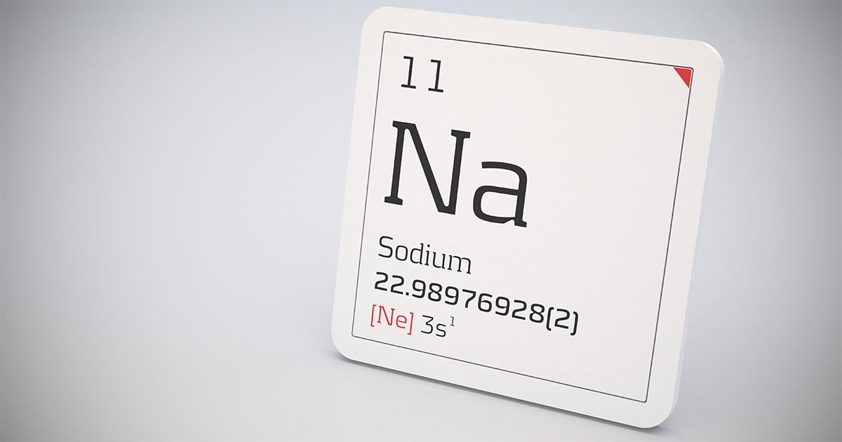
    hyponatraemia
    An example of hyponatraemia.

    Sodium is the main osmotically active particle in the extracellular fluid (ECF), so is the main determining factor of the ECF volume. Any disease process that alters the patient’s ECF volume will lead to hyponatraemia, such as:

    • dehydration
    • polyuria
    • polydipsia
    • vomiting
    • diarrhoea
    • cardiac diseases
    • pleural or peritoneal effusion

    The modulators of water and sodium balance are also different, so should be thought of as different processes.

    Water balance is modulated by thirst and antidiuretic hormone, and the effect of this is to maintain normal serum osmolality and serum sodium concentration.

    Modulators of sodium balance aim to maintain normal ECF volume. It adjusts this by altering the amount of renal sodium excretion; an expansion of ECF volume will lead to an increased sodium excretion, while a reduction in ECF volume will lead to increased sodium retention.

    Rate and magnitude

    The clinical signs of hyponatraemia are both dependent on the magnitude of the decrease and the rate at which it developed.

    In mild or chronic patients, no visible clinical signs can exist. In severe (lower than 125mEq/L) and acute cases, clinical signs exhibited are typically neurological, reflecting cerebral oedema. Possibilities include:

    • lethargy
    • anorexia
    • weakness
    • incoordination
    • disorientation
    • seizures
    • coma

    Patients with acute hyponatraemia – for example, water intoxication – are more likely to show clinical signs, compared to those with chronic hyponatraemia, because the brain takes time (at least 24 to 48 hours) to produce idiogenic osmoles, osmotically active molecules that help shift free water out of brain cells.

    Therefore, any acute hyponatraemia that develops within a 24 to 48-hour period tend to show clinical signs, whereas chronic cases are less likely.

    • Next week’s blog will look into the different causes of hyponatraemia and how they result in sodium loss.
  • Blood gas analysis, pt 4: respiratory acidosis and alkalosis

    Blood gas analysis, pt 4: respiratory acidosis and alkalosis

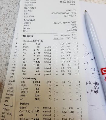
    Assessing the respiratory component is simple. A quick glance at the partial pressure of carbon dioxide (PCO2) level can tell you whether a respiratory acidosis or alkalosis is present.

    If the PCO2 level is elevated (respiratory acidosis) then either a primary respiratory acidosis is present, or it is the result of a compensatory response to a metabolic alkalosis.

    Similarly, if the PCO2 level is low (respiratory alkalosis) then it could either be a primary respiratory alkalosis, or compensation to metabolic acidosis has occurred.

    The respiratory component should always be assessed before the metabolic component, due to the ability to respond to pH shifts almost immediately. This, therefore, is a more accurate reflection of the patient’s clinical disease.

    Respiratory acidosis – increased CO2

    Respiratory acidosis occurs anytime the patient is hypoventilating and not eliminating CO2 appropriately.

    As hypoventilation can be associated with hypoxia, these patients are often critical and require immediate interventions.

    Causes of respiratory acidosis include:

    • drugs (depress respiratory centre, relax thoracic muscles)
    • neuromuscular disease (for example, tick paralysis, botulism and snake envenomation)
    • upper airway obstruction
    • pleural disease (for example, pneumothorax, pleural effusion and diaphragmatic hernia)
    • gas exchange disorders (for example, pulmonary thromboembolism, pneumonia and pulmonary oedema)

    Respiratory alkalosis – loss of CO2

    Respiratory alkalosis occurs when a patient is hyperventilating – excessive loss of CO2 causes the pH to increase.

    The health effect of this is usually minimal, since, in most cases, the effect is secondary and correction of the underlying cause usually resolves this problem. The exception is when respiratory alkalosis is a primary disorder. This is usually quite rare, but can occur with brain stem trauma where the respiratory centre is affected.

    Causes of respiratory alkalosis are:

    • hyperventilation (for example, fear, pain, stress, anxiety and hyperthermia)
    • neurological (for example, head trauma/neoplasia involving the respiratory centre)

    Anticipating changes

    Correctly identifying the primary disorder is essential for anticipating the changes the patient is likely to experience. This will help identify the underlying disease, and is essential for patient monitoring and disease management.

    In the next blog, we will discuss assessment of the metabolic component.

  • A difference of opinion

    A difference of opinion

    I’m only a few short weeks into my final-year rotations at the University of Bristol’s Veterinary Referral Hospital, but I already feel like I’ve learned a lot:

    • DOPs aren’t as scary as I’d built them up to be in my head.
    • It does get easier to navigate your way around the hospital with time (and trial and error).
    • There are quite a few differences between first-opinion and second-opinion practice that I’d never really considered until now.

    Budgets

    The gift that is the NHS can certainly make us blind to the costs of routine medical procedures. Something as simple as an ultrasound, blood work and a couple of days’ hospitalisation can amass a bill that’s simply unaffordable for a lot of pet owners.

    I’ve seen a lot of cases reach the end of the road due to lack of funds, when the answer (or potential answer) was frustratingly simple, but just too much money. The reality of referral practice, however, is that if your patient has made it to you in the first place, there is likely a higher budget to play with than the average consult.

    When you’re on a certain rotation, you find yourself doing the same diagnostics every day, so it’s easy to lose track of the value of the drug you’re administering, or the probe in your hand. I think my group and I honestly balked when the cardiology team told us the cost of a standard echocardiogram at the end of our week where we’d been observing between 6 to 10 a day. Of course, it’s still important to keep costs low wherever possible, but it’s been interesting to see how larger budgets and insurance policies are broken down.

    Seeing small animal CTs, echocardiograms and neurosurgeries for the first time was an amazing experience, but I do need to keep reminding myself that the proportion of my future patients that will go on to have these sorts of procedures is incredibly small.

    Image © TungCheung / Adobe Stock

    Specialisms

    When you’re learning in a veterinary hospital, you’re constantly surrounded by leading experts in the field of everything under the sun, and sometimes it’s hard not to feel like a monkey with a stethoscope. Usually, in first opinion, there’s an assortment of different strengths throughout the practice – one vet may have intercalated in neurology and behaviour, while another may has done more CPD on exotic animal medicine. These differences are an asset to every practice and make team working an essential and valuable commodity.

    In a referral setting, these “strengths” are often extended to actual specialisms, where the vets are not only actively involved in research in a particular field, but see only animals in a certain category of illness. The need for teamwork, however, is just as paramount here – if not more so!

    After working in cardiology for a week and beginning to feel like I didn’t know anything, I can tell you that it was very refreshing to have the head of another department pop their head through the door and ask what on earth was going on with their patient’s heart. When they also didn’t know the actions of all the drugs I’d been painstakingly trying to commit to memory for the past five days, that too came as a wave of relief.

    Client communication

    Good communication and patience come hand in hand. If you’re delivering bad news to a client or talking them through a complicated diagnosis or treatment plan, that takes time – and although the average first opinion consult is only 15 minutes long, I’d say that, rather oxymoronically, there’s more time to deal with difficult situations in that scenario than in emergency referral.

    Of course, first opinion sees it’s share of emergencies as well, but for the most part vets see a disease present slowly over time and are able to prepare their clients accordingly.

    In the past couple of weeks, however, I’ve seen referral vets have to delicately balance client communication with the urgency of life-threatening conditions. Sometimes there are mere minutes to intervene after an animal enters the hospital, and vets must be very diligent and considerate when explaining this situation to an owner who may not yet grasp the severity. Owners have to have informed consent at all times, and to be prepared and supported in the event of any potential outcomes, but the sooner an animal is triaged and either treated or prepped for surgery the better.

    I think that this is where the truly brilliant vets really shine. To have compassion and humanity at the forefront, with animal welfare and haste also in mind, takes a lot of mental and emotional gymnastics. I’m honestly in awe of every vet I’ve seen both in the past few weeks and over my years seeing practice who’s had to deal with a crisis on both the animal and the human end.

    As of yet, I’m unsure if my career will lead me to first opinion or referral practice, but I can appreciate the similarities and differences between the two – despite us all starting out in the same place.

  • Intoxication: working out possible ingested dose

    Intoxication: working out possible ingested dose

    We frequently field telephone calls from owners concerned about their pet being intoxicated or having access to a toxic compound.

    These are the list of questions I always ask owners:

    What is your pet doing?

    The main reason I ask this question first is to determine if the pet’s life is in danger. If the pet is seizing, collapsed, neurological, bleeding or having difficulty breathing then they need to come into practice immediately.

    What led to the suspicion of toxic exposure?

    This can help provide useful background information.

    What is the product?

    In some situations, owners can tell you accurately over the telephone what they think they have been exposed to.

    Asking them to bring the packaging, and whatever is remaining of the toxin, with them can help determine a possible dose they have been exposed to.

    When did this occur?

    A timeline, and when they think the pet could have been exposed to the toxin, is critical as it can help put presenting clinical signs into perspective.

    I always ask if they could have had prior exposure to the toxin. An example where this may be important is with rodenticides.

    What have you done in response to this?

    Owners may have tried to address the situation themselves, using information gained from the internet.

    Attempts to induce emesis can also make pets incredibly ill and result in neurological signs.

    Have they passed faeces or vomitus with the toxin?

    If the answer is yes, ask them to bring the pet into the practice. This can help identify the toxin; some baits are coloured and can easily be seen. These samples may even be able to be sent away for further testing.

    Do you have any other pets that may have also had access to the toxin?

    Other pets that may have had access will need to be seen in practice as well. A classic example is a multi-dog household where one pet is the scavenger. Owners may neglect to inform you their other pets may have been the culprits, but did not because they assumed it was the one with the history of being a scavenger.

    Next week, we will cover the decontamination steps owners can carry out at home.

  • Handling an Addisonian crisis – part 2

    Handling an Addisonian crisis – part 2

    Managing an Addisonian crisis can be daunting, especially when the patient looks like it is about to check out and its baseline bloods show a sodium of 110mmol/L, a potassium of 8mmol/L and a glucose of 2.3mmol/L. That is enough to make anyone’s brain explode.

    The patient can be treated in many ways, but I find it useful to try to simplify and prioritise. I have outlined my thought process in the hope some of you will find it helpful.

    First 10 minutes: protect heart and manage hypoglycaemia

    • Protect the heart – calcium gluconate 10% 0.5mL/kg to 1.5mL/kg slow IV over 10 minutes to counter the effects of hyperkalaemia on cardiac electrical activity. This buys about 20 minutes of time.
    • Treat the hypoglycaemia – the dose depends on the severity, but 0.5ml/kg of 50% dextrose IV diluted 50:50 with Hartmann’s is a good place to start. This dose of dextrose will also help correct hyperkalaemia by stimulating endogenous insulin release.

    First 20 minutes: start addressing perfusion deficits

    • Create a custom IV fluid – I do not aim to increase sodium concentration at all at this stage. I am a big fan of creating custom IV fluids. I create a fluid with a same sodium concentration as the patient then use boluses of this fluid to correct signs of shock without concerns of increasing the sodium. I use Hartmann’s as my base fluid – it has the lowest sodium concentration – and add 5% dextrose to reduce the sodium concentration (you may need to remove 100ml to 200ml from the bag first). I usually run the new fluid through the electrolyte machine to check the final sodium concentration.
    • Hartmann’s contains buffers that help address metabolic acidosis (and hyperkalaemia). It also contains potassium; however, if this concentration is less than that of serum it will still help to dilute serum potassium.
    • The formula I use to create a custom sodium IV fluid bag is beyond the scope of this blog and is detailed in the fluid therapy chapter of my book, The MiniVet Guide, under hyponatraemia.

    First hour: address hyperkalaemia

    • Image © mintra / Adobe Stock
      The author warns not to rush the sodium increase in patients. Image © mintra / Adobe Stock

      If the hyperkalaemia is severe enough to warrant more aggressive management than alkalinising IV fluids, improving renal perfusion and providing a dextrose bolus (such as potassium of more than 7mmol/L to 8mmol/L) then I would use regular short acting insulin at 0.25U/kg to 0.5U/kg IV. This should always be used in combination with a bolus of dextrose at 2g of dextrose per unit of insulin or 4ml of 50% dextrose for each unit of insulin, followed by a CRI of 2.5% to 5% glucose until insulin wears off (this could be up to six hours). This should prevent hypoglycaemia.

    • I administer dexamethasone up to 0.5mg/kg IV while running the adrenocorticotropic hormone (ACTH) stimulation test. This is the only corticosteroid that can be given as it does not cross react with the ACTH stimulation test.

    Next 2 to 24 hours: correct hydration and correct hyponatraemia

    • After I have corrected perfusion deficits with my custom IV fluid, I will address hydration deficits with an appropriate fluid plan over the next 24 hours. I usually replace 50% of the hydration deficit over the first 6 hours then the remaining 50% over the following 18 hours.
    • Correction of hyponatraemia can take a couple days as sodium should only be increased by 0.5mmol/L/hr (max 12mmol/L/day). If the sodium has not increased from the initial fluids given, I would create another custom IV fluid bag with a sodium concentration 10mmol/L above that of the patient’s. I would monitor electrolytes every one to four hours, depending on response.

    Supply mineralocorticoids and glucocorticoids

    • Options for steroid supplementation include dexamethasone 0.5mg/kg IV then 0.1mg/kg IV q12hrs or IV hydrocortisone sodium succinate at 0.5mg/kg/hr. Personally I use hydrocortisone CRI, asit has equal mineralocorticoid and glucocorticoid activity. Oral steroids can be used once the patient starts eating and drinking.
    • I only use a mineralocorticoid if I see no increase in sodium after starting hydrocortisone, despite using a fluid with a higher sodium concentration than the patient.

    Addressing patients this way will generally gets them out of the crisis. One thing that I don’t do is rush the sodium increase, it can take time and I am good with that. I have seen patients develop neurological signs from sodium levels that have increased too quickly. As for the long term management; well, I will leave that to you.

  • Festive threats to four-legged family members

    Festive threats to four-legged family members

    Now well into December, and getting ever closer to Christmas, there will be an abundance of “goodies” around the house that are not so good for the four-legged family members.

    Image: © Freeimages/s22k.
    Jordan advises colleagues to ensure clients keep an eye on their pets during the festivities. Image: © Freeimages/s22k.

    Most owners are aware of the dangers of chocolate and so are likely to rush down to the vet on Boxing Day when their Labrador has broken into the tin of Heroes – but what other festive dangers are there that owners are not so clued up on?

    Pudding

    The festive favourite Christmas pudding, while enjoyable for us, can be lethal for pets. It is the raisins, currants and sultanas that may lurk within that are poisonous.

    While the mechanism of toxicity is poorly understood, we know it can cause acute kidney failure in dogs.

    The treatment protocol for poisoning by raisins, etc, is similar to general suspected poisoning with an unknown agent: give an emetic (to make the animal vomit), an adsorbent (for example, activated charcoal) and supportive care (IV fluids) to protect the kidneys for a few days.

    Antifreeze

    Ethylene glycol, more commonly known as antifreeze, is extremely toxic to pets, with consumption of tiny amounts resulting in crystal formation in the kidneys, and so acute renal failure. Antifreeze smells and tastes sweet, so pets are often attracted to it. For this reason, it is best for owners to ensure it is kept in a secure container, well away from pets.

    Early symptoms include neurological signs, such as wobbliness, twitching, seizures and depression. As the poisoning progresses, the animal will have cardiac and respiratory signs, such as increased heart and respiratory rate. It’s vitally important owners look out for these signs if their pets have had any possible access to antifreeze (for example, if they’ve got into the garage or if a cat has been missing for a few days and potentially been locked in someone’s shed).

    The antidote for ethylene glycol poisoning is ethanol, because it replaces ethylene glycol as a substrate for the enzyme that breaks it down into toxic products. More specifically, lab-grade ethanol is best.

    Sweetener

    Xylitol is a sugar-free sweeter that is used in chewing gum, cakes and sweets, particularly diabetic foods. Xylitol causes insulin release in the body, resulting in hypoglycaemia and, later, liver damage.

    Symptoms include vomiting and signs of hypoglycaemia: lethargy, weakness, collapse and, if it progresses, seizures and coma. Unlike ethylene glycol, there isn’t a specific antidote, but supportive care, including fluids and liver protectants, is vital. The prognosis for xylitol toxicity is good if the animal’s hypoglycaemia is corrected quickly, so it is essential for owners that suspect this to contact their vet immediately. If you’re trying to determine whether xylitol is in a product, it is often listed as a food additive, code E967.

    Decorations?

    And don’t forget the potential foreign body too – if the cat has been playing with the tinsel and managed to swallow some or the dog has ingested a few tasty looking baubles.

    Enjoy the festive season, but advise owners to keep an eye on their pets this Christmas.