Tag: Mycoplasma Bovis Infection

  • Highlighting oesophageal foreign bodies on radiographs

    Highlighting oesophageal foreign bodies on radiographs

    before
    Lateral feline chest radiograph before contrast.

    QUICK TIP: Need to know if there is an oesophageal foreign body but can’t be certain on radiographs?

    We have all been in the scenario where we are unsure whether there is an oesophageal foreign body on the radiographs we have just taken.

    You might think of using a contrast medium to help, and the first that always comes to mind is barium. However, my personal first choice is a iodinated contrast medium – urografin, for example.

    Advantages

    Why? Their use is typically limited to myelograms or intravenous contrast studies but they can be given orally as well, for the assessment of oesophageal foreign bodies.

    after
    Lateral feline chest radiograph after contrast.

    The advantage of using this over barium is that if this dye is accidentally aspirated it does not cause pneumonia like barium can.

    How to

    Using iodinated contrast medium is simple:

    • Given orally non-diluted, Dogs: 5-10ml, Cats: 5ml – you can give more if necessary
    • Immediately repeat the radiographs
    • If there is anything in the oesophagus, it will be highlighted

    Tip of the Week author Gerardo Poli is the author of The MiniVet Guide to Companion Animal Medicine – now available in the UK.

  • Systemic antibiotics – a brief guide for new grads

    Systemic antibiotics – a brief guide for new grads

    A lot of information is available regarding different antibiotics and, for the newest generation of vets, the pressure to use them correctly and responsibly is greater than ever.

    One of main challenges when you start clinical practice is knowing the most appropriate antibiotic for common presenting conditions.

    Below is a rough guide for antibiotic selection according to body system. However, make sure you stick to the following rules:

    1. Limit antibiotic use to animals that actually require them – resist the urge to dispense them due to pressure from owners or when you feel there is nothing else to turn to.
    2. What is the likely type of bacteria you are aiming to target (such as anaerobes, Gram-positives and Gram-negatives)? Collect samples from lesions/discharge or effusions/blood and urine, and see if there is evidence of bacteria under the microscope.
    3. Use the most narrow spectrum antibiotic as possible.
    4. Perform a culture and sensitivity whenever possible – especially if a case does not respond to your first line antibiotic.
    5. Avoid using fluoroquinolones, third and fourth generation cephalosporins and amikacin without evidence of resistance from culture and sensitivity results.
    6. Use an appropriate dosage regime and make sure the owners have the capacity to administer them accordingly.

    Skin

    • Try topical chlorhexidine alone if surface pyoderma
    • Clindamycin
    • Cephalexin
    • Amoxicillin-clavulanic acid

    Upper respiratory tract

    • Doxycycline
    • Amoxicillin-clavulanic acid

    Lower respiratory tract

    • Amoxicillin-clavulanic acid
    • Ampicillin

    GI tract

    • Metronidazole (research questions the use of antibiotics for diarrhoea cases)
    • Tylosin (chronic diarrhoea)

    Urogenital tract

    • Remember that cystitis in cats is often stress-related rather than due to infection
    • Amoxicillin-clavulanic acid
    • Trimethoprim-sulpha (penetrates the prostate)

    Mastitis

    • Amoxicillin-clavulanic acid


  • Thoracentesis, part 2: sample work

    Thoracentesis, part 2: sample work

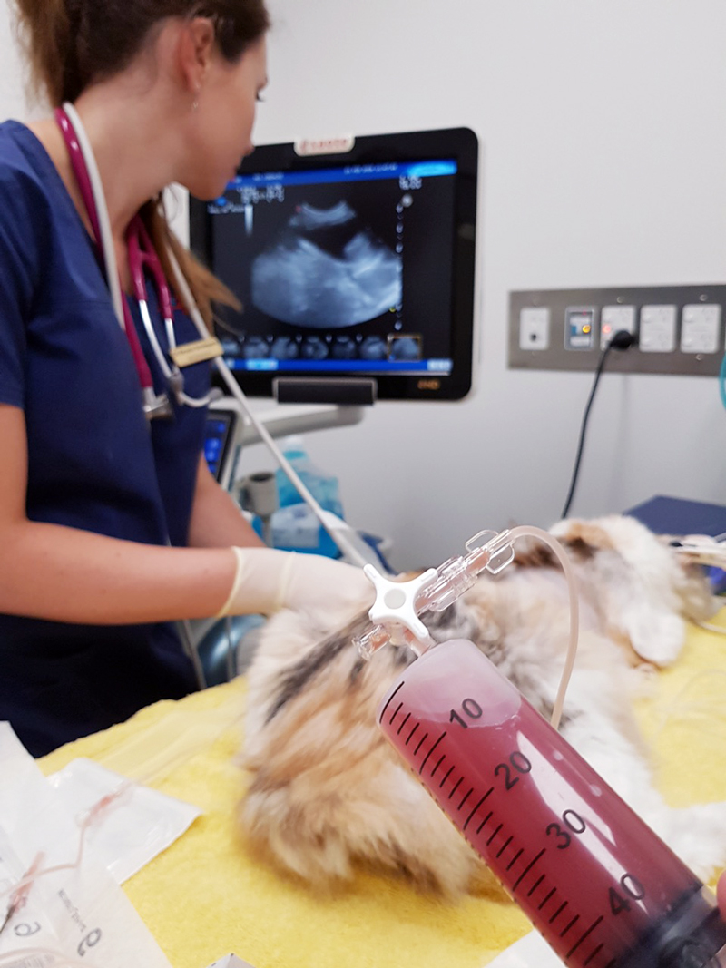
    Last week we gave some hints and tips about how to perform a thoracocentesis. This week we look at what to do with the sample you collected and where to go to next.

    Looking at the sample is not enough, there are several things you need to do to make sure you are getting the most information from the collected sample. This includes:

    • Fluid cell counts
    • Total protein assessment
    • Packed cell volume
    • Glucose
    • Lactate (if it is an exudate)
    • In-house cytology
    • Collect a sample for culture and sensitivity, and also external cytology assessment

    With this information you can narrow down your list of differentials; often enough it can give you a diagnosis.

    Here is the list I use. Note, it is not exhaustive and assumes you have taken three-view thoracic radiographs as part of the initial diagnosis.

    Transudate

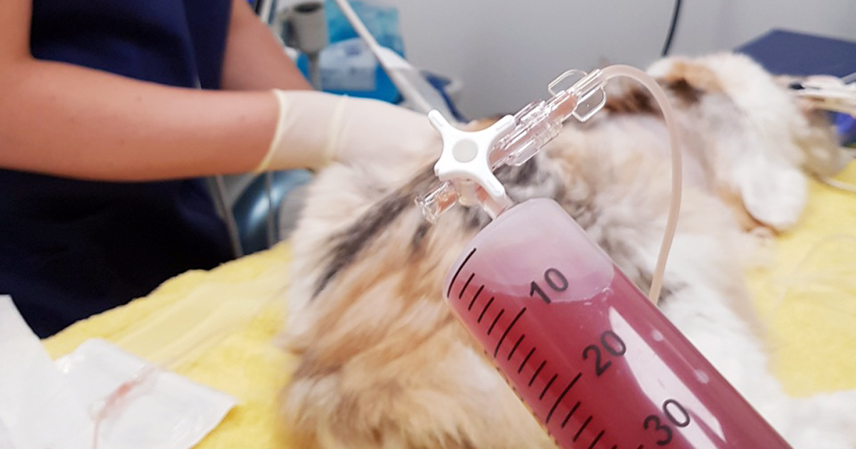
    • Haemorrhagic effusion.
      Haemorrhagic effusion.

      Clear appearance – characterised by low protein and low cellularity

    • Transudates are caused by reduced oncotic pressure
    • Total nucleated cell counts = <0.5x10e9/L
    • Total protein = <25g/L

    Differentials to consider

    • Liver disease
    • Protein-losing nephropathy
    • Protein-losing enteropathy

    Additional diagnostics

    • Cytology and culture of fluid
    • Haematology and biochemistry
    • +/- dynamic liver testing
    • Urinalysis, urine protein/creatinine ratio, culture and sensitivity

    Modified transudate

    • Yellow/serosanguinous/cloudy appearance
    • Caused by increased hydrostatic pressure leading to passive leakage of proteins and fluid into the pleural space
    • Total nucleated cell counts = 3.5-5x10e9/L
    • Total protein = variable, ~25-50g/L

    Differentials to consider

    • Increased capillary hydrostatic pressure and pericardial disease
    • Diaphragmatic hernia
    • Neoplasia
    • Lymphatic obstruction, such as neoplasia, diaphragmatic hernia and abscess
    • Increased permeability of vessels (blood and lymphatics), such as FIP

    Additional diagnostics

    • Cytology and culture of fluid
    • Haematology and biochemistry
    • Cardiac auscultation and ultrasound
    • +/- CT

    Exudate

    • Turbid appearance – Very proteinaceous liquid, froths when shaken
    • Fluid is a mix of plasma and inflammatory mediators, and is caused by either septic or aseptic inflammation
    • Total nucleated cell counts = >3.0x10e9/L
    • Total protein = >30g/L

    Aseptic exudate

    • Non-degenerate neutrophils and activated mesothelial cells predominate
    • Non-infectious cause

    Differentials

    • Inflammation: FIP (can have high globulins), liver disease, lung torsion and hernia
    • Neoplasia

    Additional diagnostics

    • Haematology and biochemistry
    • Cytology and culture of fluid
    • +/- ultrasound/CT
    • Further testing for FIP

    Septic exudate

    • Degenerate neutrophils predominate: nuclear swelling and pale staining
    • Intracellular or extracelluar microorganisms
    • Culture and sensitivity: aerobic and anaerobic
    • Pleural fluid [glucose] < serum [glucose]
    • Pleural fluid [lactate] > serum [lactate]

    Differentials to consider

    • Ruptured abscess
    • Foreign body inhalation or penetrating injury
    • Fungal infection

    Additional diagnostics

    • Haematology and biochemistry
    • Cytology and culture of fluid
    • +/- ultrasound/CT

    Chyle

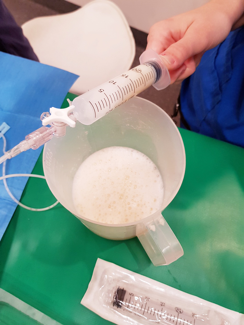
    Thoracocentesis-Chyle
    Chyle.

    Opaque (milky) to pink.

    Differentials to consider

    • Rupture or obstruction of lymphatic flow
    • Neoplasia, traumatic and idiopathic
    • Secondary to heart failure (especially in cats)
    • Pseudochyle (usually formed by lymphoma)

    Additional diagnostics

    • CBC and biochemistry
    • Cytology and culture of fluid
    • Fluid [TAG] > serum
    • Large number of lymphocytes and other inflammatory cells
    • +/- ultrasound/CT

    Haemorrhage

    • Red blood cells
    • True haemorrhagic; for example, not iatrogenic: should not see platelets or erythophagocytosis on smears and sample should not clot
    • Time frame
    • Assess history
    • Compare fluid PCV/total protein (TP) to peripheral PCV/TP:
    1. <1% – non-significant
    2. 1% to 20% – neoplasia, trauma, pneumonia
    3. >50% – haemothorax
    • Other tips:
    1. If PCV/TP is similar = recent bleed, if PCV is low and TP normal = chronic
    2. If PCV is increasing or is higher than peripheral then active bleeding
    3. Presence of erythrophagocytosis = chronic

    Differentials to Consider

    • Trauma
    • Neoplasia
    • Coagulopathies
    • Ruptured granuloma

    Diagnostics

    • Activated clotting time, activated partial thromboplastin time, prothrombin time, blood smear and other coagulation tests, see “coagulopathy”
    • Blood smear
    • CBC and biochemistry
    • +/- ultrasound/CT

    Good luck with your next thoracocentesis. I hope this information was useful.

  • Blood gas analysis, pt 7: evaluating oxygenation and ventilation

    Blood gas analysis, pt 7: evaluating oxygenation and ventilation

    In patients with respiratory compromise, it is important to look at the respiratory components of the blood gas to determine both oxygenation ability and adequacy of ventilation.

    To assess oxygenation, the partial pressure of arterial oxygen (PaO2) to fraction of inspired oxygen (FiO2) ratio, and alveolar-arterial gradient (A-a gradient) can be used. Conversely, the partial pressure of carbon dioxide (PCO2) is what dictates the adequacy of ventilation.

    Before going further, it should be noted only arterial samples can be used to evaluate oxygenation. Peripheral venous blood samples are rarely useful for oxygenation assessments, as the values are highly influenced by local factors and the degree of occlusion of the vessel from which the sample was collected from.

    Ventilation, conversely, can be assessed from either venous or arterial samples. The PCO2 values from the arterial samples are usually about 5mmHg to 10mmHg lower than the corresponding venous sample.

    Abnormalities

    Oxygenation and ventilation are not interchangeable terms. Adequate ventilation does not necessarily equate to adequate oxygenation, and vice versa.

    Oxygenation is determined by efficiency of oxygen absorption into the blood stream, after passing through the lungs, then delivered to the tissue. It is dependent on both ventilation (V) and perfusion (Q).

    For proper oxygenation to occur, matching ventilation and perfusion must occur at the level of the alveoli. When a V/Q mismatch occurs, blood is not properly oxygenated after passing through the lungs, referred to as venous admixture.

    Three forms of V/Q abnormalities exist:

    • Low V/Q can be caused by:
      • pneumonia
      • asthma
      • pulmonary oedema
      • inflammation
      • pulmonary thromboembolism
      • pulmonary neoplasia
    • Absent V/Q means blood has been diverted from parts of the lungs that are inadequately ventilated, such as those caused by atelectasis and alveolar collapse secondary to severe pleural effusion.
    • Diffusion impairment is caused by an increased distance between the gas exchange surfaces and pulmonary arterioles (rare in small animals). Pulmonary fibrosis and chronic obstructive pulmonary disease are among some of these causes where it can be severe enough to cause V/Q mismatch and, therefore, decreased PaO2.

    PaO2 to FiO2 ratio

    One way to determine the adequacy of oxygenation is by looking at the PaO2 to FiO2 ratio.

    FiO2 represents the percentage of the oxygen the patient is breathing. Room air is 21% oxygen, so the FiO2 is 0.21. PaO2 is normally about five times that of FiO2; therefore, the normal PaO2 of a patient with no pulmonary disease breathing room air should be about 100mmHg (5 × 21%).

    A normal PaO2 to FiO2 ratio is between 300 and 500. A number lower than 200 implies significant pulmonary disease or, possibly, acute respiratory distress syndrome. This ratio is particularly important in assessing patients requiring supplemental oxygen. An example is a congestive heart failure patient on bilateral nasal oxygen line (approximately FiO2 of 40%), with a PaO2 value of 80mmHg. The PaO2 appears to be adequate until the ratio is calculated (80/0.4 = 200). The change in FiO2 ratio is often more important than the PaO2 as a lone value.

    A-a gradient

    Once hypoxia has been established, the A-a gradient will help determine whether it is caused by ventilation failure or an underlying pulmonary disease. “A” stands for alveoli and “a” stands for arterial concentration of oxygen.

    A = [FiO2 × (Pb-PH20)] – (PaCO2/0.8) – Pb is the barometric pressure (760mmHg at sea level), and PH20 is saturated water vapour pressure, which is 50. The 0.8 is the respiratory quotient and affixed number.

    The simplified formula assumes the patients are breathing room air (FiO2 of 0.21) and at sea level, and the formula can be simplified to: A = 150 – (PaCO2/0.8)

    A normal A-a gradient should be less than 15. Abnormally high values indicate pulmonary parenchymal disease or an underlying heart disease, while a normal A-a gradient indicates the cause of hypoxia is likely secondary to ventilation failure.

    The ‘120 rule’

    Ventilation is particularly important to assess in animals with respiratory compromise, as it represents the entire mechanics of breathing.

    PCO2 is not only representative of the efficiency of ventilation, but also cellular metabolism and perfusion. Low PCO2, or hyperventilation, is rarely of any significance as aforementioned in the respiratory alkalosis section. High PCO2, however, is indicative of the lungs’ reduced ability to adequately shift air and can be caused by neurologic diseases, spinal cord injury, upper airway disease, trauma to the thoracic wall or muscles, and drugs that can cause respiratory depression.

    Ventilation must be assessed in light of oxygenation, as both are often affected by each other. An example of this would be a hypoxic animal (low PaO2) with a compensatory hyperventilation (low PCO2). The “120 rule” will help determine whether lung function is adequate.

    If the value you get, by adding the PaO2 and PaCO2, is greater or equal to 120, lung function is adequate. If the value is lower than or equal to 120, lung function is abnormal. This can only be calculated from patients breathing room air and at sea level.

    Ventilation adequacy

    Aside from looking at values on a blood gas, it is equally important to monitor the patient to determine whether the ventilation effort is sustainable.

    Animals with a significantly increased respiratory effort – despite normal blood gas values – are at risk of respiratory exhaustion and indicative of mechanical ventilation intervention.

    From these respiratory components of the blood gas value, clinicians should be able to determine the adequacy of ventilation, whether hypoxia is present and, if it is, whether the underlying cause is a result of ventilation failure or a possible underlying pulmonary or heart disease.

    Once oxygen supplementation has been implanted, the PaO2 to FiO2 ratio will help clinicians decide whether the response is adequate or mechanical ventilation is required.

    As with all laboratory measurements, it is extremely important to assess the patient itself. Non-sustainable respiratory efforts, in the face of normal blood gas parameters, is still an indication for mechanical ventilation.

  • Blood gas analysis, pt 4: respiratory acidosis and alkalosis

    Blood gas analysis, pt 4: respiratory acidosis and alkalosis

    Assessing the respiratory component is simple. A quick glance at the partial pressure of carbon dioxide (PCO2) level can tell you whether a respiratory acidosis or alkalosis is present.

    If the PCO2 level is elevated (respiratory acidosis) then either a primary respiratory acidosis is present, or it is the result of a compensatory response to a metabolic alkalosis.

    Similarly, if the PCO2 level is low (respiratory alkalosis) then it could either be a primary respiratory alkalosis, or compensation to metabolic acidosis has occurred.

    The respiratory component should always be assessed before the metabolic component, due to the ability to respond to pH shifts almost immediately. This, therefore, is a more accurate reflection of the patient’s clinical disease.

    Respiratory acidosis – increased CO2

    Respiratory acidosis occurs anytime the patient is hypoventilating and not eliminating CO2 appropriately.

    As hypoventilation can be associated with hypoxia, these patients are often critical and require immediate interventions.

    Causes of respiratory acidosis include:

    • drugs (depress respiratory centre, relax thoracic muscles)
    • neuromuscular disease (for example, tick paralysis, botulism and snake envenomation)
    • upper airway obstruction
    • pleural disease (for example, pneumothorax, pleural effusion and diaphragmatic hernia)
    • gas exchange disorders (for example, pulmonary thromboembolism, pneumonia and pulmonary oedema)

    Respiratory alkalosis – loss of CO2

    Respiratory alkalosis occurs when a patient is hyperventilating – excessive loss of CO2 causes the pH to increase.

    The health effect of this is usually minimal, since, in most cases, the effect is secondary and correction of the underlying cause usually resolves this problem. The exception is when respiratory alkalosis is a primary disorder. This is usually quite rare, but can occur with brain stem trauma where the respiratory centre is affected.

    Causes of respiratory alkalosis are:

    • hyperventilation (for example, fear, pain, stress, anxiety and hyperthermia)
    • neurological (for example, head trauma/neoplasia involving the respiratory centre)

    Anticipating changes

    Correctly identifying the primary disorder is essential for anticipating the changes the patient is likely to experience. This will help identify the underlying disease, and is essential for patient monitoring and disease management.

    In the next blog, we will discuss assessment of the metabolic component.

  • Emesis: a thing of the past?

    Emesis: a thing of the past?

    Until I started researching this Tip of The Week, I did not know the medical profession has abandoned the routine use of emesis in oral poisoning.

    This is based on multiple medical literatures that have proven emesis induction does not influence the clinical severity of poisoning, the length of hospitalisation and the clinical outcome or mortality.

    Although the rationale for inducing emesis is obvious, it is not necessarily evidence based. It is also dependent on satisfying a few large assumptions, all of which are untrue:

    • Emesis is a very effective way of removing gastric contents.
    • No separation exists of poison from its vehicle while inside the acidic environment of the stomach.
    • Poison is not absorbed through the stomach wall.

    Ineffective method

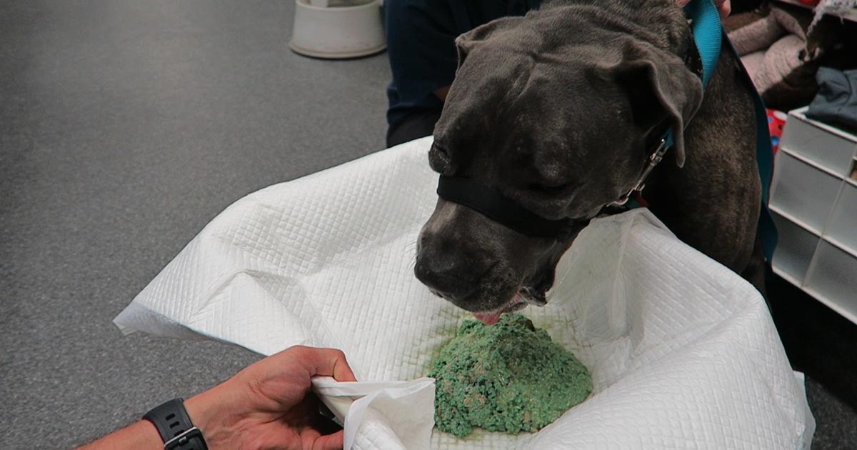
    snail bait
    Snail bait ingestions: this patient ate 500g of snail bait containing metaldehyde.

    Emesis induction is an ineffective way of clearing stomach contents. A review of the effectiveness of induced emesis, with both human and canine participants, showed at 30 minutes post-ingestion of non-absorbable markers, the recovery rate averaged between 17.5% and 52.1%, but never exceeded 62%.

    In fasted puppies, this was even lower at 2% to 31%, despite inducing emesis immediately after marker administration. These have been confirmed by the presence of poisonous materials in the stomach of dead patients, despite effective emesis induction until clear fluid was brought up.

    The clinical outcome only improves if the systemic exposure of a toxicant is reduced by more than half. However, considering animals rarely practice restraint, the ingested amount is unlikely to be exactly the lethal dose and no more. Therefore, even reducing the ingested toxic dose by 62% is unlikely to make a clinical difference.

    Furthermore, most patients rarely present within 30 minutes of ingesting a toxicant, thus further reducing its efficiency.

    The absorption conundrum

    Some may argue the retrieval of metaldehyde or anticoagulant rodenticide granules from vomitus is indicative of reducing the toxicant dose. This could be true, but only if emesis was induced immediately after ingesting the poison.

    The poison itself is colourless and has a different absorption characteristic to the coloured vehicle (granule); therefore, the presence of granule only serves to confirm ingestion, but is of no indication whether the poison has already been absorbed.

    Contraindications

    Many well-recognised absolute contraindications also exist to inducing vomiting:

    • Ingestion of oils, which includes waxes that melt to oil in the internal body environment, as this poses a high risk of lipoid and bacterial pneumonia. This is of significant veterinary significance, as wax is routinely used in rodenticide baits.
    • Ingestion of hydrocarbons and other volatile substances, or caustic or corrosive substances.
    • When the mental status is altered – for example, hyperexcitable or depressed mental state.
    • Where the patient is at risk of seizures (seizures can be induced by emesis).
    • Increased intracranial pressure.
    • Risk of intracranial or cerebral haemorrhage – for example, thrombocytopenia or abnormal clotting parameters.

    Other less severe, yet important, reasons include:

    • delays administration of more effective treatment, such as activated charcoal, antidote or other treatments
    • risk of aspiration pneumonia
    • hypochloraemia in recurrent emetic patients
    • significant CNS and respiratory depression from apomorphine
    • rare, but reported, complications such as cerebral haemorrhage, oesophageal tear/ rupture, hiatal hernia, gastric rupture, pneumothorax and pneumomediastinum
    • legal implications – for example, if the product information clearly states emesis should not be induced

    A place for everything

    Emesis induction is not a benign procedure. It still has its place in certain circumstances, but its use in the routine management of oral poisonings may need to be reconsidered – especially if it means delaying administration of a more effective treatment, such as activated charcoal.

    So, after all this, how do I tackle this information? It is a bit hard to swallow. My clinical experience is emesis is generally safe, especially in canine patients using apomorphine. So, I still feel some merit exists in reducing the amount of toxicant in the stomach if you have a chance – and in some situations, you don’t know until you try.

    Emesis after ingestion of a toxic dose of chocolate can be incredibly rewarding, even six hours after ingestion, leading to patients not developing clinical signs at all.

    Overall, I am biased by my personal successes with emesis, so still feel a time and place exist for emesis induction. But I now stop and question my decision to induce emesis, whereas I did not hesitate before.

    • Some drugs listed are used under the cascade.
  • Linear foreign bodies, part 2: tips for diagnosing with ultrasound

    Linear foreign bodies, part 2: tips for diagnosing with ultrasound

    Following on from the previous post where we discussed tips on how to diagnose a linear foreign body on a radiograph, this post sees us cover how to diagnose it on ultrasound.

    If used by an experienced ultrasonographer who knows what to look for, ultrasound can be a highly sensitive and specific diagnostic test.

    What do we look for?

    1. Remember not all patients will have intestinal dilation as the linear foreign body may be only causing a partial obstruction. Alternatively, it could be occluding the gastric outflow completely.
    2. Intestinal plication, which looks like intestinal loops bunching up on each other around the tether.
    3. A central discrete hyperechoic line running along the middle of the bunching intestine. This bright line is the tether. Often when looking closely enough, the tether will have distal acoustic shadowing as the ultrasound pulses cannot pass through it.
    4. The leading aboral segment and the trailing adoral anchor will have acoustic shadowing.
    5. The adjacent mesentery is often hyperechoic compared to other areas in the abdomen, indicating inflammation.
    6. Gastric dilation with fluid is often seen if the anchor is in the pylorus, as it causes an outflow obstruction.
    7. Free abdominal fluid may be visible and a sample should be collected for assessment. If bacteria can be demonstrated in one of the following ways:
      1. By visualising free or intracellular bacteria under the microscope.
      2. By finding that the glucose is lower (lower than 20mg/dL) and the lactate is higher (2mmol/L) in the abdominal fluid sample compared to peripheral blood then this indicates perforation of the gastrointestinal tract has occurred and septic peritonitis is present.

    In the third and final post, we will cover things to consider when deciding whether to perform the exploratory laparotomy yourself, or if you should transfer the patient to a referral facility for surgery.

  • Linear foreign bodies, part 1: tips for diagnosing with radiography

    Linear foreign bodies, part 1: tips for diagnosing with radiography

    Linear foreign bodies can be tricky to diagnose, compared to normal foreign bodies, for many reasons. Mostly because you often don’t see the classic obstructive pattern appearance on radiographs or ultrasound.

    In this short blog series, we are going to cover some hints and tips that can make diagnosing a patient with a linear foreign body easier. Then, we’ll discuss things that should be considered when deciding whether you are the right person to take the patient to surgery…

    So, let’s start with radiography.

    1. Not all patients with a linear foreign body will be completely obstructed. This means you won’t always visualise classic intestinal dilation. In fact, it has been reported that up to 50% of patients with a linear foreign body will not have an obstructive pattern present on radiographs.
    2. Look for the characteristic small “comma shaped” gas pattern. This is caused by plication and bunching of the small intestine around the tether.
    3. The small intestine can appear to be bunched up in one area, rather than spaced out around the abdomen. However, obese animals – especially cats – can have “pseudo-bunching” due to large amounts of abdominal fat bunching the intestine together.
    4. Loss of serosal detail is often seen due to inflammation surrounding the affected intestine.
    5. Always include a left lateral radiograph in your series. Gastric contents will fall to the fundus on the left of the abdomen and gas will raise to the pylorus, which will highlight the foreign body anchor in the pylorus.
    6. Perform thoracic radiographs to assess for aspiration pneumonia and a potential oesophageal component of the linear foreign body. If aspiration is present then you know you will need to continue antibiotic therapy postoperatively.

    In the next post, we cover some key points for diagnosing linear foreign bodies on ultrasound…