In part one of this series we discussed the important questions to ask when taking a history from owners of dogs and cats that are having seizures. In this part, we look at the differential diagnoses for these cases.
There are many ways to classify the different causes of seizures, but the simplest is as follows:
Structural – where intracranial pathology is causing the seizures.
Reactive – where an extracranial issue is causing a seizure response in a normal brain.
Idiopathic – a diagnosis of exclusion where we are unable to identify a reason for the disturbances in brain activity.
Structural
Intracranial differential diagnoses include:
inflammatory processes (meningoencephalitis), such as steroid responsive meningitis-arteritis
viral diseases (for example, distemper)
metabolic storage diseases
neoplasia
vascular accidents involving clots or bleeds
hydrocephalus
trauma
Reactive
Extracranial differentials include:
hepatic encephalopathy due to hepatic failure or a portosystemic shunt
various toxicities, such as lead, chocolate, caffeine, ethylene glycol, parasiticides and slug/snail bait
metabolic issues, such as hypoglycaemia, hypocalcaemia and thiamine deficiency
Idiopathic
If diagnostic investigations (including advanced imagery, such as MRI) are unable to identify an underlying cause of recurrent seizures, this is referred to as idiopathic epilepsy.
To break down this list of differentials into a more relevant and concise list is to consider the most common differentials according to signalment.
Belief #2: if you can’t afford to care for an animal then you shouldn’t have one
You, young doctor or nurse – living in your privileged society of excess, with your years of study, working in an industry that exists largely to minimise animal suffering – have certain expectations of what “taking care of an animal” looks like.
Of course, we should always strive to do the best we can for any animal in our care, but we need to learn to put judgement aside when an owner’s abilities don’t stretch to meet our expectations.
Saying “if you can’t afford to care for a pet then you shouldn’t be allowed to own one” is a subjective and biased opinion.
What defines an appropriate level of “caring for your animal” is based to a large degree on your background, prior experiences with animals and financial circumstances.
Where is the line?
Let’s agree that if someone takes an animal in your care, it is his or her responsibility to meet its basic needs – make sure it doesn’t starve, freeze or cook to death; don’t cause it unnecessary suffering through wilful abuse or neglect; and satisfy at least some basic emotional needs of connection, and freedom from fear and pain.
Beyond that, where is the line?
You could argue provision for core vaccinations should make the cut into what constitutes basic levels of care, but I can assure you many unvaccinated animals around the world live very happy lives.
I’ve seen many colleagues spit venom about “careless owners” who “don’t deserve to own an animal” when a dog presents with a pyometra, so should we make neutering a minimum requirement of pet ownership?
Should every pet owner have the means to pay for at least full bloods and an ultrasound? What about major life-saving surgery? An MRI? Chemotherapy? Specialist referral?
If a line in the sand does exist, then it’s poorly defined – and, like the line between beach and ocean, it’s a line that will shift with every wave, tide, and storm.
Collision course
So, how does this belief hold you back?
If you subscribe to this way of thinking, you’ll be on a constant collision course with a large part of the population. It’s a mindset that will set you up for daily antagonistic interactions with the people who present their animals to you, instead of creating those all-important relationships that were mentioned in part one.
None of this will benefit your patients, your clients, or yourself. To put it simply: you’ll only make yourself resentful and miserable.
Burden of care
Where things do become complicated is when individuals expect a high level of veterinary care without being willing to pay for it.
When the burden of care – and blame for lack of provision of its gold standard – are shifted solely on to the shoulders of the veterinary community, the relationship starts falling apart.
This, of course, is rubbish – and should be treated as such.
It’s a problem that vets who have the capacity to provide high levels of care will grapple with, and can be the topic of many more blogs.
For the purpose of this post, let’s just say we should always aim to help – to the best of our abilities, and with a consistent level of caring and lack of judgement – any person who presents their animal into our care, despite their individual expectations and capacity of what constitutes a good level of care.
On my latest EMS placement at an equine hospital, I’ve seen a number of surgeries – some done under general anaesthesia (GA) and others under standing sedation.
After getting over the fact a horse can stand half asleep while having its face drilled into and not really seem to care, I started wondering about the pros and cons of both approaches.
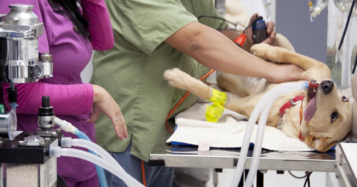
“Sinus surgery to remove a bony mass – that is me in the pink scrubs holding the head,” says Jordan.
Generally, standing sedation is accompanied by less haemorrhage and, therefore, increased visibility – in sinus surgery, for example. It also eliminates many risks associated with general anaesthesia. However, asepsis may be harder to maintain (for example, if the horse moves and the surgical site comes into contact with something that isn’t sterile, such as the stocks).
Lower costs
For the client, procedures conducted under standing sedation would be much cheaper than the costs incurred from general anaesthesia.
During general anaesthesia, atelectasis contributes to the risks from an intraoperative point of view, as well as myositis and cardiac concerns (of which the risk can be considerably reduced by the use of acepromazine in the premedication protocol).
A risk of injury also exists during recovery and knockdown, such as worsening incomplete fractures or other self-inflicted wounds, which can, to some degree, be prevented by carefully assisted knockdown and paying careful attention during recovery with the use of ropes.
Achieving optimal sedation for standing surgery can, in some cases, be difficult. For example, the horse must be adequately sedated, but not so much it is swaying; this can be an issue for intricate surgeries, but may be more of a problem for diagnostic imaging (such as MRI or bone scintigraphy).
In these cases, I have seen morphine used – opposed to the usual sedative culprits, such as detomidine, butorphanol and xylazine – and it seems to achieve sedation without so much swaying.
Choosing correct method
The choice of standing sedation versus GA depends on the type of surgery required, but a number of procedures can be done using either method.
Last week, I saw tie-back surgeries (prosthetic laryngoplasties to correct laryngeal hemiplegia) done both ways, which made for an interesting comparison. The standing tie-back was considerably quicker, taking into account the time for knockdown and recovery, as well as surgical time.
Both tie-backs were followed by a laser hobday procedure (ventriculoectomy), meaning both procedures were conducted under the same sedation in the standing horse, whereas the tie-back performed after GA had to be followed later the same day, after the horse had recovered sufficiently to undergo standing sedation for the laser.
The second tie-back was a repeat of a previously failed procedure, hence GA was chosen to allow removal of the first prosthesis.
The standing technique is still being tweaked, but, despite reports of postoperative infection in more cases than ideal, the easier access to the laryngeal cartilages while standing – and the avoidance of further risks associated with GA – contribute to continued work to perfect this method.
Some surgeries, however, can still only be done properly via GA. Colic surgery, for example, requires significant abdominal access and, often, examination of the gastrointestinal tract. It is also highly recommended septic joint surgery and lavage is conducted under GA to ensure optimal sterility on closure of the joint.
Conclusion
Having now seen both types of surgery in the horse, it’s astonishing how quick standing surgery can be, and how much goes into the preparation and recovery for GA – even for the shortest of procedures. In one surgery, division of the aryepiglottic fold, causing epiglottic entrapment, took a matter of minutes – if you didn’t count the couple of hours total taken for premedication, knockdown and recovery from GA.
The choice very much depends on the procedure, and is assessed for each case. I do, however, think the advantages to standing surgery are significant and look forward to seeing more standing techniques developed in the future.
Prolapsed gland of the third eyelid, or cherry eye as it is sometimes known. Image by Joel Mills (CC BY-SA 3.0) via Wikipedia.
A nice six-year-old Labrador with a history of possible trauma, complete forelimb muscle atrophy and proprioceptive deficits was brought to see me.
Radial paralysis was high up on the list of differentials, but the poor chap had not shown any response to conservative management.
I noticed it had mild anisocoria, slight ptosis and third eyelid prolapse, indicating the less obvious Horner’s syndrome. A bell rang in my mind of a case I recalled seeing as a student – it revealed itself to be indicative of a brachial plexus injury/tumour.