Tag: Metabolic

  • Seizures, part 3: management

    Seizures, part 3: management

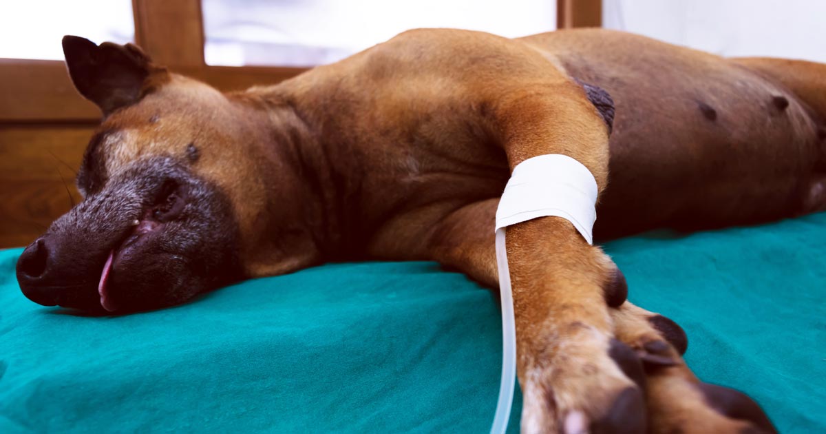
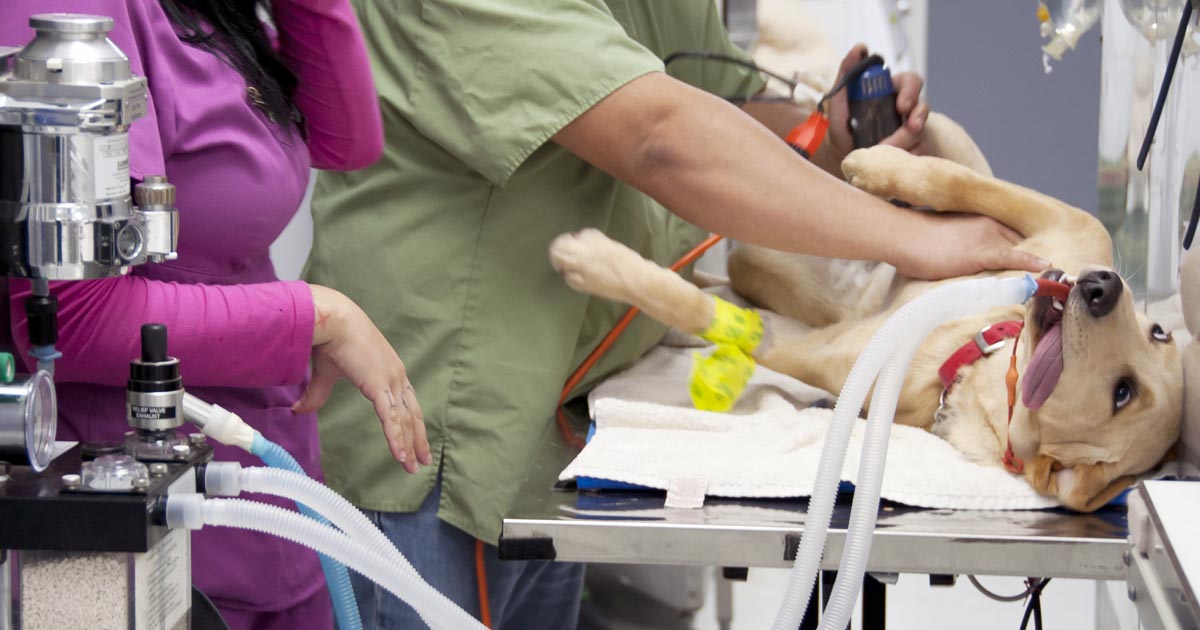
    In the third and final part of this series, we look at managing seizures in pets, both in an emergency setting and in the longer term.

    When presented with a patient in status epilepticus, appropriate emergency treatment begins with:

    • Providing oxygen therapy.
    • Placing an IV catheter, if possible.
    • Administering diazepam as an 0.5mg/kg to 1mg/kg IV bolus, rectally at 2mg/kg or intranasally at 0.5mg/kg.
    • Intubating, if required to maintain a patent airway.
    • Cooling, if hyperthermic.
    • Giving mannitol at 0.5mg/kg to 1mg/kg slowly IV if seizure activity lasts more than 15 minutes or there is any reason to suspect cerebral oedema.
    • Collecting full bloods – test glucose, electrolyte and calcium levels first.
    • If on phenobarbital, collecting a sample for baseline testing.

    It is important to remember patients may continue to paddle slowly after seizure activity has finished, but if the eyelids are twitching, they are still seizing.

    If the seizures are controlled by these first steps, give a 4mg/kg dose of phenobarbital and commence supportive therapy with IV fluids, including correction of any electrolyte and metabolic derangements.

    If at first…

    If these first emergency steps fail to get the seizures under control, the following steps can be attempted:

    1. Diazepam 0.5mg/kg to 1mg/kg IV bolus – can be repeated every five minutes for up to three doses.
    2. Propofol 2mg/kg to 4mg/kg IV titrated to effect to stop motor activity.
    3. Phenobarbital slow IV 4mg/kg if already on maintenance therapy, mg/kg to 8mg/kg if not – can be repeated at 20 to 30 minute intervals.
    4. +/- Diazepam (or midazolam) continuous rate infusion (CRI) at 0.5mg/kg/hr.
    5. Propofol CRI following titrated dose, at 0.2mg/kg to 0.5mg/kg/min – continue for six hours then wean down slowly over next six hours.
    6. Levetiracetam at 20mg/kg to 60mg/kg IV titrated can be used instead of propofol (this is safer if hepatic disease is present).

    Ongoing treatment

    The recommendations for when to start long-term treatment are summarised as follows:

    1. Structural lesion present or prior history of brain disease or injury.
    2. Acute repetitive seizures or status epilepticus (ictal event ≥5 minutes or ≥3 or more generalised seizures within a 24-hour period).
    3. ≥2 or more seizure events within a six-month period.
    4. Prolonged severe, or unusual postictal periods.

    Chronic therapy in patients with ongoing seizures aims to reduce the frequency to an acceptable and manageable level. The drug used to achieve this is often down to clinician preference; one or a combination of the following can be used:

    • Phenobarbital – solo and combination therapy, drug monitoring is required along with regular monitoring of liver enzymes and function is particularly important.
    • Potassium bromide (not cats) – combination therapy, drug monitoring is required and can cause pancreatitis.
    • Imepitoin – solo or combination therapy, does not require drug monitoring.
    • Levetiracetam – solo or combination therapy, does not require drug monitoring.

    Regular testing of blood levels of anti-epileptics is important, although it does not indicate whether the drug should be working or not, it does help provide additional information when investigating when control is inadequate and to prevent toxic side effects.

  • Seizures, part 2: the differentials

    Seizures, part 2: the differentials

    In part one of this series we discussed the important questions to ask when taking a history from owners of dogs and cats that are having seizures. In this part, we look at the differential diagnoses for these cases.

    There are many ways to classify the different causes of seizures, but the simplest is as follows:

    • Structural – where intracranial pathology is causing the seizures.
    • Reactive – where an extracranial issue is causing a seizure response in a normal brain.
    • Idiopathic – a diagnosis of exclusion where we are unable to identify a reason for the disturbances in brain activity.

    Structural

    Intracranial differential diagnoses include:

    • inflammatory processes (meningoencephalitis), such as steroid responsive meningitis-arteritis
    • viral diseases (for example, distemper)
    • metabolic storage diseases
    • neoplasia
    • vascular accidents involving clots or bleeds
    • hydrocephalus
    • trauma

    Reactive

    Extracranial differentials include:

    • hepatic encephalopathy due to hepatic failure or a portosystemic shunt
    • various toxicities, such as lead, chocolate, caffeine, ethylene glycol, parasiticides and slug/snail bait
    • metabolic issues, such as hypoglycaemia, hypocalcaemia and thiamine deficiency

    Idiopathic

    If diagnostic investigations (including advanced imagery, such as MRI) are unable to identify an underlying cause of recurrent seizures, this is referred to as idiopathic epilepsy.

    To break down this list of differentials into a more relevant and concise list is to consider the most common differentials according to signalment.

    In dogs less than a year old:

    • portosystemic shunts
    • inflammatory conditions of the brain
    • distemper
    • hydrocephalus or storage disease
    • toxicity

    In dogs one to five years old:

    • idiopathic epilepsy
    • inflammatory
    • toxicity
    • cerebral neoplasia

    In dogs of five years or older:

    • cerebral neoplasia
    • inflammatory
    • toxicity
    • idiopathic epilepsy
    • metabolic disease
    • vascular issues

    In cats:

    • toxoplasmosis
    • FIP, FeLV and FIV
    • audiogenic reflex seizures (older cats)
    • neoplasia
    • trauma
    • toxins
  • Blood gas analysis, pt 6: compensatory response

    Blood gas analysis, pt 6: compensatory response

    Simple acid-base disorders are compensated by predictable compensatory changes. The primary disorder shifts the pH, while the compensatory mechanisms aim to normalise the pH and bring it back to neutral.

    This is achieved by attempting to normalise the bicarbonate (HCO3-) to partial pressure of CO2 (PCO2) ratio in a paralleled manner.

    For example, an increase in HCO3– (metabolic alkalosis) is compensated by an increase in PCO2 (respiratory acidosis). Similarly, a respiratory alkalosis (decrease in PCO2) is compensated by a metabolic acidosis (decrease in HCO3-).

    Ruling out secondary process

    However, before jumping to the conclusion an opposing change is the result of compensation, we must rule out the presence of a secondary process. This can only be determined by calculation (Table 1).

    Table 1. Calculating compensatory change
    Component Expected compensation
    Metabolic acidosis
    ↓HCO3 (↓BE)
    per 1mEq/L ↓ in HCO3 = ↓ PCO2 of 0.7mmHg
    Metabolic alkalosis
    ↑HCO3 (↑BE)
    per 1mEq/L ↑ in HCO3 = ↑ PCO2 of 0.7mmHg
    Respiratory acidosis (acute)
    ↑PCO2
    per 1mmHg ↑ PCO2 = ↑ 0.15mEq/L HCO3
    Respiratory acidosis (chronic)
    ↑PCO2
    per 1mmHg ↑ PCO2 = ↑ 0.35mEq/L HCO3
    Respiratory alkalosis (acute)
    ↓PCO2
    per 1mmHg ↓ PCO2 = ↓ 0.25mEq/L HCO3
    Respiratory alkalosis (chronic)
    ↓PCO2
    per 1mmHg ↓ PCO2 = ↓ 0.55mEq/L HCO3

    By comparing the reported to what the calculated compensatory change should be, you can determine whether the patient’s reported value is due to compensation or a separate disorder – for example, multiple primary acid-base disorders (a mixed acid-base disorder).

    An example of a mixed disturbance could be a hyperventilating (respiratory alkalosis) dog with renal failure (metabolic acidosis).

    The level of decrease in PCO2 change is in excess of the calculated compensation for the metabolic acidosis, therefore confirming a mixed acid-base disturbance. In fact, the most common causes of hyperventilation – pain, fear and excitement – often complicate blood gas analysis.

    Another example of a mixed disorder could be a patient with traumatic haemothorax experiencing both lactic acidosis (hypoperfusion) and hypoventilation (respiratory acidosis) due to pleural space disease.

    Waiting game

    Another thing to keep in mind is compensation takes time – respiratory processes take approximately 8 to 12 hours, while metabolic processes take one to three days.

    The lungs are able to alter PCO2 levels relatively quickly by adjusting the rate of ventilation. The kidneys, on the other hand, take a longer time to adjust the pH, as the change in rate of absorption and excretion of HCO3– takes much longer in comparison.

    Regardless of the rate, physiologic compensation for a primary acid-base disturbance is almost never able to return pH to neutral.

    Summary

    A simple acid-base disorder should be suspected when the patient’s reported values are similar to the calculated compensation value, and a mixed acid-base disorder when the values fall outside the calculated range.

    Another hint that a mixed acid-base disturbance is present is if the pH falls within the normal reference range, but the HCO3– or the PCO2 are not; or if the HCO3– and PCO2 are in opposite directions as opposed to being parallel.

    Remember, the body can never overcompensate nor return the pH to neutral.

  • Blood gas analysis, pt 5: metabolic acidosis and alkalosis

    Blood gas analysis, pt 5: metabolic acidosis and alkalosis

    Base excess (BE) and bicarbonate (HCO3-) represent the metabolic components of the acid base equation. In general, both components will change in the same direction.

    Decreased HCO3– and BE indicate either a primary metabolic acidosis or a metabolic compensation for a chronic respiratory alkalosis. Elevated HCO3– and BE indicate either a primary metabolic alkalosis or a metabolic compensation for a chronic respiratory acidosis.

    The exception to this rule arises when a patient hypoventilates or hyperventilates.

    Carbonic acid equation

    CO2 + H2O ↔ H2CO3 ↔ HCO3– + H+

    When a patient hypoventilates, CO2 will increase as a result of reduced expiration, so a shift to the right of the equilibrium will occur. The shift to the right will increase the bicarbonate levels proportional to the increase in CO2.

    The opposite occurs when a patient hyperventilates; the equilibrium shifts to the left, so a decrease in HCO3– is present.

    Since HCO3– is not independent to the patient’s respiratory status, it is an inaccurate way of measuring the metabolic component in patients with respiratory changes. For this reason, the BE value is the preferred.

    The BE represents the amount of acid, or base, needed to titrate 1L of the blood sample until the pH reaches exactly 7.4, with the assumption the blood sample is equilibrated to a partial pressure of CO2 of 40mmHg (the middle of the reference range) and the patient’s body temperature is normal.

    Possible causes

    The possible causes of the primary disease are:

    Metabolic acidosis

    • lactic acidosis – shock and poor perfusion
    • renal failure – reduced hydrogen ion (H+) excretion and increased loss of HCO3
    • diabetic ketoacidosis – ketone acids
    • gastrointestinal (GI) losses – loss of HCO3– through vomiting and diarrhoea

    Metabolic alkalosis

    • GI outflow obstructions – loss of H+ and chloride via vomiting
    • reduced chloride levels and resultant poor perfusion – body attempts to reabsorb water and sodium to increase intravascular volume, but inadvertently also reabsorbs HCO3– in the process, despite existing alkalosis
    • refeeding syndrome
    • severe hypokalaemia:
      • transcellular shift – potassium ions leave and H+ ions enter the cell
      • transcellular shift in cells of proximal tubules → intracellular acidosis → promotes ammonium production and excretion
      • H+ excretion in the proximal and distal tubules increases → further reabsorption of HCO3– and net acid excretion
    • renal insufficiency
    • diuretic therapy (contraction alkalosis – loss of bodily fluids that do not contain HCO3-; this causes the extracellular volume to contract around a fixed quantity of HCO3-, resulting in a rise in the concentration of HCO3– without an actual increase in HCO3– levels)

    Next step

    After ruling out the differential causes of either respiratory or metabolic acidosis/alkalosis, the next step is to determine whether a compensatory response is present and, if so, if this is adequate or whether a true mixed acid-base disorder exists.

  • Blood gas analysis, pt 4: respiratory acidosis and alkalosis

    Blood gas analysis, pt 4: respiratory acidosis and alkalosis

    Assessing the respiratory component is simple. A quick glance at the partial pressure of carbon dioxide (PCO2) level can tell you whether a respiratory acidosis or alkalosis is present.

    If the PCO2 level is elevated (respiratory acidosis) then either a primary respiratory acidosis is present, or it is the result of a compensatory response to a metabolic alkalosis.

    Similarly, if the PCO2 level is low (respiratory alkalosis) then it could either be a primary respiratory alkalosis, or compensation to metabolic acidosis has occurred.

    The respiratory component should always be assessed before the metabolic component, due to the ability to respond to pH shifts almost immediately. This, therefore, is a more accurate reflection of the patient’s clinical disease.

    Respiratory acidosis – increased CO2

    Respiratory acidosis occurs anytime the patient is hypoventilating and not eliminating CO2 appropriately.

    As hypoventilation can be associated with hypoxia, these patients are often critical and require immediate interventions.

    Causes of respiratory acidosis include:

    • drugs (depress respiratory centre, relax thoracic muscles)
    • neuromuscular disease (for example, tick paralysis, botulism and snake envenomation)
    • upper airway obstruction
    • pleural disease (for example, pneumothorax, pleural effusion and diaphragmatic hernia)
    • gas exchange disorders (for example, pulmonary thromboembolism, pneumonia and pulmonary oedema)

    Respiratory alkalosis – loss of CO2

    Respiratory alkalosis occurs when a patient is hyperventilating – excessive loss of CO2 causes the pH to increase.

    The health effect of this is usually minimal, since, in most cases, the effect is secondary and correction of the underlying cause usually resolves this problem. The exception is when respiratory alkalosis is a primary disorder. This is usually quite rare, but can occur with brain stem trauma where the respiratory centre is affected.

    Causes of respiratory alkalosis are:

    • hyperventilation (for example, fear, pain, stress, anxiety and hyperthermia)
    • neurological (for example, head trauma/neoplasia involving the respiratory centre)

    Anticipating changes

    Correctly identifying the primary disorder is essential for anticipating the changes the patient is likely to experience. This will help identify the underlying disease, and is essential for patient monitoring and disease management.

    In the next blog, we will discuss assessment of the metabolic component.

  • Blood gas analysis, pt 3: interpreting pH

    Blood gas analysis, pt 3: interpreting pH

    After taking note of the direction of the pH shift – acidaemia or alkalaemia – it is important to determine the primary and secondary causes.

    If an acidaemia is present (pH less than 7.35), an underlying respiratory or metabolic acidosis, or both, must exist. Similarly, if an alkalaemia is present (pH more than 7.45), an underlying respiratory or metabolic alkalosis, or both, must be present.

    This is usually very simple, with the exception of cases presenting with a normal pH (between 7.35 and 7.45, slightly higher for cats).

    Cases with normal pH

    uparrowFor cases with a normal pH, we need to determine which category it falls into:

    1. No acid-base disturbance
      • Both respiratory and metabolic components are within the normal reference range.
    2. Complete compensation for the acid-base disturbance
      • This requires specific calculations that will be discussed in a later blog.
      • This cannot be determined by glancing at the figures alone.
    3. Two opposing acid-base disturbances (a mixed disorder), which are cancelling the effect of each other out in terms of pH.
      • Both the respiratory and metabolic components will be outside of their reference range, going in the opposite direction to each other.

    Determining primary disorder

    Since these animals are within the normal pH range – particularly those with complete compensation – how can you tell which is the primary disease process?

    A golden rule of thumb is: even with maximal compensation, the pH will still usually move in the same direction as the primary problem.

    Therefore, if the pH lies towards the acidaemic side of the mid-point of the pH range (less than 7.4), the primary disease process is an acidosis. By the same token, if the pH lies towards the alkalaemic side of the midpoint of the pH range (more than 7.4), it will have a primary alkalosis disorder.

    The reason behind this is the body does not usually overcompensate for an acid-base disturbance.

    Secondary disorder

    downarrowOnce the primary disorder has been identified, we need to look at whether a secondary disorder is also present and, if so, whether this is the result of compensation or a true mixed process.

    To determine whether compensation occurred, you need to understand the timeline for when compensation usually occurs.

    With respiratory compensation, this typically starts immediately, but may take up to 8 to 12 hours to occur. This is because adjusting levels of CO2 is relatively easy, with the change of respiratory rate and patterns.

    On the other hand, metabolic compensations take approximately one to three days to occur, since renal excretion of hydrogen ions or retention of bicarbonate takes longer.

    Variation magnitude

    If the magnitude of the observed variation is compatible with compensation alone (this requires calculation), a compensatory mechanism is likely. A mixed process (mixed acid-base disorder) is present if the magnitude:

    • does not correspond to the clinical status of the patient
    • falls outside of the compensation time frame
    • is outside of the expected magnitude of compensation

    All other causes for why the acid base is moving in the opposite direction must be ruled out before determining a secondary process is present.

    Once the primary and, if present, secondary disorder are determined, the next step is to determine the cause of the respiratory and metabolic acidosis and alkalosis.

  • Handling an Addisonian crisis – part 2

    Handling an Addisonian crisis – part 2

    Managing an Addisonian crisis can be daunting, especially when the patient looks like it is about to check out and its baseline bloods show a sodium of 110mmol/L, a potassium of 8mmol/L and a glucose of 2.3mmol/L. That is enough to make anyone’s brain explode.

    The patient can be treated in many ways, but I find it useful to try to simplify and prioritise. I have outlined my thought process in the hope some of you will find it helpful.

    First 10 minutes: protect heart and manage hypoglycaemia

    • Protect the heart – calcium gluconate 10% 0.5mL/kg to 1.5mL/kg slow IV over 10 minutes to counter the effects of hyperkalaemia on cardiac electrical activity. This buys about 20 minutes of time.
    • Treat the hypoglycaemia – the dose depends on the severity, but 0.5ml/kg of 50% dextrose IV diluted 50:50 with Hartmann’s is a good place to start. This dose of dextrose will also help correct hyperkalaemia by stimulating endogenous insulin release.

    First 20 minutes: start addressing perfusion deficits

    • Create a custom IV fluid – I do not aim to increase sodium concentration at all at this stage. I am a big fan of creating custom IV fluids. I create a fluid with a same sodium concentration as the patient then use boluses of this fluid to correct signs of shock without concerns of increasing the sodium. I use Hartmann’s as my base fluid – it has the lowest sodium concentration – and add 5% dextrose to reduce the sodium concentration (you may need to remove 100ml to 200ml from the bag first). I usually run the new fluid through the electrolyte machine to check the final sodium concentration.
    • Hartmann’s contains buffers that help address metabolic acidosis (and hyperkalaemia). It also contains potassium; however, if this concentration is less than that of serum it will still help to dilute serum potassium.
    • The formula I use to create a custom sodium IV fluid bag is beyond the scope of this blog and is detailed in the fluid therapy chapter of my book, The MiniVet Guide, under hyponatraemia.

    First hour: address hyperkalaemia

    • Image © mintra / Adobe Stock
      The author warns not to rush the sodium increase in patients. Image © mintra / Adobe Stock

      If the hyperkalaemia is severe enough to warrant more aggressive management than alkalinising IV fluids, improving renal perfusion and providing a dextrose bolus (such as potassium of more than 7mmol/L to 8mmol/L) then I would use regular short acting insulin at 0.25U/kg to 0.5U/kg IV. This should always be used in combination with a bolus of dextrose at 2g of dextrose per unit of insulin or 4ml of 50% dextrose for each unit of insulin, followed by a CRI of 2.5% to 5% glucose until insulin wears off (this could be up to six hours). This should prevent hypoglycaemia.

    • I administer dexamethasone up to 0.5mg/kg IV while running the adrenocorticotropic hormone (ACTH) stimulation test. This is the only corticosteroid that can be given as it does not cross react with the ACTH stimulation test.

    Next 2 to 24 hours: correct hydration and correct hyponatraemia

    • After I have corrected perfusion deficits with my custom IV fluid, I will address hydration deficits with an appropriate fluid plan over the next 24 hours. I usually replace 50% of the hydration deficit over the first 6 hours then the remaining 50% over the following 18 hours.
    • Correction of hyponatraemia can take a couple days as sodium should only be increased by 0.5mmol/L/hr (max 12mmol/L/day). If the sodium has not increased from the initial fluids given, I would create another custom IV fluid bag with a sodium concentration 10mmol/L above that of the patient’s. I would monitor electrolytes every one to four hours, depending on response.

    Supply mineralocorticoids and glucocorticoids

    • Options for steroid supplementation include dexamethasone 0.5mg/kg IV then 0.1mg/kg IV q12hrs or IV hydrocortisone sodium succinate at 0.5mg/kg/hr. Personally I use hydrocortisone CRI, asit has equal mineralocorticoid and glucocorticoid activity. Oral steroids can be used once the patient starts eating and drinking.
    • I only use a mineralocorticoid if I see no increase in sodium after starting hydrocortisone, despite using a fluid with a higher sodium concentration than the patient.

    Addressing patients this way will generally gets them out of the crisis. One thing that I don’t do is rush the sodium increase, it can take time and I am good with that. I have seen patients develop neurological signs from sodium levels that have increased too quickly. As for the long term management; well, I will leave that to you.