Tag: Liver

  • Oh, CR*P! Using point-of-care C-reactive protein tests

    Oh, CR*P! Using point-of-care C-reactive protein tests

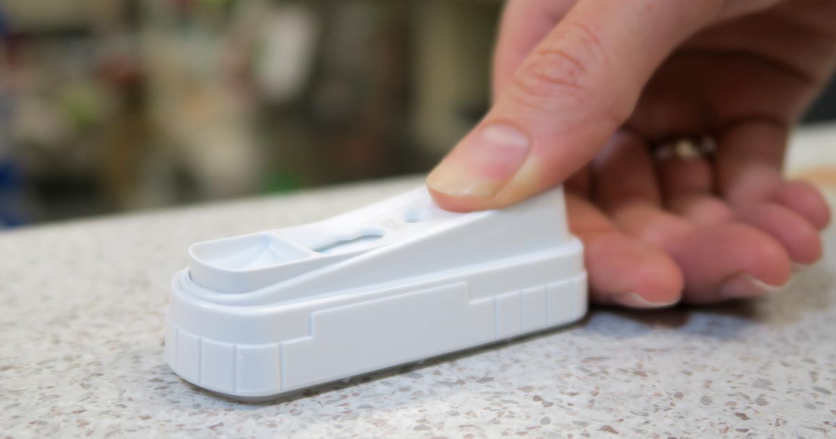
    Few companies now offer affordable point-of-care tests for canine C-reactive protein (CRP). As we did when we recently received our new box of CRP slides, you might soon be asking the question: what do we even do with this stuff?

    Here’s what we’ve learnt…

    CRP is one of the acute phase proteins produced by the liver in response to inflammation. Healthy patients have very low levels of CRP, but a systemic inflammatory condition will cause an increase in CRP within four to six hours. Conversely, increased levels will decrease rapidly on resolution of inflammation. This provides an almost real time measure of inflammation that is more responsive and reliable than the white blood cell response.

    In other words, CRP can indicate the presence of inflammation before the patient’s white blood cell count gives any clues, or before it becomes pyrexic – and, unlike the white blood cell count, stress and steroids do not affect CRP levels.

    Uses

    So, how do we use it?

    • I love it for early pickups of problems in those grey area cases: the dog seems okay on clinical examination, but something about it bothers me. A normal or mildly increased CRP test will make me sleep more easy, while a surprise high reading will prompt me to admit for full diagnostics, or at least get the patient in for a follow-up CRP the next day. Conversely, a localised problem – such as an abscess – combined with a normal CRP test might mean you can hold off on antibiotics and just recheck CRP in 24 hours.
    • It’s great for monitoring response to treatment. If my plan is working then I’d expect CRP to show a significant decrease by day two or three. If it’s not dipping by then, I need to reassess my treatment plan. Do I need to change antibiotics? Scan it again? Maybe we need to consider surgery? It can also be a good prognosticator. Research has shown failure of CRP to decrease significantly (around a 3× decrease) by around day three is generally bad news for patients with inflammatory conditions such as pancreatitis and immune-mediated haemolytic anaemia.
    • We are starting to play with it for post-surgical monitoring. Any surgery will cause inflammation with an increase in CRP levels, but in an uncomplicated postoperative period, you should expect levels to start decreasing by day three to five. A base line CRP 24 hours after surgery with a recheck on day three should pick up early signs of postoperative problems such as infection, and prompt investigation or intervention.
    • A potentially nifty use for it that we haven’t yet had the opportunity to use is in differentiating inflammatory lamenesses (arthritis, infection, injury) from a neurological causes – that is, is it arthritis or a nerve problem?

    Limitations

    • Remember, it’s very sensitive, so will increase with almost any inflammation. A mild upper respiratory infection or a bad gingivitis will likely induce some changes, so it’s important not to over-interpret (keep in mind that the magnitude of the increase in CRP does generally correspond with the severity of the inflammatory response). A pancreatitis case where the CRP fails to drop does not always mean death is looming – you may have just missed the concurrent skin disease. Always interpret CRP values in concert with your clinical examination.
    • Be aware that pregnancy and intense exercise can increase CRP values.
    • Not all serious conditions have an inflammatory component. CRP will be unchanged in most veterinary cases of heart disease; in common hormonal disease, such as adrenal disease and uncomplicated diabetes; urinary obstructions; many localised cancers; epilepsy and many others. Don’t presume that just because CRP is normal, everything is fine.
    • No similar test exists for cats.

    Sit up and say…

    My favourite way to explain how to use this test is by using its highly appropriate acronym – any unexpected increase should make you sit up and say: “Oh CR*P! What am I missing?”

  • Thoracentesis, part 2: sample work

    Thoracentesis, part 2: sample work

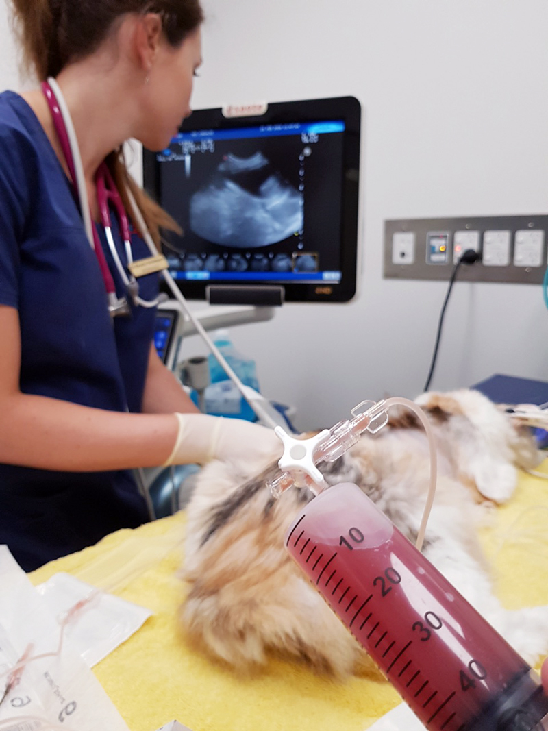
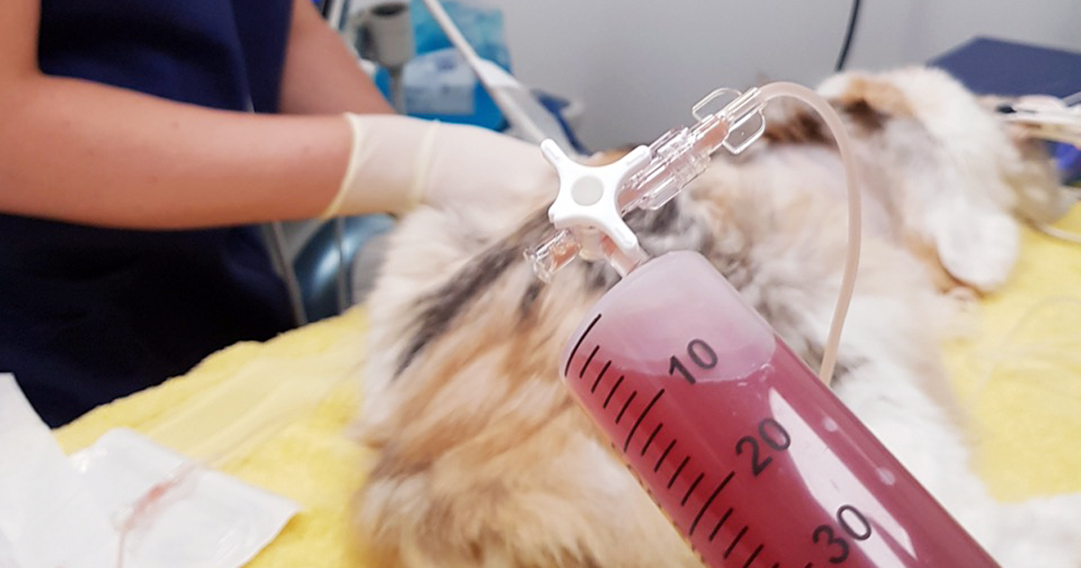
    Last week we gave some hints and tips about how to perform a thoracocentesis. This week we look at what to do with the sample you collected and where to go to next.

    Looking at the sample is not enough, there are several things you need to do to make sure you are getting the most information from the collected sample. This includes:

    • Fluid cell counts
    • Total protein assessment
    • Packed cell volume
    • Glucose
    • Lactate (if it is an exudate)
    • In-house cytology
    • Collect a sample for culture and sensitivity, and also external cytology assessment

    With this information you can narrow down your list of differentials; often enough it can give you a diagnosis.

    Here is the list I use. Note, it is not exhaustive and assumes you have taken three-view thoracic radiographs as part of the initial diagnosis.

    Transudate

    • Haemorrhagic effusion.
      Haemorrhagic effusion.

      Clear appearance – characterised by low protein and low cellularity

    • Transudates are caused by reduced oncotic pressure
    • Total nucleated cell counts = <0.5x10e9/L
    • Total protein = <25g/L

    Differentials to consider

    • Liver disease
    • Protein-losing nephropathy
    • Protein-losing enteropathy

    Additional diagnostics

    • Cytology and culture of fluid
    • Haematology and biochemistry
    • +/- dynamic liver testing
    • Urinalysis, urine protein/creatinine ratio, culture and sensitivity

    Modified transudate

    • Yellow/serosanguinous/cloudy appearance
    • Caused by increased hydrostatic pressure leading to passive leakage of proteins and fluid into the pleural space
    • Total nucleated cell counts = 3.5-5x10e9/L
    • Total protein = variable, ~25-50g/L

    Differentials to consider

    • Increased capillary hydrostatic pressure and pericardial disease
    • Diaphragmatic hernia
    • Neoplasia
    • Lymphatic obstruction, such as neoplasia, diaphragmatic hernia and abscess
    • Increased permeability of vessels (blood and lymphatics), such as FIP

    Additional diagnostics

    • Cytology and culture of fluid
    • Haematology and biochemistry
    • Cardiac auscultation and ultrasound
    • +/- CT

    Exudate

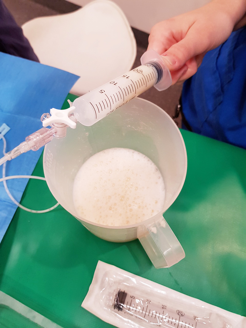
    • Turbid appearance – Very proteinaceous liquid, froths when shaken
    • Fluid is a mix of plasma and inflammatory mediators, and is caused by either septic or aseptic inflammation
    • Total nucleated cell counts = >3.0x10e9/L
    • Total protein = >30g/L

    Aseptic exudate

    • Non-degenerate neutrophils and activated mesothelial cells predominate
    • Non-infectious cause

    Differentials

    • Inflammation: FIP (can have high globulins), liver disease, lung torsion and hernia
    • Neoplasia

    Additional diagnostics

    • Haematology and biochemistry
    • Cytology and culture of fluid
    • +/- ultrasound/CT
    • Further testing for FIP

    Septic exudate

    • Degenerate neutrophils predominate: nuclear swelling and pale staining
    • Intracellular or extracelluar microorganisms
    • Culture and sensitivity: aerobic and anaerobic
    • Pleural fluid [glucose] < serum [glucose]
    • Pleural fluid [lactate] > serum [lactate]

    Differentials to consider

    • Ruptured abscess
    • Foreign body inhalation or penetrating injury
    • Fungal infection

    Additional diagnostics

    • Haematology and biochemistry
    • Cytology and culture of fluid
    • +/- ultrasound/CT

    Chyle

    Thoracocentesis-Chyle
    Chyle.

    Opaque (milky) to pink.

    Differentials to consider

    • Rupture or obstruction of lymphatic flow
    • Neoplasia, traumatic and idiopathic
    • Secondary to heart failure (especially in cats)
    • Pseudochyle (usually formed by lymphoma)

    Additional diagnostics

    • CBC and biochemistry
    • Cytology and culture of fluid
    • Fluid [TAG] > serum
    • Large number of lymphocytes and other inflammatory cells
    • +/- ultrasound/CT

    Haemorrhage

    • Red blood cells
    • True haemorrhagic; for example, not iatrogenic: should not see platelets or erythophagocytosis on smears and sample should not clot
    • Time frame
    • Assess history
    • Compare fluid PCV/total protein (TP) to peripheral PCV/TP:
    1. <1% – non-significant
    2. 1% to 20% – neoplasia, trauma, pneumonia
    3. >50% – haemothorax
    • Other tips:
    1. If PCV/TP is similar = recent bleed, if PCV is low and TP normal = chronic
    2. If PCV is increasing or is higher than peripheral then active bleeding
    3. Presence of erythrophagocytosis = chronic

    Differentials to Consider

    • Trauma
    • Neoplasia
    • Coagulopathies
    • Ruptured granuloma

    Diagnostics

    • Activated clotting time, activated partial thromboplastin time, prothrombin time, blood smear and other coagulation tests, see “coagulopathy”
    • Blood smear
    • CBC and biochemistry
    • +/- ultrasound/CT

    Good luck with your next thoracocentesis. I hope this information was useful.

  • Seizures, part 3: management

    Seizures, part 3: management

    In the third and final part of this series, we look at managing seizures in pets, both in an emergency setting and in the longer term.

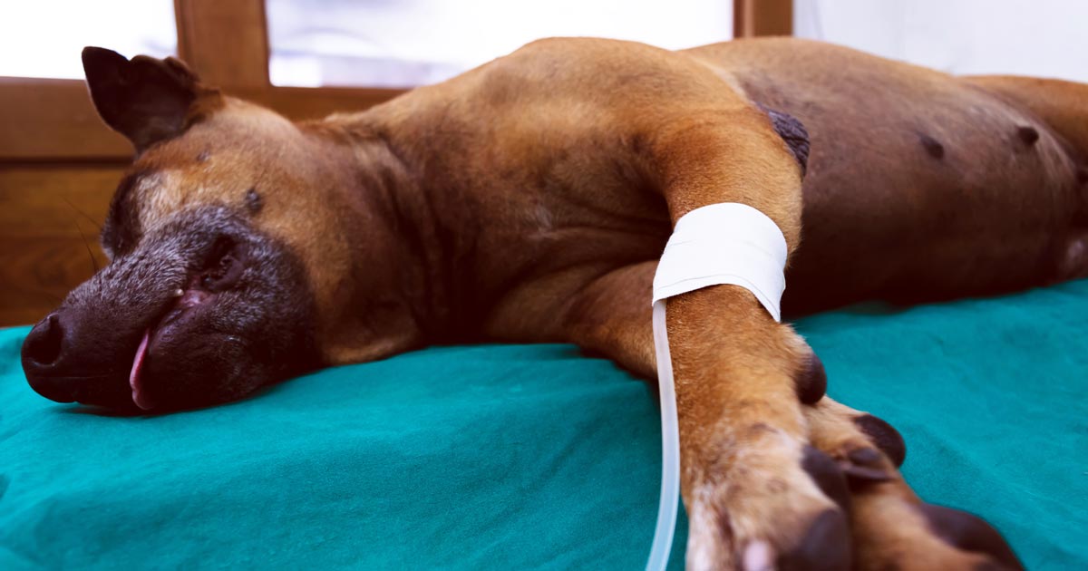
    When presented with a patient in status epilepticus, appropriate emergency treatment begins with:

    • Providing oxygen therapy.
    • Placing an IV catheter, if possible.
    • Administering diazepam as an 0.5mg/kg to 1mg/kg IV bolus, rectally at 2mg/kg or intranasally at 0.5mg/kg.
    • Intubating, if required to maintain a patent airway.
    • Cooling, if hyperthermic.
    • Giving mannitol at 0.5mg/kg to 1mg/kg slowly IV if seizure activity lasts more than 15 minutes or there is any reason to suspect cerebral oedema.
    • Collecting full bloods – test glucose, electrolyte and calcium levels first.
    • If on phenobarbital, collecting a sample for baseline testing.

    It is important to remember patients may continue to paddle slowly after seizure activity has finished, but if the eyelids are twitching, they are still seizing.

    If the seizures are controlled by these first steps, give a 4mg/kg dose of phenobarbital and commence supportive therapy with IV fluids, including correction of any electrolyte and metabolic derangements.

    If at first…

    If these first emergency steps fail to get the seizures under control, the following steps can be attempted:

    1. Diazepam 0.5mg/kg to 1mg/kg IV bolus – can be repeated every five minutes for up to three doses.
    2. Propofol 2mg/kg to 4mg/kg IV titrated to effect to stop motor activity.
    3. Phenobarbital slow IV 4mg/kg if already on maintenance therapy, mg/kg to 8mg/kg if not – can be repeated at 20 to 30 minute intervals.
    4. +/- Diazepam (or midazolam) continuous rate infusion (CRI) at 0.5mg/kg/hr.
    5. Propofol CRI following titrated dose, at 0.2mg/kg to 0.5mg/kg/min – continue for six hours then wean down slowly over next six hours.
    6. Levetiracetam at 20mg/kg to 60mg/kg IV titrated can be used instead of propofol (this is safer if hepatic disease is present).

    Ongoing treatment

    The recommendations for when to start long-term treatment are summarised as follows:

    1. Structural lesion present or prior history of brain disease or injury.
    2. Acute repetitive seizures or status epilepticus (ictal event ≥5 minutes or ≥3 or more generalised seizures within a 24-hour period).
    3. ≥2 or more seizure events within a six-month period.
    4. Prolonged severe, or unusual postictal periods.

    Chronic therapy in patients with ongoing seizures aims to reduce the frequency to an acceptable and manageable level. The drug used to achieve this is often down to clinician preference; one or a combination of the following can be used:

    • Phenobarbital – solo and combination therapy, drug monitoring is required along with regular monitoring of liver enzymes and function is particularly important.
    • Potassium bromide (not cats) – combination therapy, drug monitoring is required and can cause pancreatitis.
    • Imepitoin – solo or combination therapy, does not require drug monitoring.
    • Levetiracetam – solo or combination therapy, does not require drug monitoring.

    Regular testing of blood levels of anti-epileptics is important, although it does not indicate whether the drug should be working or not, it does help provide additional information when investigating when control is inadequate and to prevent toxic side effects.

  • Hypoglycaemia

    Hypoglycaemia

    Blood glucose is an important parameter that should be included in every “emergency database”.

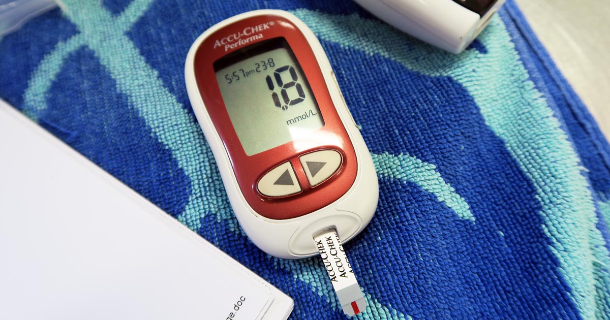
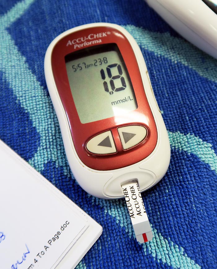
    Hypoglycaemia is considered when blood glucose levels drop below 3.5mmol/L or 63mg/dL. Symptoms can start as being vague, such as lethargy and weakness, then progress to tremoring and seizures.

    One important point is that, in an emergency setting, although reduced food intake or starvation is written in text books, unless the patient is very young or a very small size it is not a common cause of hypoglycaemia.

    The liver has a fairly substantial capacity to continue to produce glucose during periods of reduced eating or starvation.

    Common causes

    Hypo
    A blood glucose meter showing a blood glucose level of 1.8mmol/L.

    The common causes of hypoglycaemia I see in an emergency setting are:

    • sepsis: bacteria consumes glucose
    • hypoadrenocorticism: lack of cortisol
    • insulin overdose: excessive intracellular shift
    • insulinoma: malignant insulin secreting neoplasia of the pancreas
    • hepatic insufficiency: reduce production

    Treatment is fairly straightforward and the impact is often dramatic – 0.5ml/kg to 1ml/kg of 50% dextrose diluted 50:50 with saline given slow IV over a couple minutes (to reduce the risk of haemolysis).

    As the list of possible causes shows, a one-off dose of glucose is often not enough.

    Glucose supplementation often needs to be continued as a 2.5% continuous rate infusion (CRI), with frequent blood glucose monitoring and adjustments made to the rate as necessary.

    The CRI will need to be continued, as the hypoglycaemia will often continue to occur until the primary disease process is identified and appropriately addressed.

    Emergency database

    It is not uncommon to read or hear the term emergency database. This contains a number of blood parameters performed, which include:

    • blood glucose
    • alanine aminotransferase
    • lactate
    • blood urea nitrogen
    • PCV
    • total protein or total solids
    • activated clotting time
    • acid-base balance
    • electrolytes
  • Abdominal ultrasound, pt 3: scanning procedure

    Abdominal ultrasound, pt 3: scanning procedure

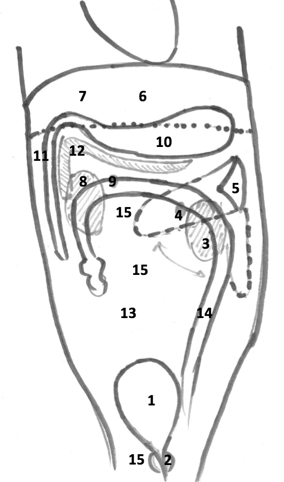
    I cannot stress enough how crucial it is to develop a systematic approach to doing ultrasounds. Not only will this approach help develop an anatomic mind map, it will also safeguard you from accidentally overlooking organs.

    Suggested scanning procedureWhen examining each individual organ, the same theory applies. This will ensure you look at each organ in its completeness, reducing the chance of an abnormality being missed.

    The only way to improve your ability to track through organs is by repetition; repeating and practising the same manoeuvres over and over again. In time, you will notice you will be able to detect subtle changes you wouldn’t have been able to before.

    Suggested scanning procedure

    Starting at the bladder and moving anti-clockwise:

    1. bladder
    2. prostate/uterus
    3. left kidney
    4. left adrenal
    5. spleen
    6. liver
    7. gall bladder
    8. right kidney
    9. right adrenal
    10. stomach
    11. duodenum and pylorus
    12. pancreas
    13. small intestine
    14. large intestine
    15. lymph nodes
      • mesenteric lymph node
      • iliac lymph node
  • Lipaemia – the bane of biochemistry

    Lipaemia – the bane of biochemistry

    Last week we covered haemolysed samples – this week we’re looking at lipaemic samples.

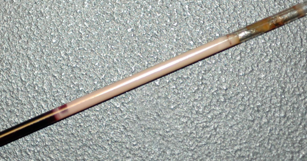
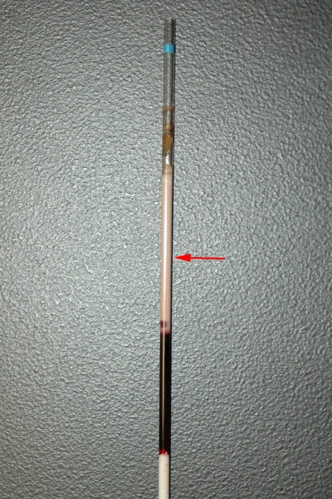
    Lipaemic samples are caused by an excess of lipoproteins in the blood, creating a milky/turbid appearance that interferes with multiple biochemical tests and can even cause haemolysis of red blood cells.

    lipaemic sample
    A severely lipaemic sample (red arrow). IMAGE: eClinPath.com (CC BY-NC-SA 4.0).

    Lipaemia can follow recent ingestion of a meal – especially one high in fat. Although not pathognomonic for any diseases, its presence can help increase the suspicion of certain diseases, including:

    • pancreatitis
    • diabetes mellitus
    • hypothyroidism
    • hyperadrenocorticism
    • primary hyperlipidaemia (in some specific breeds, such as the miniature schnauzer)

    It warrants further investigation in patients that have been ill and inappetent.

    Irksome interpretations

    Lipaemia can dramatically impact laboratory testing and is often troublesome in critically ill patients, making interpretation of biochemistry particularly difficult, if not impossible.

    Lipaemia can affect different analysers in different ways, most commonly causing:

    • Falsely increased calcium, phosphorus, bilirubin, glucose and total protein (via refractometer) and some liver parameters such as alkaline phosphatase, alanine aminotransferase, aspartate aminotransferase, haemoglobin concentration, and mean corpuscular haemoglobin concentration.
    • Falsely decreased sodium, potassium, chloride, albumin and bicarbonate.

    Tube tips

    Assessment of a centrifuged haematocrit tube before running a biochemistry panel can help reduce wasted biochemistry consumables.

    If the sample is lipaemic in the haematocrit tube then maybe try some of the following tips.

    • If blood tests are planned in advance, try fasting the patient beforehand for 12 to 24 hours.
    • Repeat sampling a couple of hours later may yield a less lipaemic sample.
    • Collecting and centrifuging a larger amount of blood (3ml to 5ml, for example) can sometimes yield enough clear sample between the lipid layer and red blood cells.
    • Refrigeration of the sample can help the separation.
    • Extract lipids using polar solvents, such as polyethylene glycol.
    • Centrifugation at higher than normal speeds (if possible) can also assist in clearing the layer.
  • Abdominal radiography, part 2

    Abdominal radiography, part 2

    Last week’s tips (Abdominal radiography, part 1) were about taking appropriate images.

    Now, here are some tips on interpreting those images.

    Interpretation

    abdominal x-ray
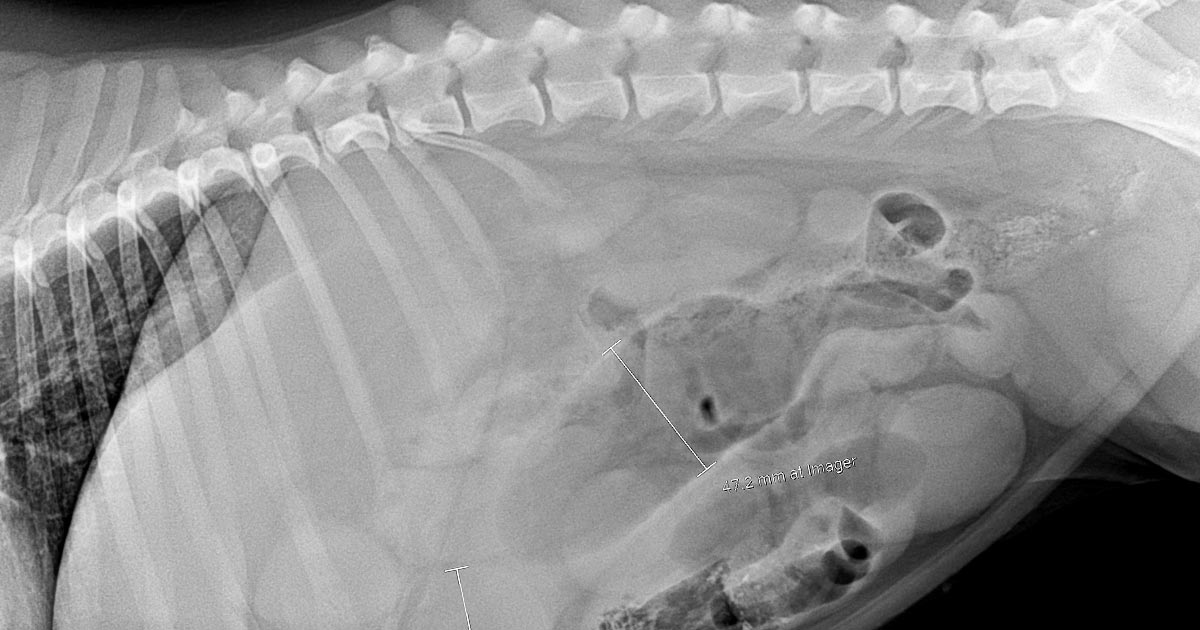
    Abdominal x-ray (click to zoom).

    I often find there is too much to look at and it gets confusing with overlapping organs. I like to step back and look from a distance; sometimes, this gives me an overview of the image first.

    Next, I use a systematic approach, starting with extra-abdominal structures and working inwards.

    Then I assess the main organs – liver, spleen, kidneys, bladder and prostatic region – and assess for a uterus. Once I have identified and assessed those, I look at the gastrointestinal tract (GIT).

    What part of the GIT is it?

    This can be the most confusing part. I start with the stomach, assess size and position, then identify and track the colon from caecum to rectum. Once I’m happy I have identified the stomach and colon then everything else with gas in it is likely to be small intestinal.

    Enema and a walk

    If there is too much faeces, or if the gas is colonic, I administer a suppository enema to facilitate defecation. This can dramatically clear up a confusing x-ray and move gas.

    Repeat the study

    If I am still concerned about an obstruction, but it is not obvious, I either transition to an ultrasound or repeat the abdominal study after a couple of hours of IV fluids and pain relief.

    I find, once a patient is rehydrated, the GIT starts to move; gas and faeces shift and things can look surprisingly different in a couple of hours. If it remains the same, or worsens, that also provides valuable information.

    Consider high GIT and partial obstructions

    Don’t forget pyloric, high duodenal and partial obstructions that don’t give you the classic small intestinal dilation. The absence of a radiopaque foreign body or gas dilated small intestines do not rule out an obstruction.

  • Using canine pancreas-specific lipase test

    Using canine pancreas-specific lipase test

    Accurately diagnosing pancreatitis in dogs is so tedious and difficult, we used to wish for a single test that could save all that hassle – hence the arrival of the pancreas-specific lipase (PL) test, designed to help vets worldwide with this difficult situation.

    So, how do I use this test? I don’t use it to diagnose pancreatitis, but to help rule out acute pancreatitis.

    Stay with me…

    Standard diagnosis

    SNAP CPL
    The pancreas-specific lipase test.

    First, let me go back to the basics. Historically, pancreatitis is diagnosed based on a combination of supportive evidence from medical history:

    • physical examination findings
    • complete blood count
    • biochemical analysis
    • abdominal ultrasound
    • occasional histology

    The reason to do all these is because no parameter is sensitive or specific enough.

    Ruling it out

    PL is exclusively of pancreatic origin, which inherently gives PL tests a high sensitivity (reported to be between 85% to 95% in acute pancreatitis) and thus has a high negative predictive value (the probability subjects with a negative screening test truly don’t have the disease). This makes this test useful in ruling out acute pancreatitis.

    Notice how I specifically mentioned acute pancreatitis? Chronic pancreatitis is often associated with fibrosis and atrophy, so is not usually associated with leakage of pancreatic acinar cell enzymes, and thus makes this test insensitive to diagnosing chronic cases.

    Looking at specificity, PL tends to have a variable specificity – between 59% and 98% depending on the test methods used – meaning it has a modest positive predictive value (the probability subjects with a positive screening test have the disease).

    False positives

    In a prospective study assessing the diagnostic accuracy of SNAP PL and Spec PL tests, Haworth et al (2014) found 40% of the 38 dogs presenting with acute abdominal disease gave a “false positive” result when used to diagnose pancreatitis. This supports the fact disease in organs closely associated with the pancreas or the pancreatic ducts – liver or duodenum, for example – can cause concurrent mild inflammation of the pancreas without causing clinical pancreatitis itself.

    Personally, I have removed foreign bodies several times from patients who were referred for suspected pancreatitis.

    My take on the SNAP PL is a negative is highly unlikely to be pancreatitis, which, in itself, is useful information as it helps rule out a common disease, but a positive can still be caused by many other abdominal inflammatory diseases and an ultrasound performed by a skilled operator is still needed.

  • RCVS council election manifesto: Thomas Lonsdale

    RCVS council election manifesto: Thomas Lonsdale

    THOMAS LONSDALE

    Thomas Lonsdale.

    BVetMed, MRCVS

    PO Box 6096, Windsor Delivery Centre, NSW 2756, Australia.

    T +61 2 4577 7061

    M +61 437 2928 00

    E tom@rawmeatybones.com

    PROPOSERS: Roger Meacock, Andrew Stephens

    1980s – woke from vet-school induced stupor to realisation junk pet-food industry relies on bogus science and negligent vet “profession”.

    1991 – Blew whistle on junk pet food cult.

    1993 – Preventive dentistry PGCVS.

    1994 – Feeding versus nutrition, Aust Vet Practice.

    1994 – Cybernetic hypothesis, J Vet Dent (postulates ecological theory of health and disease as extension of Gaia Hypothesis).

    1994-7 – Junk pet food cult brought four disciplinary actions before New South Wales vet board.

    1995 – Periodontal disease and leucopaenia, JSAP.

    2001Raw Meaty Bones: Promote Health published.

    2004 – Nominated for ACVSc award.

    2014 – Most supportive vet award; FOI research: junk pet food grease in seven Australian vet schools.

    2015 – Science death experiment.

    Manifesto

    Pompous, arrogant, mouthing incantations, the vet high priests worship at the altar of bogus science. Founded on fallacy, they oversee the junk food poisoning of pets, betrayal of consumers and brainwashing of vet students.

    They must be stopped.

    Morgan Spurlock embarked on a risky experiment. For 30 days he ate junk food at every meal. He gained 11kg, his liver turned to fat, cholesterol shot up and he doubled his risk of heart failure. Fortunately for Spurlock, he escaped addiction to junk food, followed his doctor’s orders and stopped the experiment.

    The medical profession tells us carbohydrate-laden junk food injures health; that periodontal inflammation and obesity are precursors of systemic disease and early death. By contrast, the junk pet food industry controls the veterinary agenda. Vet schools deliver industry-funded propaganda on diabetes, periodontal disease and obesity – while simultaneously ensuring pious mumbo-jumbo obscures the despicable, lamentable truth.

    Vet “experts” jet about the world spruiking the latest concoctions; extolling the alleged benefits of elaborate treatments instead of declaring that junk food contaminates all aspects of vet science, teaching and practice. According to them, natural food, as determined by evolution, is dangerous while their paymaster’s industrial junk represents the pinnacle of excellence.

    For 20 consecutive RCVS elections, I’ve called for our “self-regulating” profession to act with integrity and honour. Alas, the high priests refuse even to consider. Time, then, for the courts to decide. I recommend legal proceedings against the RCVS, junk food companies, veterinary schools and individuals in respect to animal cruelty, breach of contract, theft and fraud.

    Please vote in support; instruct your lawyers and brief the media. Pets, pet owners and the wider community need our help. Thank you.

  • Festive threats to four-legged family members

    Festive threats to four-legged family members

    Now well into December, and getting ever closer to Christmas, there will be an abundance of “goodies” around the house that are not so good for the four-legged family members.

    Image: © Freeimages/s22k.
    Jordan advises colleagues to ensure clients keep an eye on their pets during the festivities. Image: © Freeimages/s22k.

    Most owners are aware of the dangers of chocolate and so are likely to rush down to the vet on Boxing Day when their Labrador has broken into the tin of Heroes – but what other festive dangers are there that owners are not so clued up on?

    Pudding

    The festive favourite Christmas pudding, while enjoyable for us, can be lethal for pets. It is the raisins, currants and sultanas that may lurk within that are poisonous.

    While the mechanism of toxicity is poorly understood, we know it can cause acute kidney failure in dogs.

    The treatment protocol for poisoning by raisins, etc, is similar to general suspected poisoning with an unknown agent: give an emetic (to make the animal vomit), an adsorbent (for example, activated charcoal) and supportive care (IV fluids) to protect the kidneys for a few days.

    Antifreeze

    Ethylene glycol, more commonly known as antifreeze, is extremely toxic to pets, with consumption of tiny amounts resulting in crystal formation in the kidneys, and so acute renal failure. Antifreeze smells and tastes sweet, so pets are often attracted to it. For this reason, it is best for owners to ensure it is kept in a secure container, well away from pets.

    Early symptoms include neurological signs, such as wobbliness, twitching, seizures and depression. As the poisoning progresses, the animal will have cardiac and respiratory signs, such as increased heart and respiratory rate. It’s vitally important owners look out for these signs if their pets have had any possible access to antifreeze (for example, if they’ve got into the garage or if a cat has been missing for a few days and potentially been locked in someone’s shed).

    The antidote for ethylene glycol poisoning is ethanol, because it replaces ethylene glycol as a substrate for the enzyme that breaks it down into toxic products. More specifically, lab-grade ethanol is best.

    Sweetener

    Xylitol is a sugar-free sweeter that is used in chewing gum, cakes and sweets, particularly diabetic foods. Xylitol causes insulin release in the body, resulting in hypoglycaemia and, later, liver damage.

    Symptoms include vomiting and signs of hypoglycaemia: lethargy, weakness, collapse and, if it progresses, seizures and coma. Unlike ethylene glycol, there isn’t a specific antidote, but supportive care, including fluids and liver protectants, is vital. The prognosis for xylitol toxicity is good if the animal’s hypoglycaemia is corrected quickly, so it is essential for owners that suspect this to contact their vet immediately. If you’re trying to determine whether xylitol is in a product, it is often listed as a food additive, code E967.

    Decorations?

    And don’t forget the potential foreign body too – if the cat has been playing with the tinsel and managed to swallow some or the dog has ingested a few tasty looking baubles.

    Enjoy the festive season, but advise owners to keep an eye on their pets this Christmas.