Tag: lesions

  • Immunotherapy in cats

    Immunotherapy in cats

    Although “off-licence”, immunotherapy is well worth considering in our pruritic feline friends.

    Feline head pruritus secondary to food allergy
    Feline head pruritus, a result of food allergy. Image by Caroldermoid. Licensed under CC BY-SA 3.0 via Wikimedia Commons.

    Of course we need to rule out all other causes of pruritus first:

    • Ectoparasites: As well as all the usual suspects, don’t forget Demodex can occasionally cause ventral alopecia and pruritus (that’s the funny flat form, not the cigar shaped one).
    • Food allergies: Let’s face it, food allergy is tricky to pursue in cats – and if they have a partly outdoor lifestyle, hypoallergenic diets are of not of any use.

    Case study

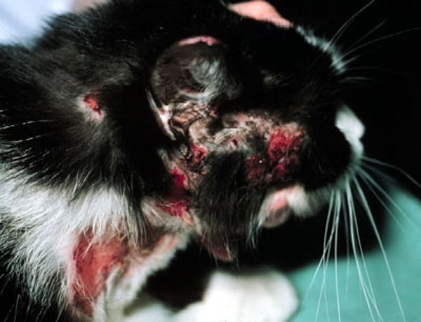
    I recently saw a case that had typical excoriation lesions around the head and neck.

    The owner was able to keep the cat indoors on Purina HA Hypoallergenic for six weeks, and was happy to dose monthly with Stronghold. This, together with a good response to steroids, was highly suggestive of atopy.

    Serum IgE testing produced a range of high levels to pollens, house dust mite and moulds.

    Our friend has recently started on immunotherapy injections and is now starting to show signs of a good response. With the feline response to immunotherapy reported to be higher than that in dogs, this may be something well worth considering in cats.

  • Treatment of cat ringworm carriers

    Treatment of cat ringworm carriers

    Ringworm under UV
    Ringworm discovered using a Wood’s lamp (ultraviolet light). Image courtesy DermVet Skin and Ear Clinic.

    Cats, particularly long-haired varieties, can be carriers of ringworm but show no clinical signs at all.

    Diagnosis in the absence of skin lesions is made by either Wood’s lamp over the entire body (only 50-60% of positives will fluoresce), or by taking coat brushings and incubating the hairs, follicles and debris in a dermatophyte test medium (DTM).

    Once a cat is diagnosed as a carrier, treatment is the same as for those with lesions: oral itraconazole.

    Even after consecutive negative results, the cat should always be considered a carrier, as it’s so difficult to eradicate ringworm from the environment. Treatment is still considered worthwhile, however, and effective cleaning will reduce/prevent further outbreaks.

  • Role of bacterial colonisation in allergic skin disease

    Beagle
    Pruritus: the unpleasant skin sensation that frequently provokes scratching. Image ©iStock.com/olaser

    As spring warms up, my own nose and sinuses have detected increasing pollen levels, and there’s been a corresponding increase in pruritic patients coming across the threshold.

    I think the role of pyoderma in allergic skin disease is well established and accepted. However, it is not uncommon to see dogs with no obvious skin lesions that are markedly pruritic, and cytology often reveals large numbers of cocci without the correspondingly high levels of inflammatory cells.

    In patients with reduced barrier function, this overgrowth can contribute significantly to the levels of pruritus. I find a significant number of dogs’ pruritus reduces by at least 50%, if not more, with appropriate antibiotic and shampoo therapy when overgrowth is identified by cytology.

    I usually repeat the cytology in around 2 to 3 weeks to check for resolution, although, of course, given the relapsing nature of the condition periodic treatment is required.

    Reference
    Rosenkrantz W (2010). Proceedings of the NAVC Conference 2010.

  • Don’t remove a mass without knowing what it is

    I came across a paper that advised we should know exactly what a mass is before surgery. The rationale being as follows (Ettinger, 2015):

    “Do something: aspirate or biopsy, and treat appropriately.

    Needle biopsy
    Vet performing a needle biopsy on a cocker spaniel. Image ©iStock.com/Paolo_Toffanin

    “Why diagnose early? Obtaining a definitive diagnosis with cytology or biopsy early and before excision will lead to improved patient outcomes for superficial masses. Surgery is likely curative for the majority of superficial tumours when detected early, when they are small – especially benign lesions and locally invasive tumours with a low probability of metastasis. If tumours are removed with complete surgical margins, the prognosis is often good with no additional treatments needed.

    “Pet owners need to be aware of the ‘pea’ size requirement to have masses evaluated, and veterinarians must measure and document the size of the mass to compare growth.

    “If more than 1cm (or the size of large pea) and present for a month, the mass should be aspirated or biopsied.

    “Knowing the tumour type prior to the first surgery will increase success of a curative-intent surgery.”

    In all honesty, I rarely do this for masses booked in for surgery and I suspect as lipomatosus, and for masses that visually appear consistent for histiocytoma – but there are tips here I will follow in the future.

    Reference
    Ettinger S (2015). Top Ten Oncology Mistakes and How to Avoid Them, North American Veterinary Conference: Small Animal & Exotics Proceedings, Gainsville, Florida.

  • Buy a dental x-ray machine

    Dental radiograph showing periodontal disease in a 2-year-old cat.
    A dental radiograph showing periodontal disease in a 2-year-old cat (note bone loss), by mariposavet. Licensed under CC BY 2.0 via Flickr.

    If I am being perfectly honest, dentistry has never filled me with excitement.

    That said, attending a number of sessions on dentistry at the North American Veterinary Conference this year was fascinating and convinced me of the value of buying a dental x-ray machine.

    Goldstein (2015), to name but one speaker, evaluated the use of radiography in assessing dental conditions in cats and convinced me of the need.

    In another talk the value in assessing canine lesions was discussed (Lewis, 2015) – definitely something to bring up at our practice’s next clinical effectiveness meeting.

    References
    Goldstein, G.S. (2015). Dental pathology case presentations, clinical and radiology: interactice discussion of what’s new, North American Veterinary Conference, Orlando 2015.

    Lewis, J.R. (2015). Interpreting canine dental radiographs: learning what you were never taught, North American Veterinary Conference, Orlando 2015.

  • Sterile pyogranulomatous cysts are worth bearing in mind

    Interdigital cyst
    An interdigital cyst in the paw of an English bull terrier – image by Nottingham Vet School (CC BY-NC-SA 2.0) via Flickr.

    I recently saw a very large, and very friendly bull mastiff that presented with what appeared to be typical interdigital cysts. These had been chronically recurrent and previous histopathology indicated a pyogranulomatous reaction.

    However his lesions responded poorly to three weeks’ of cephalexin antibiotic therapy and, in fact, he developed several lesions on his other feet.

    Cytology revealed mild bacterial colonisation, and it occurred to me this might be an immune-mediated reaction (his blood work and TSH/T4 were all within normal limits) – and, hey presto, we got a rapid clinical response to steroids.

    In spite of being under control he has also developed an acute erythematous otitis externa, so I think a food trial is my next line. Watch this space…