Tag: Lactate

  • Using lactate measurements in general practice

    Using lactate measurements in general practice

    Several easy and affordable ways exist to measure lactate in general practice, which means the clinical applications of monitoring lactate is no longer the reserve of specialist and emergency centres.

    But why and how should you be using it in general practice?

    What is lactate again?

    When oxygen is not effectively delivered to cells throughout the body – which, in our patients, will mostly be due to hypoperfusion (for example, hypovolaemia, vasodilatory shock and cardiac disease) – cells will switch from aerobic to anaerobic metabolism to stay alive.

    Think of anaerobic metabolism as the fuel-powered generator that kicks in during a power cut – it’s not as good, but it’ll keep the lights on for a while. However, unless the power comes back on, the generator will eventually also fail and plunge you into darkness.

    Lactate is the end product of this process of anaerobic metabolism. To be clear, lactate is not the bad guy – in fact, it plays an important role in keeping the cells going until they have access to sufficient oxygen again. It’s simply the bearer of bad news.

    Why should I care?

    Anaerobic metabolism – similar to a fuel generator being used during a power cut. Image © bildlove / Adobe Stock
    Anaerobic metabolism – similar to a fuel generator being used during a power cut. Image © bildlove / Adobe Stock

    Because lactate is the harbinger of doom; it’s the leading horseman of the apocalypse…

    When lactate is high, you should stop whatever else you are doing and pay attention to the patient in question – this patient is probably surviving on anaerobic metabolism. It is critically ill and possibly heading for a long walk in the great park in the sky…

    How should I use it?

    It is valuable in any patient with any serious illness or injury. Things that should make you consider checking lactate would be:

    • slow capillary refill time
    • any mucous membrane colour other than a nice, healthy pink
    • increased heart rate
    • weak or bounding pulses
    • significant dehydration
    • depressed mentation
    • history of major trauma
    • major infections
    • significant blood loss
    • any disease that has the potential to progress into a life-threatening condition

    …basically, any animal sick enough to make you worry about it possibly dying.

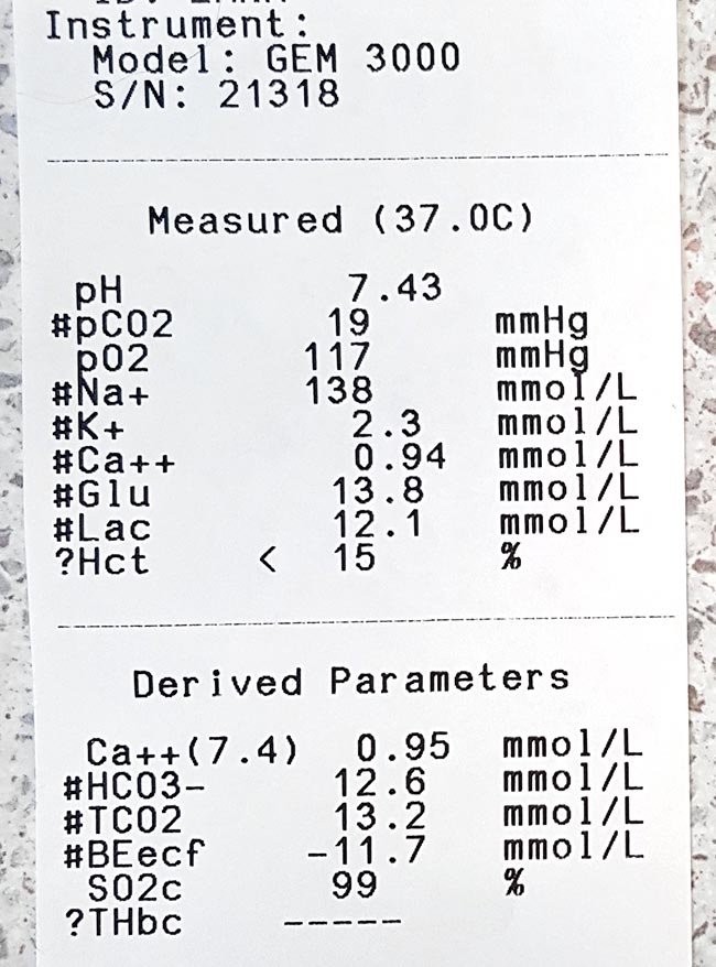
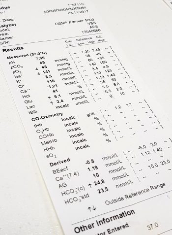
    Run it with your initial diagnostics to get a baseline level, and run it within 10 minutes of taking the sample.

    What do I do about my results?
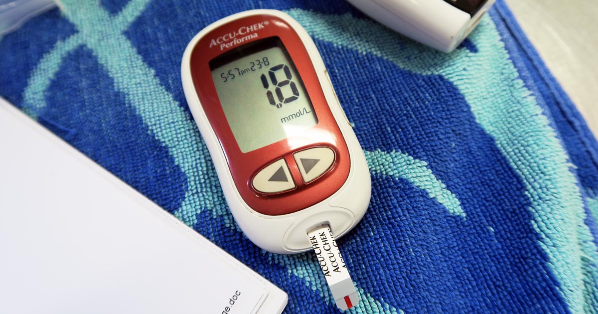
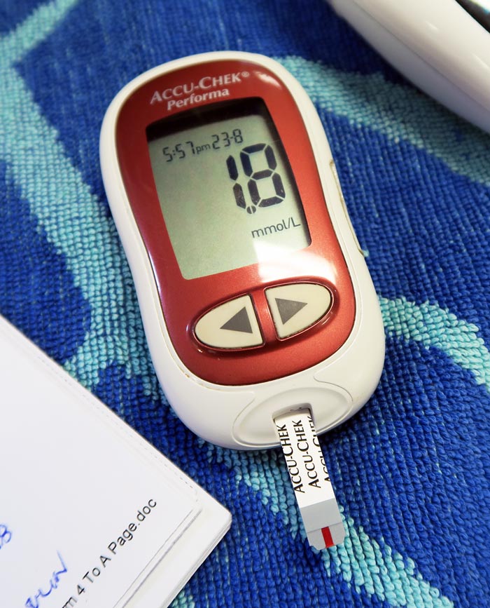
    Normal range

    What do my results mean?

    < 2.5  ?

    3-4 ? ?

    4-6  ? ?

    > 6  ? ?

    (levels in mmol/L)

    If it’s within the normal range then check regularly – ideally until your patient is well on its way to a full recovery.

    Lactate levels may start increasing in response to hypoperfusion before the patient starts showing overt signs of deterioration – the fuel in the generator hasn’t run out yet – which makes it a very useful monitoring tool to detect problems early on.

    We find a six to eight-hourly check in very sick patients will pick up deterioration fast enough to give you time to react, while a twice-a-day check in more stable animals will suffice.

    Elevated

    If it is increased at any point, you need to focus immediately on trying to reduce it.

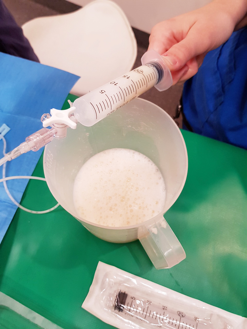
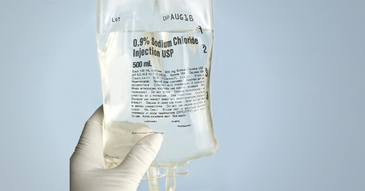
    This usually means starting with fluid boluses for shock. Recheck lactate one hour after initiating the appropriate therapy. Your goal is for the lactate value to be reduced by approximately 50% within one to three hours (ideally one hour) of initiating therapy. If it’s coming down nicely then keep checking every two to three hours.

    You want it to be back to normal within 24 hours (48 hours max).

    Nothing working?

    If it has not decreased as expected – or, especially, if it increases despite treatment – it means things are going seriously wrong. At this stage you need to:

    • Devote all your attention on trying to find and correct the underlying cause while you adjust your emergency therapy to address hypoperfusion.
    • Speak to the owners.
    • If you do not have the time, facilities or experience to deal with shock cases, you need to consider referring the patient to a specialist centre urgently for stabilisation, if this is an option available to you.

    Remember…

    Intense exercise, muscle tremors and seizures are associated with anaerobic muscle activity and can, therefore, cause significant increases in lactate levels (the cause of the “deep burn” when you’re dying in that CrossFit class). This can also occur when a patient resists when you are taking blood, or starts trembling in fear the moment it walks into the clinic, which can cause misleading lactate results.

    Puppies can have a higher “normal” level of plasma lactate up to seven months of age.

    Anaemia does not generally cause hyperlactataemia unless it is very severe, but by this stage you shouldn’t need lactate to tell you the patient is in serious trouble.

    Having said that, normal lactate levels that suddenly start climbing in a hospitalised “stable” anaemic patient could be the push you need towards giving that blood transfusion.

  • Ionised hypocalcaemia, pt 3: acute treatment and management

    Ionised hypocalcaemia, pt 3: acute treatment and management

    Treatment of ionised hypocalcaemia (iHCa) is reserved for patients with supportive clinical signs, then divided into acute and chronic management.

    Since the most common cases of clinical hypocalcaemia in canine and feline patients are acute to peracute cases, this blog will focus on the acute treatment and management of hypocalcaemia.

    Clinical signs

    The severity of clinical signs of iHCa is proportional to the magnitude, as well as the rate of decline in ionised calcium (iCa) concentration.

    The normal reference range for iCa is 1.2mmol/L to 1.5mmol/L in dogs and 1.1mmol/L to 1.4mmol/L in cats. Serum iCa concentrations in younger dogs and cats are, on average, 0.025mmol/L to  0.1mmol/L higher than adults.

    Mild iHCa (0.9mmol/L to 1.1mmol/L) – as seen in critically ill dogs and cats with diabetic ketoacidosis, acute pancreatitis, protein-losing enteropathies, sepsis, trauma, tumour lysis syndrome or urethral obstructions – often has no observable clinical signs.

    Moderately (0.8mmol/L to 0.9mmol/L) to severely (lower than 0.8mmol/L) affected animals – in the case of eclampsia and those with parathyroid disease – often display severe signs.

    Early signs of iHCa are often non-specific, and include:

    • anorexia
    • rubbing of the face
    • agitation
    • restlessness
    • hypersensitivity
    • stiff and stilted gait

    As the serum iCa concentration further decreases, patients often progress to:

    • paresthesia
    • tachypnoea
    • generalised muscle fasciculations
    • cramping
    • tetany
    • seizures

    In cats, the gastrointestinal system can also be affected, presenting as anorexia and vomiting.

    Treatment

    The need for treatment of hypocalcaemia is dependent on the presence of clinical signs, rather than a specific cut-off of serum concentration of iCa itself.

    Moderate to severe iHCa should always be treated. Mild hypocalcaemia, on the other hand, may not be necessary, especially if it is well tolerated. It should be remembered the threshold for development of clinical signs is variable, and treatment may benefit critical cases with an iCa concentration of less than 1.0mmol/L.

    Treatment is divided into the acute treatment phase and chronic management.

    In the tetanic phase, IV calcium is required – 10% calcium gluconate (equivalent to 9.3mg/ml) administered at 0.5ml/kg to 1.5ml/kg dosing to effect. This should be administered slowly with concurrent ECG monitoring. Infusion of calcium needs to be stopped if bradycardia develops or if shortening of the QT interval occurs.

    Some suggest calcium gluconate (diluted 1:1 with 0.9% sodium chloride) of half or the full IV dose can be given SC and repeated every six to eight hours until the patient is stable enough to receive oral supplementation. However, be aware calcium salts SC can cause severe necrosis or skin mineralisation.

    Calcium chloride should never be given SC, as it is a severe perivascular irritant.

    Correcting iCa

    Irrespective of the chronicity of the treatment, the rule of thumb is correction of calcium should not exceed 1.1mmol/L.

    Correction of iCa to normal or hypercalcaemic concentration should always be avoided, as this will result in the desensitisation of the parathyroid response, predisposing renal mineralisation and formation of urinary calculi.

    Some of the more common calcium supplementation medications – both parenteral and oral formulas – are detailed in Table 1. Supplementation of magnesium may also benefit some patients, as it is a common concurrent finding in critically ill patients with iHCa.

    Table 1. Common calcium supplementation medications
    Drug Calcium Content Dose Comment
    Parenteral calcium
    Calcium gluconate
    (10% solution)
    9.3mg/ml
    i) slow IV dosing to effect (0.5ml/kg to 1.5ml/kg); acute crisis, 50mg/kg to 150mg/kg over 20 to 30 minutes
    ii) 5mg/kg/hr to 15mg/kg/hr IV or 1,000mg/kg/day to 1,500mg/kg/day (or 42mg/kg/hr to 63mg/kg/hr)
    Stop if bradycardia or shortened QT interval occurs.
    Infusion to maintain normal Ca level
    SC calcium salts can cause severe skin necrosis/mineralisation.
    Calcium chloride
    (10% solution)
    27.2mg/ml 5mg/kg/hr to 15mg/kg/hr IV Do not give SC as severe perivascular irritant
    Oral calcium
    Calcium carbonate
    (many sizes)
    40% tablet 5mg/kg/day to 15mg/kg/day
    Calcium lactate
    (325mg, 650mg)
    13% tablet 25mg/kg/day to 50mg/kg/day
    Calcium chloride
    (powder)
    27.2% 25mg/kg/day to 50mg/kg/day May cause gastric irritation
    Calcium gluconate (many sizes) 10% 25mg/kg/day to 50mg/kg/day

    Next time…

    The next blog will look at the pathophysiology behind iHCa among critically ill animals. It will also look at the controversy regarding treatment of non-clinical iHCa cases and the prognostic indications of iCa concentrations.

  • Ionised hypocalcaemia, pt 1: introduction

    Ionised hypocalcaemia, pt 1: introduction

    Low ionised calcium (iCa) is a widely recognised electrolyte disturbance in critically ill human patients who have undergone surgery, are septic, have pancreatitis, or have sustained severe trauma or burns.

    Similar changes occur in our critical canine and feline patients, though less well documented.

    Calcium plays a vital role in a myriad of physiological processes in the body, so any deviation from the very narrowly controlled range is associated with severe repercussions.

    Low iCa has many causes; however, this three-part blog will only focus on the more common and peracute to acute causes. It will also discuss the treatment of low iCa and the controversy behind treatment of iCa in critically ill patients.

    Forms

    Calcium in the serum or plasma exists in three forms:

    • ionised or free calcium
    • protein-bound calcium
    • complexed or chelated calcium (bound to phosphate, bicarbonate, sulfate, citrate and lactate)

    iCa is the biologically active fraction of calcium and is not to be confused with total calcium (tCa). A lack of concordance exists between the two. Adjustment formulas are inaccurate, even with the correction of the tCa to serum total protein or albumin concentration, and should not be used to predict iCa.

    The normal reference range for iCa in dogs is 1.2mmol/L to 1.5mmol/L; in cats, it is 1.1mmol/L to 1.4mmol/L.

    Function

    An example of low ionised calcium.
    An example of low ionised calcium.

    Calcium is essential in maintaining normal physiological processes in the body. iCa regulates:

    • vascular tone
    • myocardial contraction
    • homeostasis

    In addition, it is needed for:

    • enzymatic reactions
    • nerve conductions
    • neuromuscular transmission
    • muscle contraction
    • hormone release
    • bone formation
    • resorption

    In critical patients, particularly those with severe trauma or sepsis, vascular tone and coagulation is particularly important. For this reason, iCa is tightly kept in a narrow range and regulated by the interactive feedback loop that involves iCa, phosphorous, parathyroid hormone, calcitriol and calcitonin.

    Diseases and causes

    Diseases commonly associated with low iCa in dogs and cats include:

    • acute kidney failure
    • acute pancreatitis
    • diabetic ketoacidosis
    • eclampsia
    • ethylene glycol intoxication
    • protein-losing enteropathies
    • sepsis
    • trauma
    • urethral obstruction
    • parathyroid diseases
    • tumour lysis syndrome

    Situations altering the fraction of extracellular calcium seen on a regular basis include:

    • acid-base disturbances
    • lactic acidosis
    • protein loss or gain
    • increased free fatty acids

    Iatrogenic causes include:

    • citrate (anticoagulant) administration during blood transfusions
    • phosphate
    • bicarbonate
    • sulfate administration

    Low iCa can also develop during cardiopulmonary resuscitation, quickly declining with increased duration.

    • Part two will go into more depth regarding the most common causes of low iCa that require acute treatment, the treatment involved, controversies surrounding treatment of non-clinical low iCa, and prognostic indications.
  • Blood transfusions, pt 1: clinical signs

    Blood transfusions, pt 1: clinical signs

    I get asked frequently when is the right time to transfuse an anaemic patient?

    The difficulty lies in the fact not all anaemic patients require blood transfusions. Just because a patient has pale mucous membranes does not mean the patient needs a transfusion.

    The term commonly brought up during the discussion is “transfusion triggers present”.

    What constitutes a “transfusion trigger”?

    A couple of different definitions exist: classically, it is the PCV or haemoglobin level at which a transfusion is indicated in an individual animal – essentially, if it gets below a certain number, transfusion is required – but it is not always that simple. Just because the PCV is 15%, it doesn’t always mean a transfusion is required.

    When the PCV drops low enough, clinical signs of reduced oxygen delivery to the tissues start to develop, these include:

    • decreased exercise tolerance
    • weakness
    • dull mentation
    • tachycardia
    • tachypnoea
    • elevated lactate levels when shock has been addressed

    Rapid or slow?

    These clinical signs are influenced by the speed at which the anaemia has developed.

    If the anaemia has occurred rapidly due to internal bleeding from trauma or a ruptured organ, these clinical signs can present in a matter of minutes, depending on how big the bleed is. This means a transfusion might be indicated when the PCV is still 25%, especially if further rapid blood loss is likely.

    If the anaemia developed over days to weeks (slow red cell destruction or anaemia of chronic disease, for example) transfusion triggers might not be present until the PCV drops below 15%, as the body has had time to compensate.

    Summary

    So, in summary, the decision-making process involves asking the questions:

    • What is the PCV?
    • How fast has the anaemia developed?
    • Are there clinical signs of reduced oxygen delivery?
    • Is further loss likely?

    When you combine the core aspects of each of the questions above, “transfusions triggers” change from absolute numbers to this:

    • PCV under 15% with clinical signs of reduced oxygen delivery.
    • Rapid PCV drop to under 20% in dogs and 15% in cats.
    • PCV under 25% and surgery or anaesthesia is required, and/or rapid ongoing blood loss is occurring.

    Blood products you should use and why will be covered in a future post.

    Note: Haemoglobin levels should also be assessed in conjunction with the PCV.

  • Dystocia, pt 2: diagnostics

    Dystocia, pt 2: diagnostics

    Part one of this series covered the stages of labour and indications dystocia is present.

    Once the bitch presents to the clinic, a few basic diagnostic checks need completing to determine the status of the bitch/queen and the fetuses.

    Physical examination

    The first is a thorough physical examination, starting with the bitch or queen:

    • Demeanour, hydration status, vital signs, mucous membrane colour, capillary refill time and temperature are important.
    • Pregnancy anaemia is not uncommon; however, for patients with a haemorrhagic discharge, it is important to know their cardiovascular status.
    • A thorough abdominal palpation should be carried out to assess comfort level and palpation for the presence of fetuses. Palpating fetuses can be difficult and cannot confirm if no fetuses are present.
    • A digital vaginal examination should be performed. Feathering response – also known as the Ferguson reflex in human medicine – is the neuroendocrine reflex where the self-sustained cycle of uterine contractions is initiated by firm pressure on the dorsal aspect of the vestibulovaginal wall. If this is absent, the patient is unlikely to progress with the parturition unaided.
    • Palpation of fetuses in the canal can help decide whether surgical management is required. Obvious fetal malposition, malposture or malpresentation, or fetopelvic disparity, will be indications of caesarean. Abnormal pelvic diameter is also another reason to not proceed with medical management. To confirm these suspicions, abdominal radiography is required.
    • Radiographs will also help determine the number of fetuses to be expected, the signs of fetal death (presence of gas surrounding the fetus) and aforementioned fetomaternal abnormalities. I always repeat radiographs after the expected number of neonates is passed, to make sure I have not miscounted at the start.

    Ultrasound

    Panel 1. Heart rate ranges to help indicate stress of fetuses

    Dogs:

    • normal – 180 to 220 beats per minute (bpm)
    • Stressed – 160bpm
    • Real concern – less than 160bpm

    Cats:

    • normal – more than 220pbm
    • fetal stress – less than 180bpm

    The second important diagnostic tool is ultrasound.

    Fetal heart rates are good indicators of fetal stress. Some heart rate ranges that can help provide information about the status of the fetuses are detailed in Panel 1. These ranges vary between sources, but are good guidelines.

    Ultrasounds can also help visualise the maturation status of the fetuses. At-term fetuses should have normal hepatic, renal and intestinal development. Intestinal peristalsis should be evident in at-term fetuses.

    Other diagnostics

    Other diagnostics may be indicated for patients, depending on the status of the bitch/queen:

    • If the patient is stable, but dystocia is present, a minimum database would include PCV/total protein, electrolytes, glucose, ionised calcium, lactate and acid-base balance.
    • Serum ionised calcium levels are important, as they influence the strength of contractions and how much supplementation is required.
    • Hypoglycaemia needs to be ruled out as a cause of dystocia, especially when large litters are involved.
    • If the patient is unstable or systemically unwell, include complete blood count, blood smears and biochemistry.
    • Physiological pregnancy anaemia can be present. The presence of regenerative response can help differentiate this from acute haemorrhage.
    • Abnormal leukocyte panel, especially with the presence of degenerative left shift, can indicate the presence of an infection – especially if toxic changes are present in the neutrophil.

    Part three will briefly look at the medical management of dystocia and when surgical intervention is required.

  • Thoracentesis, part 2: sample work

    Thoracentesis, part 2: sample work

    Last week we gave some hints and tips about how to perform a thoracocentesis. This week we look at what to do with the sample you collected and where to go to next.

    Looking at the sample is not enough, there are several things you need to do to make sure you are getting the most information from the collected sample. This includes:

    • Fluid cell counts
    • Total protein assessment
    • Packed cell volume
    • Glucose
    • Lactate (if it is an exudate)
    • In-house cytology
    • Collect a sample for culture and sensitivity, and also external cytology assessment

    With this information you can narrow down your list of differentials; often enough it can give you a diagnosis.

    Here is the list I use. Note, it is not exhaustive and assumes you have taken three-view thoracic radiographs as part of the initial diagnosis.

    Transudate

    • Haemorrhagic effusion.
      Haemorrhagic effusion.

      Clear appearance – characterised by low protein and low cellularity

    • Transudates are caused by reduced oncotic pressure
    • Total nucleated cell counts = <0.5x10e9/L
    • Total protein = <25g/L

    Differentials to consider

    • Liver disease
    • Protein-losing nephropathy
    • Protein-losing enteropathy

    Additional diagnostics

    • Cytology and culture of fluid
    • Haematology and biochemistry
    • +/- dynamic liver testing
    • Urinalysis, urine protein/creatinine ratio, culture and sensitivity

    Modified transudate

    • Yellow/serosanguinous/cloudy appearance
    • Caused by increased hydrostatic pressure leading to passive leakage of proteins and fluid into the pleural space
    • Total nucleated cell counts = 3.5-5x10e9/L
    • Total protein = variable, ~25-50g/L

    Differentials to consider

    • Increased capillary hydrostatic pressure and pericardial disease
    • Diaphragmatic hernia
    • Neoplasia
    • Lymphatic obstruction, such as neoplasia, diaphragmatic hernia and abscess
    • Increased permeability of vessels (blood and lymphatics), such as FIP

    Additional diagnostics

    • Cytology and culture of fluid
    • Haematology and biochemistry
    • Cardiac auscultation and ultrasound
    • +/- CT

    Exudate

    • Turbid appearance – Very proteinaceous liquid, froths when shaken
    • Fluid is a mix of plasma and inflammatory mediators, and is caused by either septic or aseptic inflammation
    • Total nucleated cell counts = >3.0x10e9/L
    • Total protein = >30g/L

    Aseptic exudate

    • Non-degenerate neutrophils and activated mesothelial cells predominate
    • Non-infectious cause

    Differentials

    • Inflammation: FIP (can have high globulins), liver disease, lung torsion and hernia
    • Neoplasia

    Additional diagnostics

    • Haematology and biochemistry
    • Cytology and culture of fluid
    • +/- ultrasound/CT
    • Further testing for FIP

    Septic exudate

    • Degenerate neutrophils predominate: nuclear swelling and pale staining
    • Intracellular or extracelluar microorganisms
    • Culture and sensitivity: aerobic and anaerobic
    • Pleural fluid [glucose] < serum [glucose]
    • Pleural fluid [lactate] > serum [lactate]

    Differentials to consider

    • Ruptured abscess
    • Foreign body inhalation or penetrating injury
    • Fungal infection

    Additional diagnostics

    • Haematology and biochemistry
    • Cytology and culture of fluid
    • +/- ultrasound/CT

    Chyle

    Thoracocentesis-Chyle
    Chyle.

    Opaque (milky) to pink.

    Differentials to consider

    • Rupture or obstruction of lymphatic flow
    • Neoplasia, traumatic and idiopathic
    • Secondary to heart failure (especially in cats)
    • Pseudochyle (usually formed by lymphoma)

    Additional diagnostics

    • CBC and biochemistry
    • Cytology and culture of fluid
    • Fluid [TAG] > serum
    • Large number of lymphocytes and other inflammatory cells
    • +/- ultrasound/CT

    Haemorrhage

    • Red blood cells
    • True haemorrhagic; for example, not iatrogenic: should not see platelets or erythophagocytosis on smears and sample should not clot
    • Time frame
    • Assess history
    • Compare fluid PCV/total protein (TP) to peripheral PCV/TP:
    1. <1% – non-significant
    2. 1% to 20% – neoplasia, trauma, pneumonia
    3. >50% – haemothorax
    • Other tips:
    1. If PCV/TP is similar = recent bleed, if PCV is low and TP normal = chronic
    2. If PCV is increasing or is higher than peripheral then active bleeding
    3. Presence of erythrophagocytosis = chronic

    Differentials to Consider

    • Trauma
    • Neoplasia
    • Coagulopathies
    • Ruptured granuloma

    Diagnostics

    • Activated clotting time, activated partial thromboplastin time, prothrombin time, blood smear and other coagulation tests, see “coagulopathy”
    • Blood smear
    • CBC and biochemistry
    • +/- ultrasound/CT

    Good luck with your next thoracocentesis. I hope this information was useful.

  • Hyponatraemia, pt 3: correcting a sodium concentration of 110mEq/L

    Hyponatraemia, pt 3: correcting a sodium concentration of 110mEq/L

    The amount of sodium required to increase serum sodium concentration to a desired value can be calculated from the following formula:

    Sodium deficit = 0.6 × bodyweight (kg) × (normal sodium [mEq/L] – patient sodium [mEq/L])

    Table 1. Sodium content of various fluids
    Fluid Type Sodium content (mEq/L)
    0.9% sodium chloride 154
    Normosol-R 140
    Hartmann’s solution
    130
    3% sodium chloride 513
    7.5% sodium chloride 1,300

    This sodium deficit is then replaced over “x” hours, at an average rate of 0.5mEq/L/hr.

    In hypovolaemic hyponatraemia patients – where the fluid deficits also need correcting – it is important to select a fluid where the sodium concentration is within 5mEq/L to 10mEq/L of the patient plasma sodium level.

    Table 1 shows the sodium content of various fluids. If none of the fluids listed in Table 1 are suitable – for example, the patient’s sodium level is 110 – you can make your own fluid by mixing 5% dextrose in water using the formula below:

    Volume of 5% dextrose in water to be added (ml) =

    ([current IV fluid Na+] – [desired IV fluid Na+]) × 1,000ml ÷ ([desired IV fluid Na+] – [supplemental IV fluid Na+])

    Hartmann’s example

    The most common cause of severe hyponatraemia is hypoadrenocorticism. Using an example of a severe hyponatraemia of 110mEq/L, I select Hartmann’s solution first, as it has the lowest sodium concentration. How low I dilute Hartmann’s depends on the patient’s volume status.

    If the patient requires fluid resuscitation because it is showing signs of poor perfusion – such as elevated heart rate, poor pulse quality, pale gums, prolonged capillary refill time, dull mentation, low core body temperature and elevated lactate – I aim for a sodium concentration the same as the patient. For this example, I would dilute the Hartmann’s to 110mEq/L, as then I can bolus therapy this without elevating the patient’s sodium concentration.

    So, aiming for 110mEq/L, the volume of 5% dextrose in water (D5W) required to dilute Hartmann’s is:

    = ([130 – 110] × 1,000) ÷ (110 – 0)

    = (20 × 1,000) ÷ 110

    = 181ml of D5W.

    This volume may not fit in the bag, so I remove 150ml from the Hartmann’s bag first and insert 850ml into the equation:

    = ([130 – 110] × 850) ÷ (110 – 0)

    = (20 × 850) ÷ 110

    = 154ml of D5W to be added to the bag for a total volume of 1,054ml with a sodium concentration of 110mEq/L.

    TIP:

    Electrolytes can be used on custom solutions to check the final sodium concentration. It will be a couple of mEq/L above or below, due to variations in each Hartmann’s bag.

    I bolus with this 110mEq/L of custom solution for correct perfusion, reassess the patient and sodium concentration – and make a solution between 5mEq/L and 10mEq/L higher – and administer at much slower rates with repeated monitoring.

    The treatment of hypervolaemic hyponatraemic patients will not be discussed here, as it revolves around treating the underlying medical condition.

    Conclusion

    Hyponatraemia is a common and potentially life-threatening change in our critical patients.

    It is crucial to establish whether this is an acute or chronic change, to avoid development of osmotic demyelination syndrome. If I have any doubt about the timeline, I treat as a chronic change and increase slowly.

  • Hypoglycaemia

    Hypoglycaemia

    Blood glucose is an important parameter that should be included in every “emergency database”.

    Hypoglycaemia is considered when blood glucose levels drop below 3.5mmol/L or 63mg/dL. Symptoms can start as being vague, such as lethargy and weakness, then progress to tremoring and seizures.

    One important point is that, in an emergency setting, although reduced food intake or starvation is written in text books, unless the patient is very young or a very small size it is not a common cause of hypoglycaemia.

    The liver has a fairly substantial capacity to continue to produce glucose during periods of reduced eating or starvation.

    Common causes

    Hypo
    A blood glucose meter showing a blood glucose level of 1.8mmol/L.

    The common causes of hypoglycaemia I see in an emergency setting are:

    • sepsis: bacteria consumes glucose
    • hypoadrenocorticism: lack of cortisol
    • insulin overdose: excessive intracellular shift
    • insulinoma: malignant insulin secreting neoplasia of the pancreas
    • hepatic insufficiency: reduce production

    Treatment is fairly straightforward and the impact is often dramatic – 0.5ml/kg to 1ml/kg of 50% dextrose diluted 50:50 with saline given slow IV over a couple minutes (to reduce the risk of haemolysis).

    As the list of possible causes shows, a one-off dose of glucose is often not enough.

    Glucose supplementation often needs to be continued as a 2.5% continuous rate infusion (CRI), with frequent blood glucose monitoring and adjustments made to the rate as necessary.

    The CRI will need to be continued, as the hypoglycaemia will often continue to occur until the primary disease process is identified and appropriately addressed.

    Emergency database

    It is not uncommon to read or hear the term emergency database. This contains a number of blood parameters performed, which include:

    • blood glucose
    • alanine aminotransferase
    • lactate
    • blood urea nitrogen
    • PCV
    • total protein or total solids
    • activated clotting time
    • acid-base balance
    • electrolytes
  • Blood gas analysis, pt 1: why everyone needs to know about it

    Blood gas analysis, pt 1: why everyone needs to know about it

    For those of you who have received referral histories from emergency or specialists hospitals, blood gas analysis is probably no stranger to you. For those who have never heard of them before, fear not – you are in for a treat.

    In my emergency hospital, the blood gas analyser is arguably one of the most frequently used bench top lab machines, second only to centrifuge, and for good reasons…

    Acid-base disturbances are common in critically ill and emergency patients, and it can help determine the severity of their condition and sometimes provide the answer. Tracking changes in blood gas parameters can provide information about the patient’s response to your interventions.

    blood-gas-analyser_output
    Blood gas analysis can help assess the severity of a patient’s condition and help guide your diagnostic plan.

    The information gained from pulse oximetry is very limited in patients with severe respiratory compromise, and the only way to accurately assess their oxygenation and/or ventilation status is by looking at their blood gas status.

    So what does the blood gas analysis actually measure?

    Most blood gas panels assess the pH of the blood, partial pressure of oxygen (PO2) and partial pressure of carbon dioxide (PCO2). From these, the machine is able to derive the percentage of haemoglobin saturated with oxygen (SO2), bicarbonate (HCO3) concentration and base excess of the extracellular fluid (BEecf).

    In most machines, they are also able to measure other parameters, such as electrolytes (Na, K, Ca, Cl), glucose and lactate.

    While arterial blood gas samples are required for determining the ability of the body to oxygenate the haemoglobin, venous samples are suitable for determining the ventilation status, assessing acid base balance, electrolytes, glucose and lactate levels.

    So how can this help as a point-of-care test?

    As mentioned previously, blood gas analysis can help assess the severity of a patient’s condition and help guide your diagnostic plan. It can also provide a diagnosis (such as diabetic ketoacidosis, typical hypoadrenocorticism and high gastrointestinal obstructions).

    The changes in these parameters over time can be essential in managing critical patients in the emergency setting; it will help guide you in developing an appropriate IV fluid therapy regime and fluid choice, address the patient’s oxygenation and/or ventilation needs, correct any electrolyte and glucose abnormalities, and – although fallen out of favour – the administration of sodium-bicarbonate therapy.

    In upcoming blogs, I will teach you how to interpret the blood gas results. At the end of this, I hope everyone will incorporate blood gas analysis as their standard point-of-care test for the better assessment and management of patients.

    If given the choice between a biochemistry and a blood gas panel in a critical patient, I would hands down select blood gas every time.

  • Focus on GDV, part 4: the recovery

    Focus on GDV, part 4: the recovery

    Postoperatively, gastric dilatation-volvulus (GDV) patients remain in our intensive care unit for at least two to three days.

    Monitoring includes standard general physical examination parameters, invasive arterial blood pressures, ECG, urine output via urinary catheter and pain scoring.

    I repeat PCV/total protein, lactate, blood gas and activated clotting times (ACT) immediately postoperatively and then every 8-12 hours, depending on abnormalities and patient progress.

    GDV recovery
    Patient recovering in the pet intensive care unit. As well as standard monitoring parameters, GDV patients have constant ECG, arterial blood pressure and urine output monitoring to enable the early detection and correction of abnormalities.

    I always repeat these blood tests postoperatively, as IV fluids given during the resuscitation and intraoperative period often cause derangements. I use the results to guide my fluid therapy, but also take it with a grain of salt.

    IV fluids

    I generally continue a balanced and buffered crystalloid. The rate depends on blood pressures, urine output and assessment of general physical examination parameters for perfusion and hydration, but I try to avoid fluid overload and reduce the IV fluids postoperatively as soon as possible.

    Coagulopathy

    Prolonged clotting times are frequently seen as a result of consumption in a dog with GDV. However, one should note it can also occur as the result of haemodilution.

    As the underlying disease process has been corrected, and haemostasis achieved during surgery, I usually monitor ACTs, but may not necessarily treat with blood products as prolonged ACTs do not always translate to clinical bleeding. Unless clinical evidence of bleeding exists, I generally hold off treatment and monitor.

    Hypoproteinaemia

    Low total protein is also common. This is generally due to haemodilution from fluid resuscitation. However, a low total protein does not mean oedema will develop, or that it requires management. I generally track the protein levels, use conservative fluid therapy and try to correct it by instituting enteral nutrition as soon as possible.

    Electrolyte imbalances

    Hypokalaemia is a common complication of fluid therapy. This can be rectified with potassium supplementation in the IV fluids.

    Hyperlactataemia

    If present post-surgery, this is usually corrected with a fluid bolus. However, I always assess for other things that may affect oxygen delivery to the tissues, such as poor cardiac output (arrthymias), hypoxaemia (respiratory disease) and anaemia (from surgical blood loss).

    Arrhythmias

    Ventricular arrhythmias are common post-surgery. Accelerated idioventricular rhythms are the most common cause, especially if a splenectomy was performed.

    arrhythmia
    Ventricular premature contractions are common postoperative arrhythmia.

    Before reaching for anti-arrythmia medications, first check and correct:

    • electrolyte abnormalities
    • hypoxaemia
    • pain control
    • hypovolaemia or hypotension

    If they are still present, despite correction of the above, consider treating the rhythm if:

    • multifocal beats (ventricular premature contractions of various sizes)
    • overall rate greater than 190 beats per minute
    • R-on-T phenomenon
    • low blood pressure during a run of ventricular premature contractions

    I start with a bolus 2mg/kg lidocaine IV and start a constant-rate infusion of 50ug/kg/min to 75ug/kg/min.

    Anaemia

    It is common to have a mild anaemia post-surgery, due to a combination of blood loss and haemodilution. In the absence of transfusion triggers – such as increased heart rate, increased respiratory rate or hyperlactataemia – it does not require treatment.

    Vomiting

    Anti-emetics are the first line of medication. Non-prokinetic anti-emetics, such as maropitant and ondansetron, can be used immediately; otherwise, after 12 hours, metoclopramide can also be used postoperatively. If the patient remains nauseous despite these medications, the placement of a nasogastric tube can ease nausea by removing static gastric fluid.

    Excessive pain relief may also contribute to the nauseous state.

    Pain relief

    I mostly rely on potent-pure opioid agonists, such as fentanyl constant-rate infusions and patches. This is generally sufficient for most patients. Ketamine is occasionally used.

    • Some drugs listed in this article are used under the cascade.