Tag: Ketoacidosis

  • Ionised hypocalcaemia, pt 3: acute treatment and management

    Ionised hypocalcaemia, pt 3: acute treatment and management

    Treatment of ionised hypocalcaemia (iHCa) is reserved for patients with supportive clinical signs, then divided into acute and chronic management.

    Since the most common cases of clinical hypocalcaemia in canine and feline patients are acute to peracute cases, this blog will focus on the acute treatment and management of hypocalcaemia.

    Clinical signs

    The severity of clinical signs of iHCa is proportional to the magnitude, as well as the rate of decline in ionised calcium (iCa) concentration.

    The normal reference range for iCa is 1.2mmol/L to 1.5mmol/L in dogs and 1.1mmol/L to 1.4mmol/L in cats. Serum iCa concentrations in younger dogs and cats are, on average, 0.025mmol/L to  0.1mmol/L higher than adults.

    Mild iHCa (0.9mmol/L to 1.1mmol/L) – as seen in critically ill dogs and cats with diabetic ketoacidosis, acute pancreatitis, protein-losing enteropathies, sepsis, trauma, tumour lysis syndrome or urethral obstructions – often has no observable clinical signs.

    Moderately (0.8mmol/L to 0.9mmol/L) to severely (lower than 0.8mmol/L) affected animals – in the case of eclampsia and those with parathyroid disease – often display severe signs.

    Early signs of iHCa are often non-specific, and include:

    • anorexia
    • rubbing of the face
    • agitation
    • restlessness
    • hypersensitivity
    • stiff and stilted gait

    As the serum iCa concentration further decreases, patients often progress to:

    • paresthesia
    • tachypnoea
    • generalised muscle fasciculations
    • cramping
    • tetany
    • seizures

    In cats, the gastrointestinal system can also be affected, presenting as anorexia and vomiting.

    Treatment

    The need for treatment of hypocalcaemia is dependent on the presence of clinical signs, rather than a specific cut-off of serum concentration of iCa itself.

    Moderate to severe iHCa should always be treated. Mild hypocalcaemia, on the other hand, may not be necessary, especially if it is well tolerated. It should be remembered the threshold for development of clinical signs is variable, and treatment may benefit critical cases with an iCa concentration of less than 1.0mmol/L.

    Treatment is divided into the acute treatment phase and chronic management.

    In the tetanic phase, IV calcium is required – 10% calcium gluconate (equivalent to 9.3mg/ml) administered at 0.5ml/kg to 1.5ml/kg dosing to effect. This should be administered slowly with concurrent ECG monitoring. Infusion of calcium needs to be stopped if bradycardia develops or if shortening of the QT interval occurs.

    Some suggest calcium gluconate (diluted 1:1 with 0.9% sodium chloride) of half or the full IV dose can be given SC and repeated every six to eight hours until the patient is stable enough to receive oral supplementation. However, be aware calcium salts SC can cause severe necrosis or skin mineralisation.

    Calcium chloride should never be given SC, as it is a severe perivascular irritant.

    Correcting iCa

    Irrespective of the chronicity of the treatment, the rule of thumb is correction of calcium should not exceed 1.1mmol/L.

    Correction of iCa to normal or hypercalcaemic concentration should always be avoided, as this will result in the desensitisation of the parathyroid response, predisposing renal mineralisation and formation of urinary calculi.

    Some of the more common calcium supplementation medications – both parenteral and oral formulas – are detailed in Table 1. Supplementation of magnesium may also benefit some patients, as it is a common concurrent finding in critically ill patients with iHCa.

    Table 1. Common calcium supplementation medications
    Drug Calcium Content Dose Comment
    Parenteral calcium
    Calcium gluconate
    (10% solution)
    9.3mg/ml
    i) slow IV dosing to effect (0.5ml/kg to 1.5ml/kg); acute crisis, 50mg/kg to 150mg/kg over 20 to 30 minutes
    ii) 5mg/kg/hr to 15mg/kg/hr IV or 1,000mg/kg/day to 1,500mg/kg/day (or 42mg/kg/hr to 63mg/kg/hr)
    Stop if bradycardia or shortened QT interval occurs.
    Infusion to maintain normal Ca level
    SC calcium salts can cause severe skin necrosis/mineralisation.
    Calcium chloride
    (10% solution)
    27.2mg/ml 5mg/kg/hr to 15mg/kg/hr IV Do not give SC as severe perivascular irritant
    Oral calcium
    Calcium carbonate
    (many sizes)
    40% tablet 5mg/kg/day to 15mg/kg/day
    Calcium lactate
    (325mg, 650mg)
    13% tablet 25mg/kg/day to 50mg/kg/day
    Calcium chloride
    (powder)
    27.2% 25mg/kg/day to 50mg/kg/day May cause gastric irritation
    Calcium gluconate (many sizes) 10% 25mg/kg/day to 50mg/kg/day

    Next time…

    The next blog will look at the pathophysiology behind iHCa among critically ill animals. It will also look at the controversy regarding treatment of non-clinical iHCa cases and the prognostic indications of iCa concentrations.

  • Ionised hypocalcaemia, pt 1: introduction

    Ionised hypocalcaemia, pt 1: introduction

    Low ionised calcium (iCa) is a widely recognised electrolyte disturbance in critically ill human patients who have undergone surgery, are septic, have pancreatitis, or have sustained severe trauma or burns.

    Similar changes occur in our critical canine and feline patients, though less well documented.

    Calcium plays a vital role in a myriad of physiological processes in the body, so any deviation from the very narrowly controlled range is associated with severe repercussions.

    Low iCa has many causes; however, this three-part blog will only focus on the more common and peracute to acute causes. It will also discuss the treatment of low iCa and the controversy behind treatment of iCa in critically ill patients.

    Forms

    Calcium in the serum or plasma exists in three forms:

    • ionised or free calcium
    • protein-bound calcium
    • complexed or chelated calcium (bound to phosphate, bicarbonate, sulfate, citrate and lactate)

    iCa is the biologically active fraction of calcium and is not to be confused with total calcium (tCa). A lack of concordance exists between the two. Adjustment formulas are inaccurate, even with the correction of the tCa to serum total protein or albumin concentration, and should not be used to predict iCa.

    The normal reference range for iCa in dogs is 1.2mmol/L to 1.5mmol/L; in cats, it is 1.1mmol/L to 1.4mmol/L.

    Function

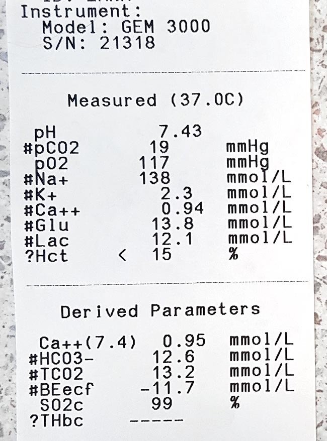
    An example of low ionised calcium.
    An example of low ionised calcium.

    Calcium is essential in maintaining normal physiological processes in the body. iCa regulates:

    • vascular tone
    • myocardial contraction
    • homeostasis

    In addition, it is needed for:

    • enzymatic reactions
    • nerve conductions
    • neuromuscular transmission
    • muscle contraction
    • hormone release
    • bone formation
    • resorption

    In critical patients, particularly those with severe trauma or sepsis, vascular tone and coagulation is particularly important. For this reason, iCa is tightly kept in a narrow range and regulated by the interactive feedback loop that involves iCa, phosphorous, parathyroid hormone, calcitriol and calcitonin.

    Diseases and causes

    Diseases commonly associated with low iCa in dogs and cats include:

    • acute kidney failure
    • acute pancreatitis
    • diabetic ketoacidosis
    • eclampsia
    • ethylene glycol intoxication
    • protein-losing enteropathies
    • sepsis
    • trauma
    • urethral obstruction
    • parathyroid diseases
    • tumour lysis syndrome

    Situations altering the fraction of extracellular calcium seen on a regular basis include:

    • acid-base disturbances
    • lactic acidosis
    • protein loss or gain
    • increased free fatty acids

    Iatrogenic causes include:

    • citrate (anticoagulant) administration during blood transfusions
    • phosphate
    • bicarbonate
    • sulfate administration

    Low iCa can also develop during cardiopulmonary resuscitation, quickly declining with increased duration.

    • Part two will go into more depth regarding the most common causes of low iCa that require acute treatment, the treatment involved, controversies surrounding treatment of non-clinical low iCa, and prognostic indications.
  • Blood gas analysis, pt 5: metabolic acidosis and alkalosis

    Blood gas analysis, pt 5: metabolic acidosis and alkalosis

    Base excess (BE) and bicarbonate (HCO3-) represent the metabolic components of the acid base equation. In general, both components will change in the same direction.

    Decreased HCO3– and BE indicate either a primary metabolic acidosis or a metabolic compensation for a chronic respiratory alkalosis. Elevated HCO3– and BE indicate either a primary metabolic alkalosis or a metabolic compensation for a chronic respiratory acidosis.

    The exception to this rule arises when a patient hypoventilates or hyperventilates.

    Carbonic acid equation

    CO2 + H2O ↔ H2CO3 ↔ HCO3– + H+

    When a patient hypoventilates, CO2 will increase as a result of reduced expiration, so a shift to the right of the equilibrium will occur. The shift to the right will increase the bicarbonate levels proportional to the increase in CO2.

    The opposite occurs when a patient hyperventilates; the equilibrium shifts to the left, so a decrease in HCO3– is present.

    Since HCO3– is not independent to the patient’s respiratory status, it is an inaccurate way of measuring the metabolic component in patients with respiratory changes. For this reason, the BE value is the preferred.

    The BE represents the amount of acid, or base, needed to titrate 1L of the blood sample until the pH reaches exactly 7.4, with the assumption the blood sample is equilibrated to a partial pressure of CO2 of 40mmHg (the middle of the reference range) and the patient’s body temperature is normal.

    Possible causes

    The possible causes of the primary disease are:

    Metabolic acidosis

    • lactic acidosis – shock and poor perfusion
    • renal failure – reduced hydrogen ion (H+) excretion and increased loss of HCO3
    • diabetic ketoacidosis – ketone acids
    • gastrointestinal (GI) losses – loss of HCO3– through vomiting and diarrhoea

    Metabolic alkalosis

    • GI outflow obstructions – loss of H+ and chloride via vomiting
    • reduced chloride levels and resultant poor perfusion – body attempts to reabsorb water and sodium to increase intravascular volume, but inadvertently also reabsorbs HCO3– in the process, despite existing alkalosis
    • refeeding syndrome
    • severe hypokalaemia:
      • transcellular shift – potassium ions leave and H+ ions enter the cell
      • transcellular shift in cells of proximal tubules → intracellular acidosis → promotes ammonium production and excretion
      • H+ excretion in the proximal and distal tubules increases → further reabsorption of HCO3– and net acid excretion
    • renal insufficiency
    • diuretic therapy (contraction alkalosis – loss of bodily fluids that do not contain HCO3-; this causes the extracellular volume to contract around a fixed quantity of HCO3-, resulting in a rise in the concentration of HCO3– without an actual increase in HCO3– levels)

    Next step

    After ruling out the differential causes of either respiratory or metabolic acidosis/alkalosis, the next step is to determine whether a compensatory response is present and, if so, if this is adequate or whether a true mixed acid-base disorder exists.

  • Blood gas analysis, pt 1: why everyone needs to know about it

    Blood gas analysis, pt 1: why everyone needs to know about it

    For those of you who have received referral histories from emergency or specialists hospitals, blood gas analysis is probably no stranger to you. For those who have never heard of them before, fear not – you are in for a treat.

    In my emergency hospital, the blood gas analyser is arguably one of the most frequently used bench top lab machines, second only to centrifuge, and for good reasons…

    Acid-base disturbances are common in critically ill and emergency patients, and it can help determine the severity of their condition and sometimes provide the answer. Tracking changes in blood gas parameters can provide information about the patient’s response to your interventions.

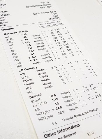
    blood-gas-analyser_output
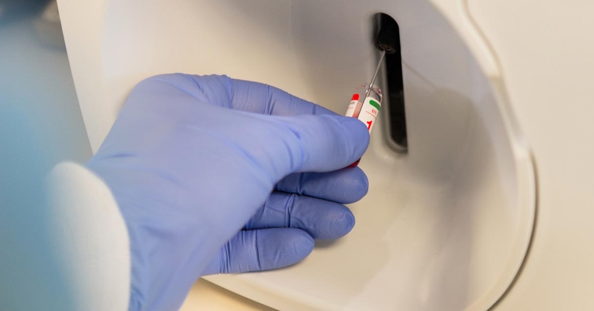
    Blood gas analysis can help assess the severity of a patient’s condition and help guide your diagnostic plan.

    The information gained from pulse oximetry is very limited in patients with severe respiratory compromise, and the only way to accurately assess their oxygenation and/or ventilation status is by looking at their blood gas status.

    So what does the blood gas analysis actually measure?

    Most blood gas panels assess the pH of the blood, partial pressure of oxygen (PO2) and partial pressure of carbon dioxide (PCO2). From these, the machine is able to derive the percentage of haemoglobin saturated with oxygen (SO2), bicarbonate (HCO3) concentration and base excess of the extracellular fluid (BEecf).

    In most machines, they are also able to measure other parameters, such as electrolytes (Na, K, Ca, Cl), glucose and lactate.

    While arterial blood gas samples are required for determining the ability of the body to oxygenate the haemoglobin, venous samples are suitable for determining the ventilation status, assessing acid base balance, electrolytes, glucose and lactate levels.

    So how can this help as a point-of-care test?

    As mentioned previously, blood gas analysis can help assess the severity of a patient’s condition and help guide your diagnostic plan. It can also provide a diagnosis (such as diabetic ketoacidosis, typical hypoadrenocorticism and high gastrointestinal obstructions).

    The changes in these parameters over time can be essential in managing critical patients in the emergency setting; it will help guide you in developing an appropriate IV fluid therapy regime and fluid choice, address the patient’s oxygenation and/or ventilation needs, correct any electrolyte and glucose abnormalities, and – although fallen out of favour – the administration of sodium-bicarbonate therapy.

    In upcoming blogs, I will teach you how to interpret the blood gas results. At the end of this, I hope everyone will incorporate blood gas analysis as their standard point-of-care test for the better assessment and management of patients.

    If given the choice between a biochemistry and a blood gas panel in a critical patient, I would hands down select blood gas every time.

  • Urinalysis: dipstick tips

    Urinalysis: dipstick tips

    Following on from July’s post entitled Urinalysis: the neglected test, let’s have a look at the dipstick – it’s a very easy part of a urinalysis and essential to perform.

    Here are some of my tips in regards to using dipsticks:

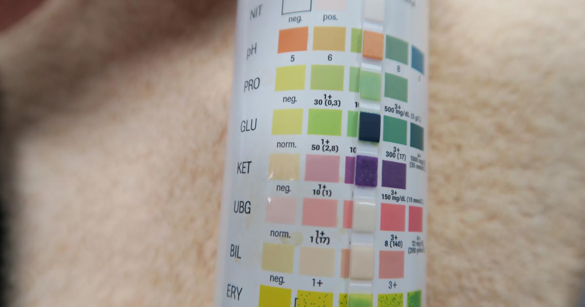
    Poli_dipstick
    Dipstick: despite the name, DON’T DIP!
    • It may sound obvious, but you should always use veterinary-specific dipsticks. Human-specific dipsticks include panels for urobilinogen, nitrates and leukocytes, which we often do not interpret in small animal patients, as they are neither sensitive nor specific.
    • DON’T DIP! Use a syringe and drop samples on to each square, leave for 10 seconds, then flick off the excess.
    • Any amount of protein in dilute urine should raise suspicion. A reasonably large amount of protein has to be present in the urine for it to be positive on a dipstick. A urine protein to creatinine ratio may be the only way to quantify the amount of protein present, but first you must rule out evidence of inflammation or haematuria via a sediment examination.
    • The ketone panel on the dipstick test is only for acetoacetate (and not beta-hydroxybutyrate), although it is extremely rare for diabetic ketoacidosis patients to not produce any acetoacetate.
    • Trace blood can be a common artefact finding, especially during a cystocentesis where needle trauma can contaminate the sample with blood.
    • In our feline patients, any hyperbilirubinuria is abnormal, but this may be normal in a dog depending on urine concentration.