Tag: Insulin

  • Hypoglycaemia

    Hypoglycaemia

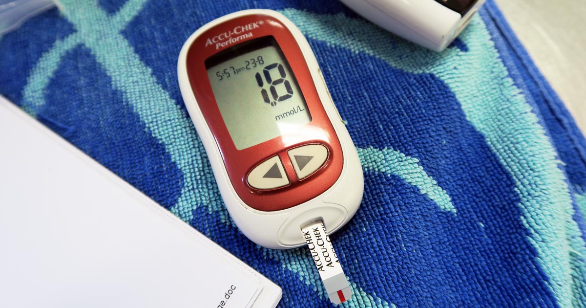
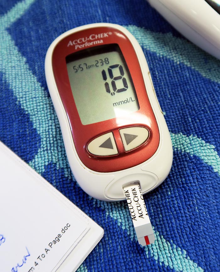
    Blood glucose is an important parameter that should be included in every “emergency database”.

    Hypoglycaemia is considered when blood glucose levels drop below 3.5mmol/L or 63mg/dL. Symptoms can start as being vague, such as lethargy and weakness, then progress to tremoring and seizures.

    One important point is that, in an emergency setting, although reduced food intake or starvation is written in text books, unless the patient is very young or a very small size it is not a common cause of hypoglycaemia.

    The liver has a fairly substantial capacity to continue to produce glucose during periods of reduced eating or starvation.

    Common causes

    Hypo
    A blood glucose meter showing a blood glucose level of 1.8mmol/L.

    The common causes of hypoglycaemia I see in an emergency setting are:

    • sepsis: bacteria consumes glucose
    • hypoadrenocorticism: lack of cortisol
    • insulin overdose: excessive intracellular shift
    • insulinoma: malignant insulin secreting neoplasia of the pancreas
    • hepatic insufficiency: reduce production

    Treatment is fairly straightforward and the impact is often dramatic – 0.5ml/kg to 1ml/kg of 50% dextrose diluted 50:50 with saline given slow IV over a couple minutes (to reduce the risk of haemolysis).

    As the list of possible causes shows, a one-off dose of glucose is often not enough.

    Glucose supplementation often needs to be continued as a 2.5% continuous rate infusion (CRI), with frequent blood glucose monitoring and adjustments made to the rate as necessary.

    The CRI will need to be continued, as the hypoglycaemia will often continue to occur until the primary disease process is identified and appropriately addressed.

    Emergency database

    It is not uncommon to read or hear the term emergency database. This contains a number of blood parameters performed, which include:

    • blood glucose
    • alanine aminotransferase
    • lactate
    • blood urea nitrogen
    • PCV
    • total protein or total solids
    • activated clotting time
    • acid-base balance
    • electrolytes
  • Blood gas analysis, pt 2: acid-base disturbances

    Blood gas analysis, pt 2: acid-base disturbances

    Acid-base disturbances are common in critical patients. These changes must be identified, as even minor deviations from the normal range can lead to significant abnormal body functions.

    Acidaemia and alkalaemia

    Acidaemia, which occurs when blood pH falls below 7.35, will lead to:

    • impedance of cardiac output
    • reduced cardiac contractility
    • a blunted response to catecholamine manifesting as hypotension
    • antagonism to insulin
    • a compensatory hyperkalaemia (extracellular movement of potassium in exchange for hydrogen ions [H+])

    Alkalaemia – blood pH above 7.45 – although less critical compared to acidosis, will result in:

    • muscle spasm
    • stuporous mentation
    • hypocalcaemia
    • hypokalaemia (intracellular movement of potassium in exchange for H+)

    As well as the aforementioned altered functions, H+ is essential for the normal function of enzymes and maintenance of normal cell structures. This is why the body maintains a very narrow pH range and uses multiple buffering mechanisms to achieve this.

    Buffering systems

    The two main buffering systems are the kidneys and lungs.

    Kidneys adjust the pH via the excretion of H+ and the uptake of bicarbonate (HCO3-), which is the primary extracellular buffer and has a linear relationship with pH.

    An increase in HCO3– concentration will result in a pH increase and vice versa. This mechanism can take hours or days from the time a shift in the pH is detected.

    The main respiratory buffer is CO2 – an acid. CO2 has an inverse relationship with pH, so an increase is equivalent to a lower pH level and vice versa. The effect of respiratory adjustments is immediate. This occurs by altering the respiratory rate to adjust CO2 levels.

    The first step towards interpretation of acid-base disturbances is identifying whether an alkalaemia or acidaemia is present. The next blog will discuss determining what is causing it – identifying the primary disorder and the compensatory mechanism employed to balance it out.

  • Christmas dangers

    Christmas dangers

    Christmas can be a busy time for vet clinics, so here is a list of common intoxications and conditions to keep an eye out on during the festive period.

    Chocolate

    • Michael Pettigrew / fotoliaNumerous online calculators can determine whether a toxic dose has been consumed and they are a great place to start.
    • I always perform emesis in patients that have ingested chocolate, even hours after ingestion as often large amounts can reside in the stomach.
    • Remember that cardiac arrhythmias can also occur in clinically normal looking patients, so perform an ECG.
    • The toxic components can be reabsorbed through the bladder wall; therefore, urinary catheterisation is a part of management of this intoxication.

    Onions

    • Onions used in roasts and on BBQ’s can cause Heinz body formation, haemolytic anaemias and pigmenturia.
    • This is not a common intoxication, but should be considered in anaemia patients and those with discoloured urine.

    Raisins

    • Commonly used in Christmas cakes and puddings. They can cause acute kidney failure, the exact mechanism of action is unknown, and there does not appear to be a dose-dependent relationship.
    • It should always be a differential for azotemic patients this time of year.
    • IV fluid induced diuresis for 48 hours is the safest way to manage raisin exposure.

    Mistletoe

    • The berries can be fatal, even if only a couple are ingested.

    Ethylene glycol

    • In colder climates, ethylene glycol can be a very common toxicity.
    • This sweet liquid is very attractive to pets and can cause acute renal failure, with the first signs being acute onset ataxia.

    Macadamia nuts

    • Macadamia nuts are common in some parts of the world. They result in joint pain in the hocks and carpus leading to weakness and ataxia.
    • Often confused with trauma and soft tissue injuries. Hyperextension of the hocks and sometimes flexion of the carpus are the clinical features.

    Xylitol

    • Xylitol is a sugar-free product used in lollies and baking.
    • In dogs, it triggers endogenous insulin to be released and a subsequent hypoglycemia develops. It can also cause hepatic failure.
    • As a general rule, I approach all intoxications as if they could be fatal as it is rare to know exactly how much of the toxic agent they have been exposed to. I consider if a patient I am treating for intoxication never develops clinical signs and wonder whether it was going to or not is the best outcome.

    Strings

    • Look under the tongue.
    • Linear foreign bodies can be difficult to diagnose. Some features on abdominal radiographs to look out for include abnormal bunching of the small intestines, and “c” and “comma” shaped gas patterns.

    Christmas meals

    • Gastroenteritis is the most common presenting condition over the Christmas period, with dietary change and indiscretion often being the culprit.
    • Bones can lead to obstructions from oral cavity to the intestines and can also cause constipation.
    • Leftover meat trimmings, often fat laden, are a common cause of pancreatitis.

    BBQ skewers

    • In some parts of the world (Australia especially) BBQs are common around Christmas time.
    • BBQ skewers can cause gastrointestinal tract perforation and septic peritonitis.
    • Because they are not radiopaque they are often difficult to diagnose.
  • Handling an Addisonian crisis – part 2

    Handling an Addisonian crisis – part 2

    Managing an Addisonian crisis can be daunting, especially when the patient looks like it is about to check out and its baseline bloods show a sodium of 110mmol/L, a potassium of 8mmol/L and a glucose of 2.3mmol/L. That is enough to make anyone’s brain explode.

    The patient can be treated in many ways, but I find it useful to try to simplify and prioritise. I have outlined my thought process in the hope some of you will find it helpful.

    First 10 minutes: protect heart and manage hypoglycaemia

    • Protect the heart – calcium gluconate 10% 0.5mL/kg to 1.5mL/kg slow IV over 10 minutes to counter the effects of hyperkalaemia on cardiac electrical activity. This buys about 20 minutes of time.
    • Treat the hypoglycaemia – the dose depends on the severity, but 0.5ml/kg of 50% dextrose IV diluted 50:50 with Hartmann’s is a good place to start. This dose of dextrose will also help correct hyperkalaemia by stimulating endogenous insulin release.

    First 20 minutes: start addressing perfusion deficits

    • Create a custom IV fluid – I do not aim to increase sodium concentration at all at this stage. I am a big fan of creating custom IV fluids. I create a fluid with a same sodium concentration as the patient then use boluses of this fluid to correct signs of shock without concerns of increasing the sodium. I use Hartmann’s as my base fluid – it has the lowest sodium concentration – and add 5% dextrose to reduce the sodium concentration (you may need to remove 100ml to 200ml from the bag first). I usually run the new fluid through the electrolyte machine to check the final sodium concentration.
    • Hartmann’s contains buffers that help address metabolic acidosis (and hyperkalaemia). It also contains potassium; however, if this concentration is less than that of serum it will still help to dilute serum potassium.
    • The formula I use to create a custom sodium IV fluid bag is beyond the scope of this blog and is detailed in the fluid therapy chapter of my book, The MiniVet Guide, under hyponatraemia.

    First hour: address hyperkalaemia

    • Image © mintra / Adobe Stock
      The author warns not to rush the sodium increase in patients. Image © mintra / Adobe Stock

      If the hyperkalaemia is severe enough to warrant more aggressive management than alkalinising IV fluids, improving renal perfusion and providing a dextrose bolus (such as potassium of more than 7mmol/L to 8mmol/L) then I would use regular short acting insulin at 0.25U/kg to 0.5U/kg IV. This should always be used in combination with a bolus of dextrose at 2g of dextrose per unit of insulin or 4ml of 50% dextrose for each unit of insulin, followed by a CRI of 2.5% to 5% glucose until insulin wears off (this could be up to six hours). This should prevent hypoglycaemia.

    • I administer dexamethasone up to 0.5mg/kg IV while running the adrenocorticotropic hormone (ACTH) stimulation test. This is the only corticosteroid that can be given as it does not cross react with the ACTH stimulation test.

    Next 2 to 24 hours: correct hydration and correct hyponatraemia

    • After I have corrected perfusion deficits with my custom IV fluid, I will address hydration deficits with an appropriate fluid plan over the next 24 hours. I usually replace 50% of the hydration deficit over the first 6 hours then the remaining 50% over the following 18 hours.
    • Correction of hyponatraemia can take a couple days as sodium should only be increased by 0.5mmol/L/hr (max 12mmol/L/day). If the sodium has not increased from the initial fluids given, I would create another custom IV fluid bag with a sodium concentration 10mmol/L above that of the patient’s. I would monitor electrolytes every one to four hours, depending on response.

    Supply mineralocorticoids and glucocorticoids

    • Options for steroid supplementation include dexamethasone 0.5mg/kg IV then 0.1mg/kg IV q12hrs or IV hydrocortisone sodium succinate at 0.5mg/kg/hr. Personally I use hydrocortisone CRI, asit has equal mineralocorticoid and glucocorticoid activity. Oral steroids can be used once the patient starts eating and drinking.
    • I only use a mineralocorticoid if I see no increase in sodium after starting hydrocortisone, despite using a fluid with a higher sodium concentration than the patient.

    Addressing patients this way will generally gets them out of the crisis. One thing that I don’t do is rush the sodium increase, it can take time and I am good with that. I have seen patients develop neurological signs from sodium levels that have increased too quickly. As for the long term management; well, I will leave that to you.

  • Festive threats to four-legged family members

    Festive threats to four-legged family members

    Now well into December, and getting ever closer to Christmas, there will be an abundance of “goodies” around the house that are not so good for the four-legged family members.

    Image: © Freeimages/s22k.
    Jordan advises colleagues to ensure clients keep an eye on their pets during the festivities. Image: © Freeimages/s22k.

    Most owners are aware of the dangers of chocolate and so are likely to rush down to the vet on Boxing Day when their Labrador has broken into the tin of Heroes – but what other festive dangers are there that owners are not so clued up on?

    Pudding

    The festive favourite Christmas pudding, while enjoyable for us, can be lethal for pets. It is the raisins, currants and sultanas that may lurk within that are poisonous.

    While the mechanism of toxicity is poorly understood, we know it can cause acute kidney failure in dogs.

    The treatment protocol for poisoning by raisins, etc, is similar to general suspected poisoning with an unknown agent: give an emetic (to make the animal vomit), an adsorbent (for example, activated charcoal) and supportive care (IV fluids) to protect the kidneys for a few days.

    Antifreeze

    Ethylene glycol, more commonly known as antifreeze, is extremely toxic to pets, with consumption of tiny amounts resulting in crystal formation in the kidneys, and so acute renal failure. Antifreeze smells and tastes sweet, so pets are often attracted to it. For this reason, it is best for owners to ensure it is kept in a secure container, well away from pets.

    Early symptoms include neurological signs, such as wobbliness, twitching, seizures and depression. As the poisoning progresses, the animal will have cardiac and respiratory signs, such as increased heart and respiratory rate. It’s vitally important owners look out for these signs if their pets have had any possible access to antifreeze (for example, if they’ve got into the garage or if a cat has been missing for a few days and potentially been locked in someone’s shed).

    The antidote for ethylene glycol poisoning is ethanol, because it replaces ethylene glycol as a substrate for the enzyme that breaks it down into toxic products. More specifically, lab-grade ethanol is best.

    Sweetener

    Xylitol is a sugar-free sweeter that is used in chewing gum, cakes and sweets, particularly diabetic foods. Xylitol causes insulin release in the body, resulting in hypoglycaemia and, later, liver damage.

    Symptoms include vomiting and signs of hypoglycaemia: lethargy, weakness, collapse and, if it progresses, seizures and coma. Unlike ethylene glycol, there isn’t a specific antidote, but supportive care, including fluids and liver protectants, is vital. The prognosis for xylitol toxicity is good if the animal’s hypoglycaemia is corrected quickly, so it is essential for owners that suspect this to contact their vet immediately. If you’re trying to determine whether xylitol is in a product, it is often listed as a food additive, code E967.

    Decorations?

    And don’t forget the potential foreign body too – if the cat has been playing with the tinsel and managed to swallow some or the dog has ingested a few tasty looking baubles.

    Enjoy the festive season, but advise owners to keep an eye on their pets this Christmas.