Tag: hypothyroidism

  • Ionised hypocalcaemia, pt 2: eclampsia

    Ionised hypocalcaemia, pt 2: eclampsia

    As discussed in part one of this blog series, a myriad of disease processes can lead to ionised hypocalcaemia (iHCa).

    Despite this, only hypocalcaemia caused by eclampsia and hypoparathyroidism (primary or iatrogenic – post-surgical parathyroidectomy) are severe enough to demand immediate parenteral calcium administration.

    Hypoparathyroidism is quite rare, so this blog will not explore the detailed pathophysiology behind this syndrome. However, it is worthwhile mentioning – aside from primary hypoparathyroidism – no other disease state requires long-term calcium supplementation.

    Eclampsia, on the other hand, is the most common cause of clinical hypocalcaemia in dogs and cats. Multiple factors can predispose animals to the development of this phenomenon, so understanding the pathophysiology behind this potentially fatal disease will not only help with future diagnosis and treatment, but also help prevent this issue.

    Periparturient occurrence

    Eclampsia – also known as puerperal tetany or periparturient hypocalcaemia – occurs in the periparturient period anywhere from the final few weeks of gestation to four weeks postpartum, with the latter being the more common time frame of manifestation.

    The serum concentration of ionised calcium (iCa) is often less than 0.9mmol/L in bitches or less than 0.8mmol/L in queens. It presents as muscle fasciculation and tetany, but not usually in seizure since most patients maintain consciousness. Exceptions occur when these patients are left untreated – these patients may develop refractory seizures, cerebral oedema and death.

    The increased muscle activity generates a lot of heat and uses a significant amount of glucose; therefore, hyperthermia and hypoglycaemia are common sequelae in patients with delayed presentations.

    Reduced iCa

    Eclampsia occurs as a result of reduced iCa in the extracellular compartment. In lactation-associated hypocalcaemia, it is the result of the body’s inability to maintain serum iCa through increased osteolytic activity and gastrointestinal calcium absorption, and reduced renal calcium excretion to compensate for the loss of calcium through milk production.

    Other factors often predispose animals to developing eclampsia. These can include poor periparturient nutrition, excessive calcium supplementation and large litter size.

    Excessive calcium supplementation in the prenatal period causes parathyroid gland atrophy, preventing parathyroid hormone release – resulting in reduced gastrointestinal calcium absorption and osteoclastic activity, and increased kidney calcium loss.

    Clinical signs

    Clinical signs can progress rapidly and become fatal if left untreated.

    In the early phases, non-specific signs can present as:

    • facial pruritus
    • hyperaesthesia
    • panting
    • tremors
    • muscle fasciculations
    • paresis
    • ataxia

    Within a few hours, these clinical signs rapidly progress to rigidity, and tonic and clonic spasms with opisthotonos. By this stage, animals will develop severe tachycardia, tachypnoea and hyperthermia. Without treatment, a high mortality rate exists.
    kitten
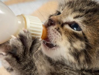
    “Early supplementation of puppies and kittens with commercial milk formula will significantly reduce the lactation demand on the dam.” Image © Dobroslav / Adobe Stock

    Patients presenting with eclampsia require immediate medical intervention, as well as concurrent supportive therapy. The acute management of clinical iHCa is the same, regardless of the cause, and will be discussed in detail in part three.

    Supportive therapies required to manage and prevent a patient relapsing in eclampsia often include active cooling and glucose supplementation. In cases that seizure, anti-seizure medications – such as diazepam and barbiturates – and mannitol for cerebral oedema may be required.

    Prevention

    Even before getting to the stage where an animal requires treatment, all effort must be taken to prevent a dam from developing hypocalcaemia. This can be easily achieved by improving the calcium content of the food during the perinatal period, as well as reducing the milk demand by early weaning kittens or puppies. This is likely particularly helpful for those with a history of eclampsia or with large litters.

    From the second half of gestation, it is recommended a commercial formulation of puppy/kitten food (1% to 1.8% calcium and 0.8% to 1.6% phosphorus) is to be fed to the dam without any additional minerals or vitamin supplementation.

    Postpartum calcium is similar to the second half of gestation, requiring a diet containing at least 1.4% calcium with a 1:1 ratio with phosphorus (most balanced growth formula for puppies and kittens).

    Less demand

    Early supplementation of puppies and kittens with commercial milk formula will significantly reduce the lactation demand on the dam. Together with this, starting at aged three to four weeks, solids can be introduced at this time. These techniques will be particularly helpful to those with a history of previous eclampsia or those with large litter sizes.

    Aside from the parenteral calcium supplementation required, other supportive therapy – such as active cooling, IV fluid therapy and glucose supplementation – may be required.

    Long term, the dam’s nutritional content of calcium must be optimal from the second half of gestation. All additional calcium or other vitamins and mineral supplementations should not occur prior to parturition.

    In the postpartum dam with a history of eclampsia or that is at risk, changing to a nutritionally balanced commercial food aim for growing puppies and kittens is ideal. Early weaning – or abrupt weaning if hypocalcaemia is severe – may be required in severe cases or those with a high risk of relapse/development.

  • Hyponatraemia, pt 2: causes

    Hyponatraemia, pt 2: causes

    The causes of hyponatraemia can be divided into three major categories, based on serum osmolality. This is further divided based on the patient’s volume status (Table 1).

    Most patients we see in clinic fall into the hypovolaemic category, except patients with diabetes mellitus.

    Table 1. Causes of hyponatraemia based on osmolality and volume status (from Guillaumin and DiBartola, 2017).
    Hypo-osmolar Hyperosmolar Normo-osmolar
    Hypovolaemic Normovolaemic Hypervolaemic
    Gastrointestinal fluid loss
    Third-space fluid losses
    Shock
    Hypoadrenocorticism (Addison’s disease)
    Renal insufficiency
    Excessive diuretic administration
    Salt-losing nephropathy
    Cerebral salt wasting syndrome
    Syndrome of inappropriate antidiuretic hormone secretion (SIADH)
    Hypotonic fluid administration
    Hypothyroidism
    Glucocorticoid insufficiency
    Psychogenic polydipsia
    Reset osmostat (SIADH type B)
    Congestive heart failure
    Acute or chronic renal failure
    Nephrotic syndrome
    Hepatic cirrhosis
    Accidental ingestion or injection of water (water intoxication)
    Hyperglycaemia
    Mannitol
    Severe azotaemia
    Hyperlipidaemia
    Hyperproteinaemia

    Common causes

    In dogs, the three most common causes of hyponatraemia are:

    • gastrointestinal (GI) fluid loss
    • third-space fluid loss
    • fluid shift from intracellular fluid to extracellular fluid (ECF) as a result of hyperglycaemia

    In cats, the three most common causes of hyponatraemia are:

    • urologic diseases
    • GI fluid loss
    • third-space fluid losses

    In most patients, more than one pathophysiologic factor is likely to be contributing to the hyponatraemia.

    Circulating volume

    Hypovolaemic patients – those with, for example, GI losses, hypoadrenocorticism, renal losses and haemorrhagic shock – have a reduced effective circulating volume. ECF contraction triggers antidiuretic hormone (ADH) secretion, which leads to increases in free water absorption and thirst, and results in dilution of the serum sodium concentration. Aldosterone secretion is reduced in hypoadrenocorticism, so an overall reduction in sodium reabsorption compounds the problem.

    Hypervolaemic patients are those with an increased fluid retention state, such as:

    • congestive heart failure (pulmonary oedema)
    • advanced hepatic failure (ascites, third-space fluid)
    • renal failure
    • free water ingestion

    Congestive heart failure patients have a reduced cardiac output and, therefore, a decreased effective circulating volume, despite the presence of the extra fluid status. Renin-angiotensin activation leads to release of ADH and aldosterone, resulting in sodium and free water reabsorption, and increased thirst. Both lead to an excess of free water retention.

    Advanced hepatic (cirrhosis) or renal failure (nephrotic syndrome) both result in hypoalbuminaemia, leading to fluid shifting into the interstitial space and third space, reducing effective circulating volumes. This leads to activation of ADH to increase free water reabsorption, to restore the circulating volume in the face of existing hypervolaemia and hyponatraemia.

    Diabetic patients

    Moderate to severe hyperglycaemic diabetic patients can be either hyperosmolar or normo-osmolar, depending on the serum blood glucose concentration. Hyponatraemia occurs when water shifts from the intracellular fluid to the ECF down the osmotic gradient, diluting the serum sodium content.

    Despite this osmotic shift, not all diabetic patients develop hyponatraemia. Glucosuria also causes also causes a renal osmotic shift, sometimes resulting in urine water loss in excess to sodium. This offsets the hyponatraemia – in some cases, hypernatraemia results.

    Treatment

    Treatment of hyponatraemia hinges on how quickly it developed and the volume status of the patient. The rule of thumb is to correct hyponatraemia slowly – not exceeding 0.5meq/L/hr – especially in chronic cases, or cases where the duration of hyponatraemia is unknown. Keeping to this rate is paramount until serum sodium concentration reaches 130meq/L.

    In acute patients with severe clinical signs, such as seizures, some clinicians may choose to use a higher rate of 1meq/L/hr to 2meq/L/hr until clinical signs resolved.

    It should be emphasised, once again, this rate should never be used in chronic patients, patients with an unknown duration of hyponatraemia, or where frequent serum sodium concentration cannot be monitored. The rapid correction of hyponatraemia can lead to osmotic demyelination syndrome (myelinolysis).

    Its effect will not be apparent until three or four days after therapy, and can result in neurological abnormalities such as:

    • weakness
    • ataxia
    • dysphagia
    • paresis
    • coma

    For that reason, frequent electrolyte measurements are required, starting hourly then once a suitable rate of increase has been established and less frequently thereafter.

    • Part 3 will look at how to correct patients with hyponatraemia.

    Reference

    Guillaumin J and DiBartola SP (2017). A quick reference on hyponatremia, Veterinary Clinics of North America: Small Animal Practice 47(2): 213-217.

  • Lipaemia – the bane of biochemistry

    Lipaemia – the bane of biochemistry

    Last week we covered haemolysed samples – this week we’re looking at lipaemic samples.

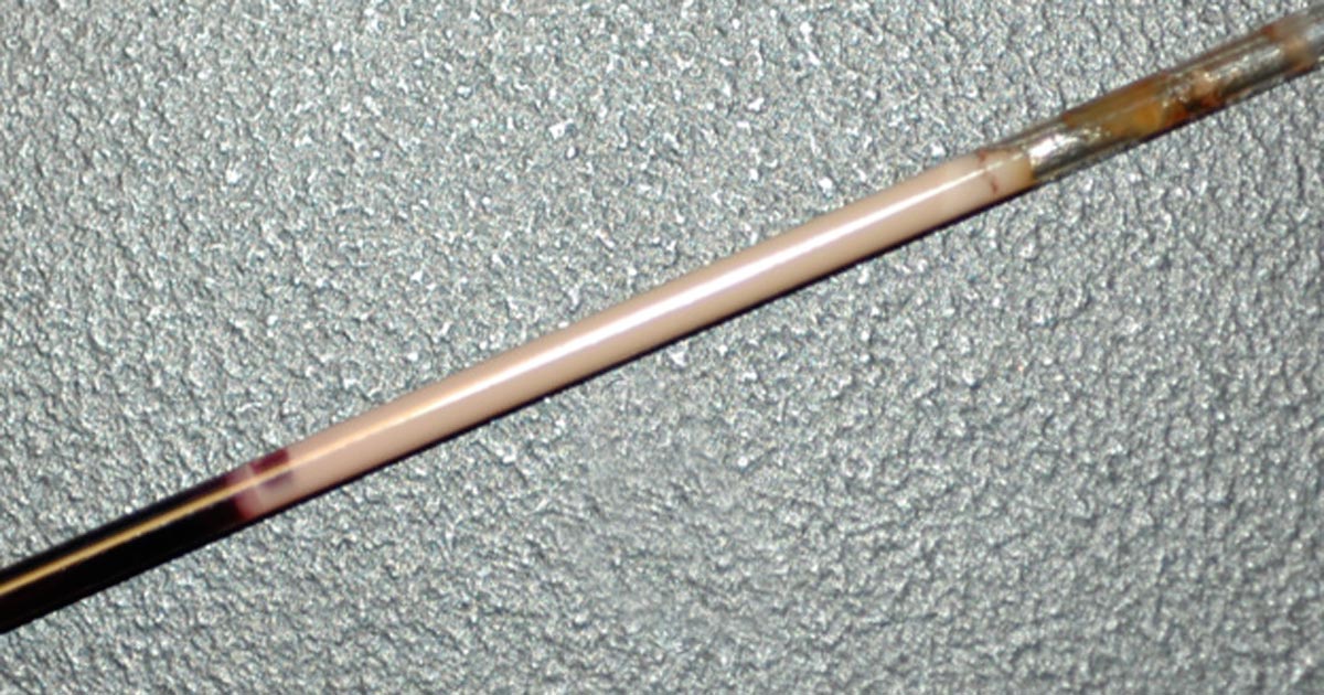
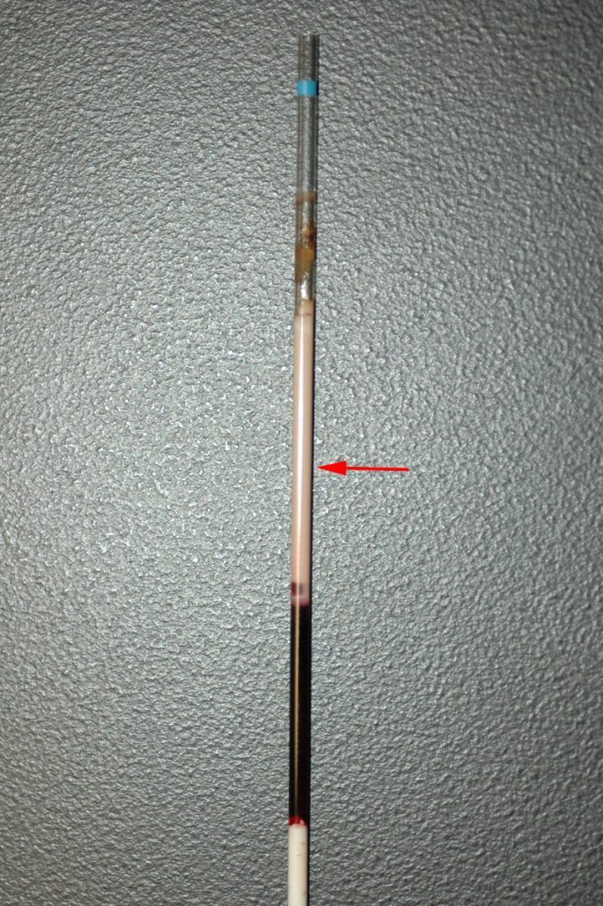
    Lipaemic samples are caused by an excess of lipoproteins in the blood, creating a milky/turbid appearance that interferes with multiple biochemical tests and can even cause haemolysis of red blood cells.

    lipaemic sample
    A severely lipaemic sample (red arrow). IMAGE: eClinPath.com (CC BY-NC-SA 4.0).

    Lipaemia can follow recent ingestion of a meal – especially one high in fat. Although not pathognomonic for any diseases, its presence can help increase the suspicion of certain diseases, including:

    • pancreatitis
    • diabetes mellitus
    • hypothyroidism
    • hyperadrenocorticism
    • primary hyperlipidaemia (in some specific breeds, such as the miniature schnauzer)

    It warrants further investigation in patients that have been ill and inappetent.

    Irksome interpretations

    Lipaemia can dramatically impact laboratory testing and is often troublesome in critically ill patients, making interpretation of biochemistry particularly difficult, if not impossible.

    Lipaemia can affect different analysers in different ways, most commonly causing:

    • Falsely increased calcium, phosphorus, bilirubin, glucose and total protein (via refractometer) and some liver parameters such as alkaline phosphatase, alanine aminotransferase, aspartate aminotransferase, haemoglobin concentration, and mean corpuscular haemoglobin concentration.
    • Falsely decreased sodium, potassium, chloride, albumin and bicarbonate.

    Tube tips

    Assessment of a centrifuged haematocrit tube before running a biochemistry panel can help reduce wasted biochemistry consumables.

    If the sample is lipaemic in the haematocrit tube then maybe try some of the following tips.

    • If blood tests are planned in advance, try fasting the patient beforehand for 12 to 24 hours.
    • Repeat sampling a couple of hours later may yield a less lipaemic sample.
    • Collecting and centrifuging a larger amount of blood (3ml to 5ml, for example) can sometimes yield enough clear sample between the lipid layer and red blood cells.
    • Refrigeration of the sample can help the separation.
    • Extract lipids using polar solvents, such as polyethylene glycol.
    • Centrifugation at higher than normal speeds (if possible) can also assist in clearing the layer.
  • Aggression as an unusual presenting sign of hypothyroidism

    Aggressive dog
    Image ©iStock.com/YuriyGreen

    I’ve not long come across a case of a middle-aged dog that had suddenly become unaccustomedly grumpy and started snapping at its poor owners.

    This was uncharacteristic, as it had no previous history of behavioural problems. There were no other obvious clinical signs – perhaps it had become lazy of late, but there was nothing to put your finger on.

    I recalled having attended a CPD event a while before when the speaker mentioned hypothyroidism leading to aggression. I thought this was worth a punt, and low and behold it had a very high thyroid-stimulating hormone level and very low thyroxine result. Our friend is now on thyroid supplementation and is much happier, although at the time of writing some two to three months into therapy, it was not completely better.

    So, it may be worthwhile considering a blood profile when presented with signs of aggression in middle-aged dogs.

  • Check tear production in cases of facial paralysis

    Schirmer tear test
    Schirmer tear test

    We recently saw a 10-year-old, West Highland white that had developed a middle ear infection and facial paralysis.

    Following treatment and further investigation, it turned out the poor little chap had concurrent hypothyroidism. Several weeks later, I noticed the eye the same side as the ear infection had developed a mucopurulent discharge and mild keratitis.

    On checking its Schirmer tear tests, this was markedly reduced for that eye, and borderline for the other. A bit of research indicated facial paralysis can affect both tear and saliva production.

    The good news is the little chap is doing well with false tear preparations.