Tag: Hypokalaemia

  • Blood gas analysis, pt 5: metabolic acidosis and alkalosis

    Blood gas analysis, pt 5: metabolic acidosis and alkalosis

    Base excess (BE) and bicarbonate (HCO3-) represent the metabolic components of the acid base equation. In general, both components will change in the same direction.

    Decreased HCO3– and BE indicate either a primary metabolic acidosis or a metabolic compensation for a chronic respiratory alkalosis. Elevated HCO3– and BE indicate either a primary metabolic alkalosis or a metabolic compensation for a chronic respiratory acidosis.

    The exception to this rule arises when a patient hypoventilates or hyperventilates.

    Carbonic acid equation

    CO2 + H2O ↔ H2CO3 ↔ HCO3– + H+

    When a patient hypoventilates, CO2 will increase as a result of reduced expiration, so a shift to the right of the equilibrium will occur. The shift to the right will increase the bicarbonate levels proportional to the increase in CO2.

    The opposite occurs when a patient hyperventilates; the equilibrium shifts to the left, so a decrease in HCO3– is present.

    Since HCO3– is not independent to the patient’s respiratory status, it is an inaccurate way of measuring the metabolic component in patients with respiratory changes. For this reason, the BE value is the preferred.

    The BE represents the amount of acid, or base, needed to titrate 1L of the blood sample until the pH reaches exactly 7.4, with the assumption the blood sample is equilibrated to a partial pressure of CO2 of 40mmHg (the middle of the reference range) and the patient’s body temperature is normal.

    Possible causes

    The possible causes of the primary disease are:

    Metabolic acidosis

    • lactic acidosis – shock and poor perfusion
    • renal failure – reduced hydrogen ion (H+) excretion and increased loss of HCO3
    • diabetic ketoacidosis – ketone acids
    • gastrointestinal (GI) losses – loss of HCO3– through vomiting and diarrhoea

    Metabolic alkalosis

    • GI outflow obstructions – loss of H+ and chloride via vomiting
    • reduced chloride levels and resultant poor perfusion – body attempts to reabsorb water and sodium to increase intravascular volume, but inadvertently also reabsorbs HCO3– in the process, despite existing alkalosis
    • refeeding syndrome
    • severe hypokalaemia:
      • transcellular shift – potassium ions leave and H+ ions enter the cell
      • transcellular shift in cells of proximal tubules → intracellular acidosis → promotes ammonium production and excretion
      • H+ excretion in the proximal and distal tubules increases → further reabsorption of HCO3– and net acid excretion
    • renal insufficiency
    • diuretic therapy (contraction alkalosis – loss of bodily fluids that do not contain HCO3-; this causes the extracellular volume to contract around a fixed quantity of HCO3-, resulting in a rise in the concentration of HCO3– without an actual increase in HCO3– levels)

    Next step

    After ruling out the differential causes of either respiratory or metabolic acidosis/alkalosis, the next step is to determine whether a compensatory response is present and, if so, if this is adequate or whether a true mixed acid-base disorder exists.

  • Blood gas analysis, pt 2: acid-base disturbances

    Blood gas analysis, pt 2: acid-base disturbances

    Acid-base disturbances are common in critical patients. These changes must be identified, as even minor deviations from the normal range can lead to significant abnormal body functions.

    Acidaemia and alkalaemia

    Acidaemia, which occurs when blood pH falls below 7.35, will lead to:

    • impedance of cardiac output
    • reduced cardiac contractility
    • a blunted response to catecholamine manifesting as hypotension
    • antagonism to insulin
    • a compensatory hyperkalaemia (extracellular movement of potassium in exchange for hydrogen ions [H+])

    Alkalaemia – blood pH above 7.45 – although less critical compared to acidosis, will result in:

    • muscle spasm
    • stuporous mentation
    • hypocalcaemia
    • hypokalaemia (intracellular movement of potassium in exchange for H+)

    As well as the aforementioned altered functions, H+ is essential for the normal function of enzymes and maintenance of normal cell structures. This is why the body maintains a very narrow pH range and uses multiple buffering mechanisms to achieve this.

    Buffering systems

    The two main buffering systems are the kidneys and lungs.

    Kidneys adjust the pH via the excretion of H+ and the uptake of bicarbonate (HCO3-), which is the primary extracellular buffer and has a linear relationship with pH.

    An increase in HCO3– concentration will result in a pH increase and vice versa. This mechanism can take hours or days from the time a shift in the pH is detected.

    The main respiratory buffer is CO2 – an acid. CO2 has an inverse relationship with pH, so an increase is equivalent to a lower pH level and vice versa. The effect of respiratory adjustments is immediate. This occurs by altering the respiratory rate to adjust CO2 levels.

    The first step towards interpretation of acid-base disturbances is identifying whether an alkalaemia or acidaemia is present. The next blog will discuss determining what is causing it – identifying the primary disorder and the compensatory mechanism employed to balance it out.

  • Focus on GDV, part 4: the recovery

    Focus on GDV, part 4: the recovery

    Postoperatively, gastric dilatation-volvulus (GDV) patients remain in our intensive care unit for at least two to three days.

    Monitoring includes standard general physical examination parameters, invasive arterial blood pressures, ECG, urine output via urinary catheter and pain scoring.

    I repeat PCV/total protein, lactate, blood gas and activated clotting times (ACT) immediately postoperatively and then every 8-12 hours, depending on abnormalities and patient progress.

    GDV recovery
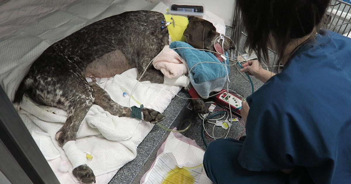
    Patient recovering in the pet intensive care unit. As well as standard monitoring parameters, GDV patients have constant ECG, arterial blood pressure and urine output monitoring to enable the early detection and correction of abnormalities.

    I always repeat these blood tests postoperatively, as IV fluids given during the resuscitation and intraoperative period often cause derangements. I use the results to guide my fluid therapy, but also take it with a grain of salt.

    IV fluids

    I generally continue a balanced and buffered crystalloid. The rate depends on blood pressures, urine output and assessment of general physical examination parameters for perfusion and hydration, but I try to avoid fluid overload and reduce the IV fluids postoperatively as soon as possible.

    Coagulopathy

    Prolonged clotting times are frequently seen as a result of consumption in a dog with GDV. However, one should note it can also occur as the result of haemodilution.

    As the underlying disease process has been corrected, and haemostasis achieved during surgery, I usually monitor ACTs, but may not necessarily treat with blood products as prolonged ACTs do not always translate to clinical bleeding. Unless clinical evidence of bleeding exists, I generally hold off treatment and monitor.

    Hypoproteinaemia

    Low total protein is also common. This is generally due to haemodilution from fluid resuscitation. However, a low total protein does not mean oedema will develop, or that it requires management. I generally track the protein levels, use conservative fluid therapy and try to correct it by instituting enteral nutrition as soon as possible.

    Electrolyte imbalances

    Hypokalaemia is a common complication of fluid therapy. This can be rectified with potassium supplementation in the IV fluids.

    Hyperlactataemia

    If present post-surgery, this is usually corrected with a fluid bolus. However, I always assess for other things that may affect oxygen delivery to the tissues, such as poor cardiac output (arrthymias), hypoxaemia (respiratory disease) and anaemia (from surgical blood loss).

    Arrhythmias

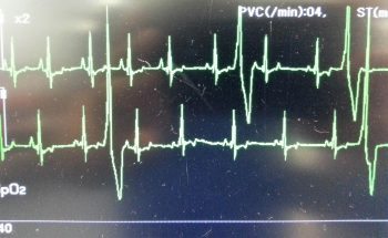
    Ventricular arrhythmias are common post-surgery. Accelerated idioventricular rhythms are the most common cause, especially if a splenectomy was performed.

    arrhythmia
    Ventricular premature contractions are common postoperative arrhythmia.

    Before reaching for anti-arrythmia medications, first check and correct:

    • electrolyte abnormalities
    • hypoxaemia
    • pain control
    • hypovolaemia or hypotension

    If they are still present, despite correction of the above, consider treating the rhythm if:

    • multifocal beats (ventricular premature contractions of various sizes)
    • overall rate greater than 190 beats per minute
    • R-on-T phenomenon
    • low blood pressure during a run of ventricular premature contractions

    I start with a bolus 2mg/kg lidocaine IV and start a constant-rate infusion of 50ug/kg/min to 75ug/kg/min.

    Anaemia

    It is common to have a mild anaemia post-surgery, due to a combination of blood loss and haemodilution. In the absence of transfusion triggers – such as increased heart rate, increased respiratory rate or hyperlactataemia – it does not require treatment.

    Vomiting

    Anti-emetics are the first line of medication. Non-prokinetic anti-emetics, such as maropitant and ondansetron, can be used immediately; otherwise, after 12 hours, metoclopramide can also be used postoperatively. If the patient remains nauseous despite these medications, the placement of a nasogastric tube can ease nausea by removing static gastric fluid.

    Excessive pain relief may also contribute to the nauseous state.

    Pain relief

    I mostly rely on potent-pure opioid agonists, such as fentanyl constant-rate infusions and patches. This is generally sufficient for most patients. Ketamine is occasionally used.

    • Some drugs listed in this article are used under the cascade.