Tag: Hypoadrenocorticism

  • Hyponatraemia, pt 3: correcting a sodium concentration of 110mEq/L

    Hyponatraemia, pt 3: correcting a sodium concentration of 110mEq/L

    The amount of sodium required to increase serum sodium concentration to a desired value can be calculated from the following formula:

    Sodium deficit = 0.6 × bodyweight (kg) × (normal sodium [mEq/L] – patient sodium [mEq/L])

    Table 1. Sodium content of various fluids
    Fluid Type Sodium content (mEq/L)
    0.9% sodium chloride 154
    Normosol-R 140
    Hartmann’s solution
    130
    3% sodium chloride 513
    7.5% sodium chloride 1,300

    This sodium deficit is then replaced over “x” hours, at an average rate of 0.5mEq/L/hr.

    In hypovolaemic hyponatraemia patients – where the fluid deficits also need correcting – it is important to select a fluid where the sodium concentration is within 5mEq/L to 10mEq/L of the patient plasma sodium level.

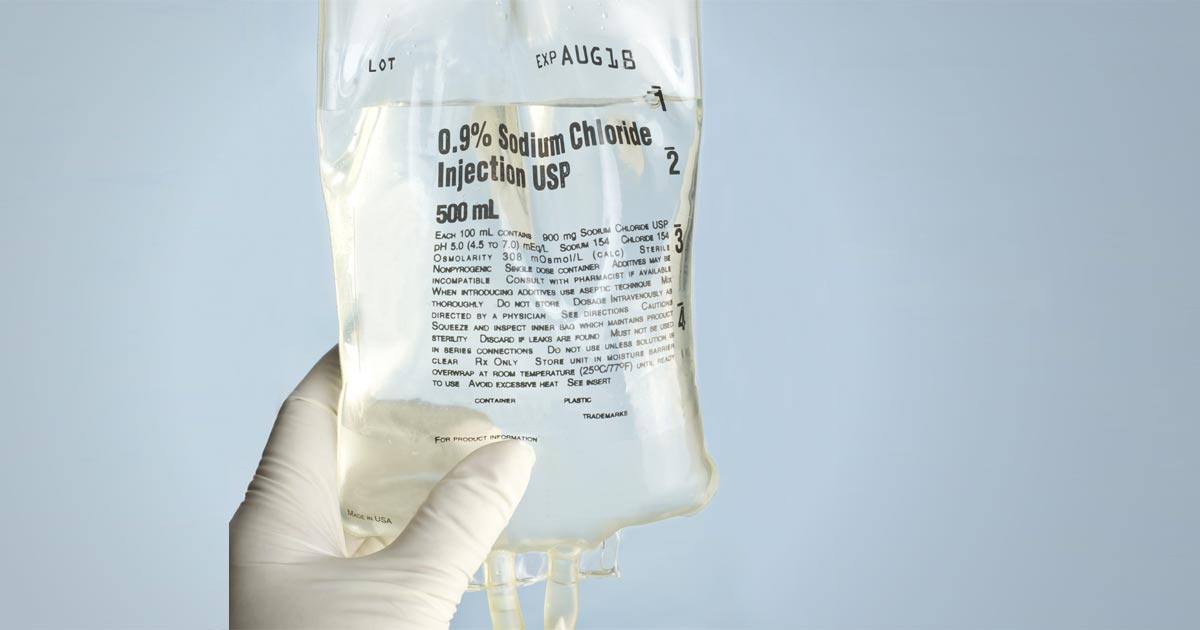
    Table 1 shows the sodium content of various fluids. If none of the fluids listed in Table 1 are suitable – for example, the patient’s sodium level is 110 – you can make your own fluid by mixing 5% dextrose in water using the formula below:

    Volume of 5% dextrose in water to be added (ml) =

    ([current IV fluid Na+] – [desired IV fluid Na+]) × 1,000ml ÷ ([desired IV fluid Na+] – [supplemental IV fluid Na+])

    Hartmann’s example

    The most common cause of severe hyponatraemia is hypoadrenocorticism. Using an example of a severe hyponatraemia of 110mEq/L, I select Hartmann’s solution first, as it has the lowest sodium concentration. How low I dilute Hartmann’s depends on the patient’s volume status.

    If the patient requires fluid resuscitation because it is showing signs of poor perfusion – such as elevated heart rate, poor pulse quality, pale gums, prolonged capillary refill time, dull mentation, low core body temperature and elevated lactate – I aim for a sodium concentration the same as the patient. For this example, I would dilute the Hartmann’s to 110mEq/L, as then I can bolus therapy this without elevating the patient’s sodium concentration.

    So, aiming for 110mEq/L, the volume of 5% dextrose in water (D5W) required to dilute Hartmann’s is:

    = ([130 – 110] × 1,000) ÷ (110 – 0)

    = (20 × 1,000) ÷ 110

    = 181ml of D5W.

    This volume may not fit in the bag, so I remove 150ml from the Hartmann’s bag first and insert 850ml into the equation:

    = ([130 – 110] × 850) ÷ (110 – 0)

    = (20 × 850) ÷ 110

    = 154ml of D5W to be added to the bag for a total volume of 1,054ml with a sodium concentration of 110mEq/L.

    TIP:

    Electrolytes can be used on custom solutions to check the final sodium concentration. It will be a couple of mEq/L above or below, due to variations in each Hartmann’s bag.

    I bolus with this 110mEq/L of custom solution for correct perfusion, reassess the patient and sodium concentration – and make a solution between 5mEq/L and 10mEq/L higher – and administer at much slower rates with repeated monitoring.

    The treatment of hypervolaemic hyponatraemic patients will not be discussed here, as it revolves around treating the underlying medical condition.

    Conclusion

    Hyponatraemia is a common and potentially life-threatening change in our critical patients.

    It is crucial to establish whether this is an acute or chronic change, to avoid development of osmotic demyelination syndrome. If I have any doubt about the timeline, I treat as a chronic change and increase slowly.
  • Hyponatraemia, pt 2: causes

    Hyponatraemia, pt 2: causes

    The causes of hyponatraemia can be divided into three major categories, based on serum osmolality. This is further divided based on the patient’s volume status (Table 1).

    Most patients we see in clinic fall into the hypovolaemic category, except patients with diabetes mellitus.

    Table 1. Causes of hyponatraemia based on osmolality and volume status (from Guillaumin and DiBartola, 2017).
    Hypo-osmolar Hyperosmolar Normo-osmolar
    Hypovolaemic Normovolaemic Hypervolaemic
    Gastrointestinal fluid loss
    Third-space fluid losses
    Shock
    Hypoadrenocorticism (Addison’s disease)
    Renal insufficiency
    Excessive diuretic administration
    Salt-losing nephropathy
    Cerebral salt wasting syndrome
    Syndrome of inappropriate antidiuretic hormone secretion (SIADH)
    Hypotonic fluid administration
    Hypothyroidism
    Glucocorticoid insufficiency
    Psychogenic polydipsia
    Reset osmostat (SIADH type B)
    Congestive heart failure
    Acute or chronic renal failure
    Nephrotic syndrome
    Hepatic cirrhosis
    Accidental ingestion or injection of water (water intoxication)
    Hyperglycaemia
    Mannitol
    Severe azotaemia
    Hyperlipidaemia
    Hyperproteinaemia

    Common causes

    In dogs, the three most common causes of hyponatraemia are:

    • gastrointestinal (GI) fluid loss
    • third-space fluid loss
    • fluid shift from intracellular fluid to extracellular fluid (ECF) as a result of hyperglycaemia

    In cats, the three most common causes of hyponatraemia are:

    • urologic diseases
    • GI fluid loss
    • third-space fluid losses

    In most patients, more than one pathophysiologic factor is likely to be contributing to the hyponatraemia.

    Circulating volume

    Hypovolaemic patients – those with, for example, GI losses, hypoadrenocorticism, renal losses and haemorrhagic shock – have a reduced effective circulating volume. ECF contraction triggers antidiuretic hormone (ADH) secretion, which leads to increases in free water absorption and thirst, and results in dilution of the serum sodium concentration. Aldosterone secretion is reduced in hypoadrenocorticism, so an overall reduction in sodium reabsorption compounds the problem.

    Hypervolaemic patients are those with an increased fluid retention state, such as:

    • congestive heart failure (pulmonary oedema)
    • advanced hepatic failure (ascites, third-space fluid)
    • renal failure
    • free water ingestion

    Congestive heart failure patients have a reduced cardiac output and, therefore, a decreased effective circulating volume, despite the presence of the extra fluid status. Renin-angiotensin activation leads to release of ADH and aldosterone, resulting in sodium and free water reabsorption, and increased thirst. Both lead to an excess of free water retention.

    Advanced hepatic (cirrhosis) or renal failure (nephrotic syndrome) both result in hypoalbuminaemia, leading to fluid shifting into the interstitial space and third space, reducing effective circulating volumes. This leads to activation of ADH to increase free water reabsorption, to restore the circulating volume in the face of existing hypervolaemia and hyponatraemia.

    Diabetic patients

    Moderate to severe hyperglycaemic diabetic patients can be either hyperosmolar or normo-osmolar, depending on the serum blood glucose concentration. Hyponatraemia occurs when water shifts from the intracellular fluid to the ECF down the osmotic gradient, diluting the serum sodium content.

    Despite this osmotic shift, not all diabetic patients develop hyponatraemia. Glucosuria also causes also causes a renal osmotic shift, sometimes resulting in urine water loss in excess to sodium. This offsets the hyponatraemia – in some cases, hypernatraemia results.

    Treatment

    Treatment of hyponatraemia hinges on how quickly it developed and the volume status of the patient. The rule of thumb is to correct hyponatraemia slowly – not exceeding 0.5meq/L/hr – especially in chronic cases, or cases where the duration of hyponatraemia is unknown. Keeping to this rate is paramount until serum sodium concentration reaches 130meq/L.

    In acute patients with severe clinical signs, such as seizures, some clinicians may choose to use a higher rate of 1meq/L/hr to 2meq/L/hr until clinical signs resolved.

    It should be emphasised, once again, this rate should never be used in chronic patients, patients with an unknown duration of hyponatraemia, or where frequent serum sodium concentration cannot be monitored. The rapid correction of hyponatraemia can lead to osmotic demyelination syndrome (myelinolysis).

    Its effect will not be apparent until three or four days after therapy, and can result in neurological abnormalities such as:

    • weakness
    • ataxia
    • dysphagia
    • paresis
    • coma

    For that reason, frequent electrolyte measurements are required, starting hourly then once a suitable rate of increase has been established and less frequently thereafter.

    • Part 3 will look at how to correct patients with hyponatraemia.

    Reference

    Guillaumin J and DiBartola SP (2017). A quick reference on hyponatremia, Veterinary Clinics of North America: Small Animal Practice 47(2): 213-217.

  • Hypoglycaemia

    Hypoglycaemia

    Blood glucose is an important parameter that should be included in every “emergency database”.

    Hypoglycaemia is considered when blood glucose levels drop below 3.5mmol/L or 63mg/dL. Symptoms can start as being vague, such as lethargy and weakness, then progress to tremoring and seizures.

    One important point is that, in an emergency setting, although reduced food intake or starvation is written in text books, unless the patient is very young or a very small size it is not a common cause of hypoglycaemia.

    The liver has a fairly substantial capacity to continue to produce glucose during periods of reduced eating or starvation.

    Common causes

    Hypo
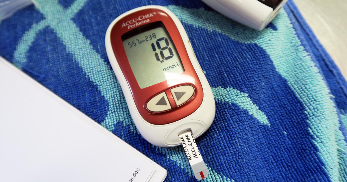
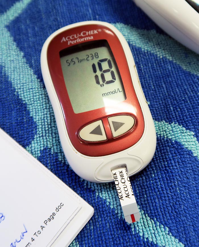
    A blood glucose meter showing a blood glucose level of 1.8mmol/L.

    The common causes of hypoglycaemia I see in an emergency setting are:

    • sepsis: bacteria consumes glucose
    • hypoadrenocorticism: lack of cortisol
    • insulin overdose: excessive intracellular shift
    • insulinoma: malignant insulin secreting neoplasia of the pancreas
    • hepatic insufficiency: reduce production

    Treatment is fairly straightforward and the impact is often dramatic – 0.5ml/kg to 1ml/kg of 50% dextrose diluted 50:50 with saline given slow IV over a couple minutes (to reduce the risk of haemolysis).

    As the list of possible causes shows, a one-off dose of glucose is often not enough.

    Glucose supplementation often needs to be continued as a 2.5% continuous rate infusion (CRI), with frequent blood glucose monitoring and adjustments made to the rate as necessary.

    The CRI will need to be continued, as the hypoglycaemia will often continue to occur until the primary disease process is identified and appropriately addressed.

    Emergency database

    It is not uncommon to read or hear the term emergency database. This contains a number of blood parameters performed, which include:

    • blood glucose
    • alanine aminotransferase
    • lactate
    • blood urea nitrogen
    • PCV
    • total protein or total solids
    • activated clotting time
    • acid-base balance
    • electrolytes
  • Idiopathic AHDS in dogs: treat with antibiotics or not?

    Idiopathic AHDS in dogs: treat with antibiotics or not?

    Idiopathic acute haemorrhagic diarrhoea syndrome (AHDS) – previously known as haemorrhagic gastroenteritis – remains the one disease where constant debate exists as to whether antibiotics should be used as part of the standard treatment.

    The logic behind using antibiotics to prevent bacterial translocation is sound, and if AHDS is truly initiated by Clostridium species or their toxins then the use of antibiotics can be justified.

    However, no knowledge exists of the true frequency of bacterial translocation in AHDS patients and conflicting evidence supports Clostridium being the initiating cause of AHDS in dogs.

    Together with new data indicating the use of antibiotic therapy in aseptic AHDS patients did not change the case outcome or time to recovery, the benefit of using antibiotics must be weighed against the very real risk of selection of antibiotic resistance and other complications associated with inappropriate antibiotic use.

    In this blog, we will explore the evidence against the use of antibiotics in AHDS.

    Cause unknown

    AHDS is characterised by an acute onset of vomiting (of less than three days’ duration) that can quickly progress to haemetamesis, and severe and malodorous haemorrhagic diarrhoea, accompanied by marked haemoconcentration that can be fatal if left untreated.

    <em>Clostridium perfringens<em>.
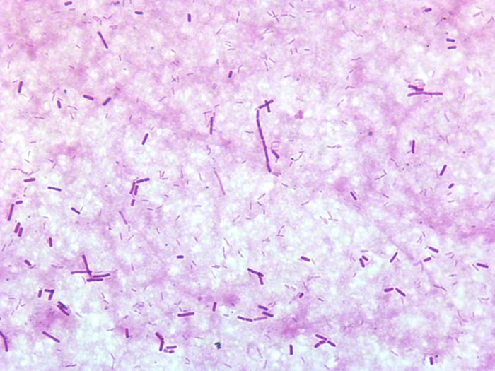
    Gram-stained Clostridium perfringens. Image © Andreas Zautner / Wikimedia Commons

    AHDS is a diagnosis of exclusion; other diseases (such as canine parvoviral enteritis, thrombocytopenia, hypoadrenocorticism, azotaemia, hepatopathy, neoplasia, intussusception, intestinal foreign body and intestinal parasitism) must be ruled out by a combination of medical history, vaccination status, complete blood count, serum biochemistry, coagulation times, diagnostic imaging and faecal testing.

    Small breed dogs – in particular, the Yorkshire terrier, miniature pinscher, miniature schnauzer and Maltese – have been found to be particularly predisposed. On average, the affected dogs were young (a median of five years old).

    The cause of AHDS is still unknown. Clostridium perfringens and its toxin has been incriminated as being the initiating cause; however, conflicting studies have refuted this claim. It is also difficult to determine whether overgrowth of Clostridium species is primary or secondary to the intestinal injury.

    Virus theory

    Another theory is viruses may have a role in AHDS’ aetiology. At this stage, only single agents had been investigated. It is possible a novel agent not yet been tested is the cause of this syndrome, or possibly the syndrome is the result of a very complex interaction between multiple organisms or their toxins.

    For the aforementioned reason, no indication exists for the use of antibiotics to treat for the underlying cause.

    Another argument behind the use of antibiotics lies in the fact most idiopathic AHDS patients have several risk factors for bacteraemia.

    Necrosis of intestinal mucosa, leading to the disruption of the gastrointestinal mucosa-blood barrier; adherence of significant numbers of bacteria to the necrotic mucosal surfaces that increases the risk of bacterial translocation; significant hypoalbuminaemia indicating substantial loss of mucosal epithelial layer; splanchnic and intestinal hypoperfusion, leading to reduced intestinal barrier function; and microbial dysbiosis all contribute to an increased risk of bacterial translocation.

    Although bacterial translocation has the potential to lead to sepsis, the true incidence of bacterial translocation needs to be established in idiopathic AHDS patients, as well as their influence on the outcome of the patients.

    Antibiotic requirement

    Antibiotics.
    Use of unnecessary antibiotics not only disrupts the protective mechanisms of a normal intestinal microflora, but also the real risk of post-antibiotic salmonellosis and Clostridium difficile-associated diarrhoea.

    Multiple studies have suggested antibiotics are not required in the treatment of aseptic idiopathic AHDS patients.

    In a prospective study of bacteraemia in AHDS dogs by Unterer et al (2015), the incidence of bacteraemia of patients with idiopathic AHDS was 11%, compared to those of healthy controls, where it was 14%.

    Transient bacterial translocation to mesenteric lymph nodes occurred in 52% of dogs undergoing elective ovariohysterectomy (Dahlinger et al, 1997), and confirmed in studies by others (Harari et al, 1993; Howe et al, 1999; Winkler et al, 2003), where portal and systemic bacteraemia was reported in clinically normal dogs.

    As long as the immune system is competent, and the functional capacity of the hepatic reticuloendothelial system is not overwhelmed, the body is usually effective at eliminating organisms from the blood.

    This is reflected in the Unterer et al (2015) study result, where – regardless of the bacteraemia status – all idiopathic AHDS dogs survived to discharge.

    In another prospective, placebo-controlled, blind study by Unterer et al (2011), idiopathic AHDS patients were either treated with amoxicillin/clavulanic acid for six days or a placebo, and no significant difference occurred between the treatment groups concerning mortality rate, duration of hospitalisation or severity of clinical signs.

    They concluded, without the evidence of sepsis, antibiotics do not appear to change the case outcome or shorten the time to recovery.

    Negative impacts

    The negative impacts of inappropriate antibiotic use are undeniable – especially in a time where resistance has become a worldwide public health concern.

    Use of unnecessary antibiotics not only disrupts the protective mechanisms of a normal intestinal microflora, but also the real risk of post-antibiotic salmonellosis and Clostridium difficile-associated diarrhoea.

    With evidence all pointing against the use of antibiotics as routine treatment of aseptic idiopathic AHDS, next time you are about to reach for antibiotics, I urge you to reconsider. Although it has taken some time to adopt and requires clear communication with clients, all vets should feel comfortable not using antibiotics for AHDS patients.

  • Blood gas analysis, pt 1: why everyone needs to know about it

    Blood gas analysis, pt 1: why everyone needs to know about it

    For those of you who have received referral histories from emergency or specialists hospitals, blood gas analysis is probably no stranger to you. For those who have never heard of them before, fear not – you are in for a treat.

    In my emergency hospital, the blood gas analyser is arguably one of the most frequently used bench top lab machines, second only to centrifuge, and for good reasons…

    Acid-base disturbances are common in critically ill and emergency patients, and it can help determine the severity of their condition and sometimes provide the answer. Tracking changes in blood gas parameters can provide information about the patient’s response to your interventions.

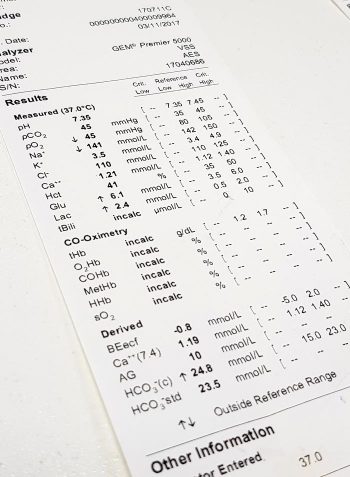
    blood-gas-analyser_output
    Blood gas analysis can help assess the severity of a patient’s condition and help guide your diagnostic plan.

    The information gained from pulse oximetry is very limited in patients with severe respiratory compromise, and the only way to accurately assess their oxygenation and/or ventilation status is by looking at their blood gas status.

    So what does the blood gas analysis actually measure?

    Most blood gas panels assess the pH of the blood, partial pressure of oxygen (PO2) and partial pressure of carbon dioxide (PCO2). From these, the machine is able to derive the percentage of haemoglobin saturated with oxygen (SO2), bicarbonate (HCO3) concentration and base excess of the extracellular fluid (BEecf).

    In most machines, they are also able to measure other parameters, such as electrolytes (Na, K, Ca, Cl), glucose and lactate.

    While arterial blood gas samples are required for determining the ability of the body to oxygenate the haemoglobin, venous samples are suitable for determining the ventilation status, assessing acid base balance, electrolytes, glucose and lactate levels.

    So how can this help as a point-of-care test?

    As mentioned previously, blood gas analysis can help assess the severity of a patient’s condition and help guide your diagnostic plan. It can also provide a diagnosis (such as diabetic ketoacidosis, typical hypoadrenocorticism and high gastrointestinal obstructions).

    The changes in these parameters over time can be essential in managing critical patients in the emergency setting; it will help guide you in developing an appropriate IV fluid therapy regime and fluid choice, address the patient’s oxygenation and/or ventilation needs, correct any electrolyte and glucose abnormalities, and – although fallen out of favour – the administration of sodium-bicarbonate therapy.

    In upcoming blogs, I will teach you how to interpret the blood gas results. At the end of this, I hope everyone will incorporate blood gas analysis as their standard point-of-care test for the better assessment and management of patients.

    If given the choice between a biochemistry and a blood gas panel in a critical patient, I would hands down select blood gas every time.

  • Handling an Addisonian crisis – part 1

    Handling an Addisonian crisis – part 1

    Addison’s disease (hypoadrenocorticism) is one of those annoying diseases that does not always play by the rules.

    One of the main reasons is the clinical signs of Addison’s disease can be frustratingly non-specific and we don’t often see the classic “low sodium, high potassium” electrolyte changes we are attuned to noticing. Therefore, it is important to recognise the early signs, or have a set of clinical signs, history and biochemistry changes that trigger the Addison’s disease alarm bells.

    Once diagnosed, we can look at the approach to stabilising a patient in an Addisonian crisis.

    Clinical signs

    The clinical signs of Addison’s disease can be vague and non-specific, these include:

    • anorexia
    • lethargy
    • weakness
    • gastrointestinal signs
    • polyuria and polydipsia

    A history of chronic intermittent vomiting and/or diarrhoea that resolves with symptomatic management would be one of the triggers.

    Another trigger is the subtle changes in blood tests. These include:

    • the absence of a stress hyperglycaemia in a sick patient (normal glucose or even a low blood glucose, for example)
    • hypercalaemia (ionised) of any degree
    • absence of stress leukogram

    These changes are often seen in isolation, so don’t expect them to all be there at the same time. We see patients all the time with inappropriately normal or low blood glucoses with gastrointestinal signs that we later diagnose with Addison’s.

    Hypercalcaemia – what’s the big deal?

    Why hype up and down about normal or low blood glucoses? Because low blood glucoses are rarely caused by inappetence or gastroenteritis alone, and the body is pretty good at maintaining glucose within normal levels (except, maybe, for very young patients and some toy breeds). Any low glucose Addison’s should be considered, and a stress hyperglycaemia is so common in ill patients that its absence makes me concerned.

    What is the big deal about hypercalcaemia? It is so tightly regulated that any elevation Addison’s is on the differential list. How does Addision’s cause these changes? Corticosteroids are released during stress or ill states, which result in a stress hyperglycaemia. They also play a role in the excretion of calcium in the urine, so preventing a hypercalcaemia.

    What about the electrolyte changes, the hyponatraemia and hyperkalaemia? When these changes are present they make diagnosis much easier, but they are not always present, such as with “atypical hypoadrenocorticism”, so the absence of that classic change does not rule out Addison’s.

    Next, we will cover the management of the Addisonian crisis.

  • SNAP cortisol test

    SNAP cortisol test

    While hyperadrenocorticism is not an uncommon incidental finding in patients presenting to our emergency clinic, hypoadrenocorticism is a lot less common. Or, possibly, more frequently underdiagnosed.

    Textbook clinical presentations combined with haematology and biochemicial changes can make diagnosis straightforward, but not all patients will present with all the classic signs.

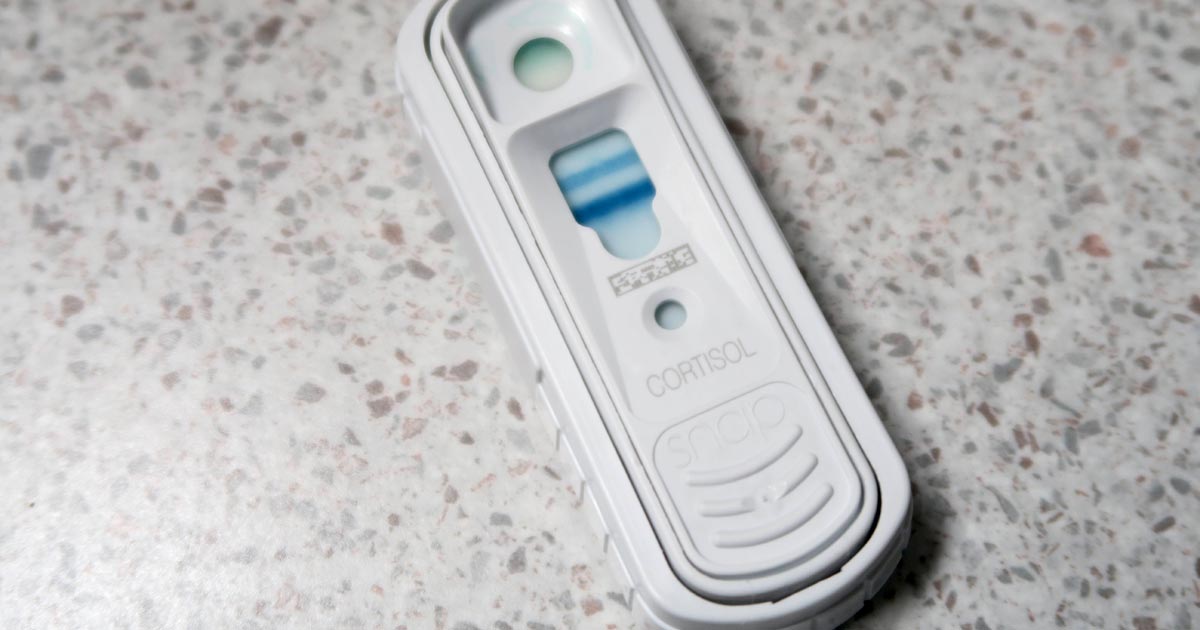
    SNAP cortisol test
    The SNAP cortisol test is a quantitative ELISA test that measures the level of serum cortisol in dogs.

    To complicate things further, hypoadrenocorticism is the great mimicker of diseases; it is often impossible to arrive at a definitive diagnosis without knowing the cortisol levels.

    The SNAP cortisol test allows clinicians to determine cortisol levels in-house – a blessing to those of us who work out-of-hours – but is not without its limitations.

    Suspicious signs

    Patients with hypoadrenocorticism often present with vague and non-specific clinical signs, but certain clinicopathological changes help raise the suspicion:

    • a decrease in sodium-to-potassium ratio (below 1:27)
    • azotaemia
    • an inappropriately low urinary specific gravity, despite evidence of dehydration or hypovolaemia
    • a leukogram unfitting to the degree of illness of the patient (a “reverse stress leukogram”- neutropenia, lymphocytosis, eosinophilia)
    • anaemia
    • hypoglycaemia
    • hypercalcaemia

    Although most Addisonian patients will not present with all these signs – especially those in the early stages of disease or those with atypical Addisonian disease (glucocorticoid insufficiency only) – any patients showing any of these haematology and biochemicial changes should have hypoadrenocorticism ruled out as part of the diagnostic plan.

    Imperfect ELISA

    The SNAP cortisol test has been advertised as an in-house assay to aid the diagnosis, treatment and management of both hyperadrenocorticism and hypoadrenocorticism, although the quality of the result is not perfect. This quantitative ELISA test measures the level of serum cortisol in dogs.

    In one study1, the SNAP cortisol test appears to have a good correlation with an external laboratory chemiluminescent assay test; however, in 12.8% of cases (5 of 39 patients), the SNAP test result could have led to a different clinical decision regarding the management of the patient.

    Since long-term Cushing’s management relies on reliable, repeatable cortisol level detection, this high level of discrepancy is unacceptable, especially when more accurate alternatives are available at external laboratories.

    Still useful

    Despite this, it is still very useful helping to assess for the presence or absence of hypoadrenocorticism, especially in an emergency setting.

    I use the SNAP cortisol to measure the resting cortisol level. If it is below 2ug/dL or in inconclusive range (between 2ug/L and 6ug/L), but the clinical picture suggests hypoadrenocorticism, I would perform an adrenocorticotropic hormone (ACTH) stimulation test and send samples to an external laboratory. If it is well above the inconclusive range, I would not perform an ACTH stimulation test.

    In summary, I think the SNAP cortisol test can be useful in helping assess for hypoadrenocorticism, but would still recommend performing an ACTH stimulation test and running the samples externally.

    However, use it with caution for hyperadrenocorticism diagnosis and its long-term management – especially when more accurate and economical alternatives are available.

  • Barking up the right tree – with Trusty Paws

    Barking up the right tree – with Trusty Paws

    Originally a charity set up by veterinary students for the homeless hounds of Glasgow in October 2014, Trusty Paws has become incredibly successful and has received a huge amount of public support.

    The Trusty Paws Clinic logo

    This success has allowed the charity to run monthly clinics at the Simon Community Scotland drop-in centre, providing free health checks, vaccinations, microchipping, and flea and worming treatment for the pets of the homeless.

    Essential supplies, such as food, coats and harnesses for the dogs, are also given out at these clinics for those in need.

    Branching out

    The success of the Glasgow clinics has led to a branch of the charity opening in London, with the first clinic taking place in November 2015.

    Run by fourth year RVC students, the clinics take place at the West London Day Centre in Marylebone, which also provides other services for the homeless.

    The expansion of the charity is excellent news for the pets of the homeless, for whom we can continue to provide the veterinary care they deserve.

    In at the deep end

    The Glasgow clinics are organised by the student committee, but health checks are conducted by other fourth year student volunteers. Last week I had the chance to get involved and, under the supervision of a volunteer vet, conducted my first full consultation that didn’t involve actors in a communication skills class.

    My patient, Bruno, wasn’t particularly well. The owner said he was not himself (he certainly looked depressed), had lost a significant amount of weight since his last visit and had a slow heart rate, in addition to some evident skin issues.

    The vet suspected Cushing’s disease, so we referred him to the local Pets’n’Vets branch that undertakes any secondary veterinary care Trusty Paws patients require. They have conducted blood tests, paid for by the charity, and have confirmed the diagnosis.

    Offering support

    While a little daunting to be thrown straight into a full consultation, I thoroughly enjoyed helping out at the clinic and would certainly encourage other students (whether in Glasgow or London) to do so in the future.

    The clients are extremely appreciative and evidently love their pets dearly, so it’s easy to see the benefits of such a charity to everyone involved.

    • If you can’t get directly involved, but wish to offer financial support, donations can be made via PayPal.
    • Alternatively, the charity has two Amazon wishlists (one for Glasgow, one for London), enabling supporters to purchase specific products that each clinic requires.