Secondary survey refers to the detailed physical examination performed after the primary survey, and should only be performed once the patient has been adequately stabilised.
It is always important to perform physical examinations systematically to avoid overlooking organ systems. This could be difficult in a stressful emergency situation, so one way to remind yourself is with the following acronym:
thorax (do this at the same time as assessing the abdominal cavity):
ensure you do both left and right sides
chest tube site
pericardial site
wet/dry/third space
S – Spine and tail
gait and posture
pain sensation
crepitus
H – Head
mentation
cognitive function
cranial nerves
external wounds/bruising
eyes – including symmetry, third eyelids, eye position, haemorrhage and detailed ophthalmological examination
ears
nose
The secondary survey will help identify any concurrent problems not seen on the primary survey.
P – Pelvis
wounds
bruising
pain
cepitus
perineum
external genitalia
L – Limbs
deformities
fractures
pain
bruising
wounds
weight bearing vs non-weight bearing
A – Arteries
all accessible superficial arteries – pulses and pressure
N – Nerves
mentation
cranial nerves
conscious proprioception
postural reflexes
peripheral spinal reflexes
withdrawal reflexes
deep pain
cutaneous trunci reflex
anal tone
Stable patient
By following the primary and secondary triage processes consistently, you should be able to quickly determine the order of criticalness of patients, institute appropriate resuscitative measures and manage life-threatening injuries. Then, with your thorough physical examination, identify any other concurrent problems not seen on the primary survey.
Overall, you have a stable patient, and are able to formulate an appropriate diagnostic and treatment plan.
Following on from part one, where we discussed that just by getting a good history and assessing the signalment a list of differentials can be narrowed down, we now look at how we can continue to narrow down the list based on the physical examination findings.
Although we now understand the way coagulation occurs in the body is different from the primary and secondary haemostasis model, it is useful to use this model when it comes to diagnosing the underlying cause.
Primary coagulopathy deficiencies – involve the platelets
ONSET: Usually more insidious, not usually enough to present with life-threatening blood loss (unless gastrointestinal).
CAUSES: Petechiae, ecchymosis, bleeding from mucosal surface.
LEADS TO: Epistaxis, gingival bleeding, haematuria, melaena.
EXAMPLE: Older dog with continuous bleeding from a lump above eye.
Pro Tip
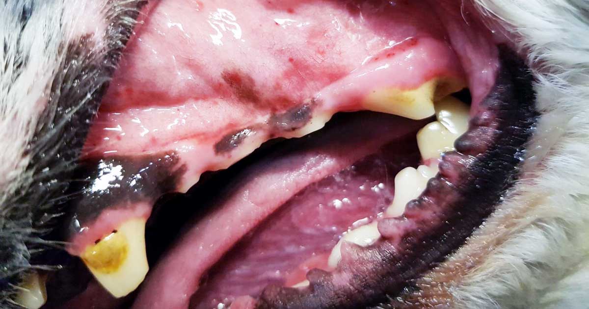
Think the three Ps: primary, platelets, petechiae
Gingival bleeding.
Secondary coagulopathy deficiencies – involve the clotting factors
ONSET: Usually more acute and present with life-threatening blood loss.
CAUSES: Bleeding into SC tissue, body cavities, muscles, joints.
LEADS TO: Haematoma, haemothorax, haemoabdomen, pulmonary haemorrhage.
EXAMPLE: Young dog coughing up blood, more likely secondary coagulopathy.
Pro Tip
Clinical signs overlap, so always rely on diagnostics to confirm your presumptive diagnosis.
Bleeding into SC tissue.
In part three, we cover what diagnostic tests you need to perform to confirm your suspicions…
Last week we discussed the causes and diagnostic pathway for investigating immune-mediated thrombocytopenia. This week we will go through the management of this condition.
Despite the fact red blood cells are not actually being destroyed, a severe anaemia can develop from blood loss due to coagulopathy – a common reason for why they present to emergency practices. The management of these patients is broken down into three main areas:
improving oxygen delivery
commencing immunosuppression
management of the underlying cause (if identified)
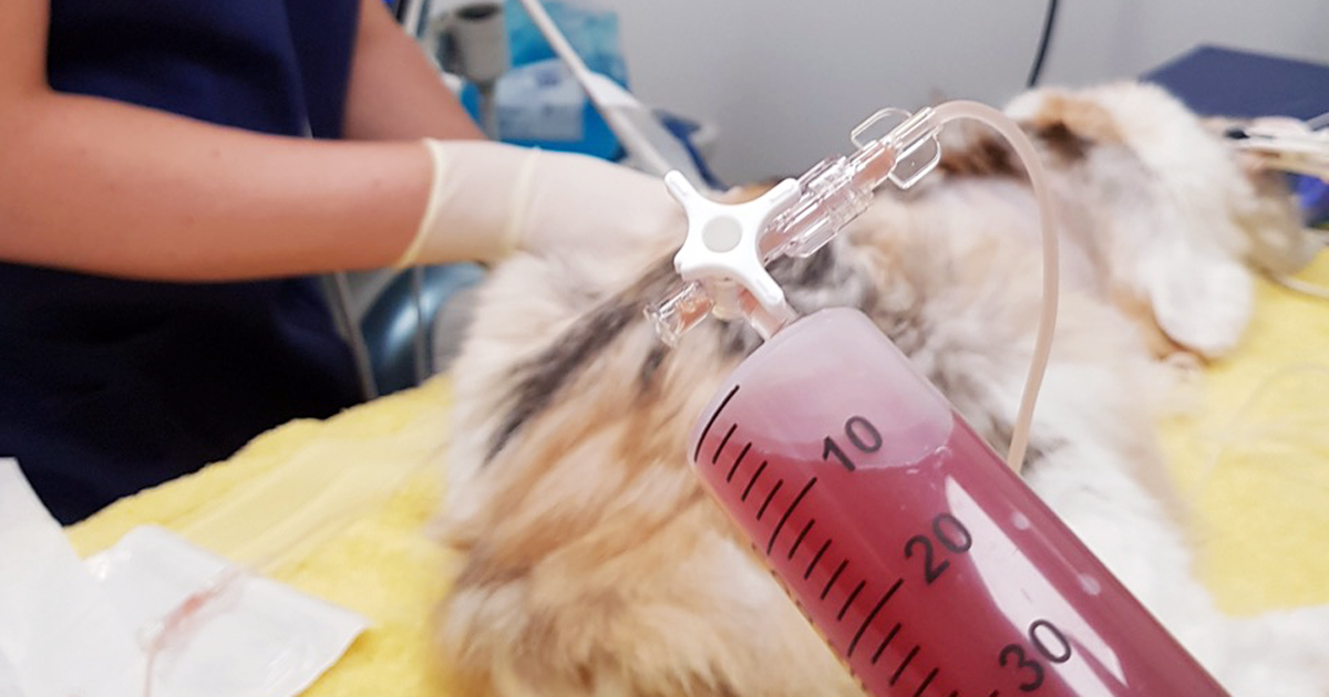
Optimising oxygen delivery in the acute phase is going to keep them alive long enough for immunosuppression to work. This is achieved through IV fluids to help improve perfusion and blood transfusions to replace red blood cells. If fresh whole blood is available, it can assist in increasing platelet numbers, but generally it is not very effective.
Platelet transfusions using platelet-rich plasma can be considered if it is available. Plasma transfusion is not effective at managing the coagulopathy as it is due to a loss of platelets, not a loss of coagulation factors.
Immunosuppression
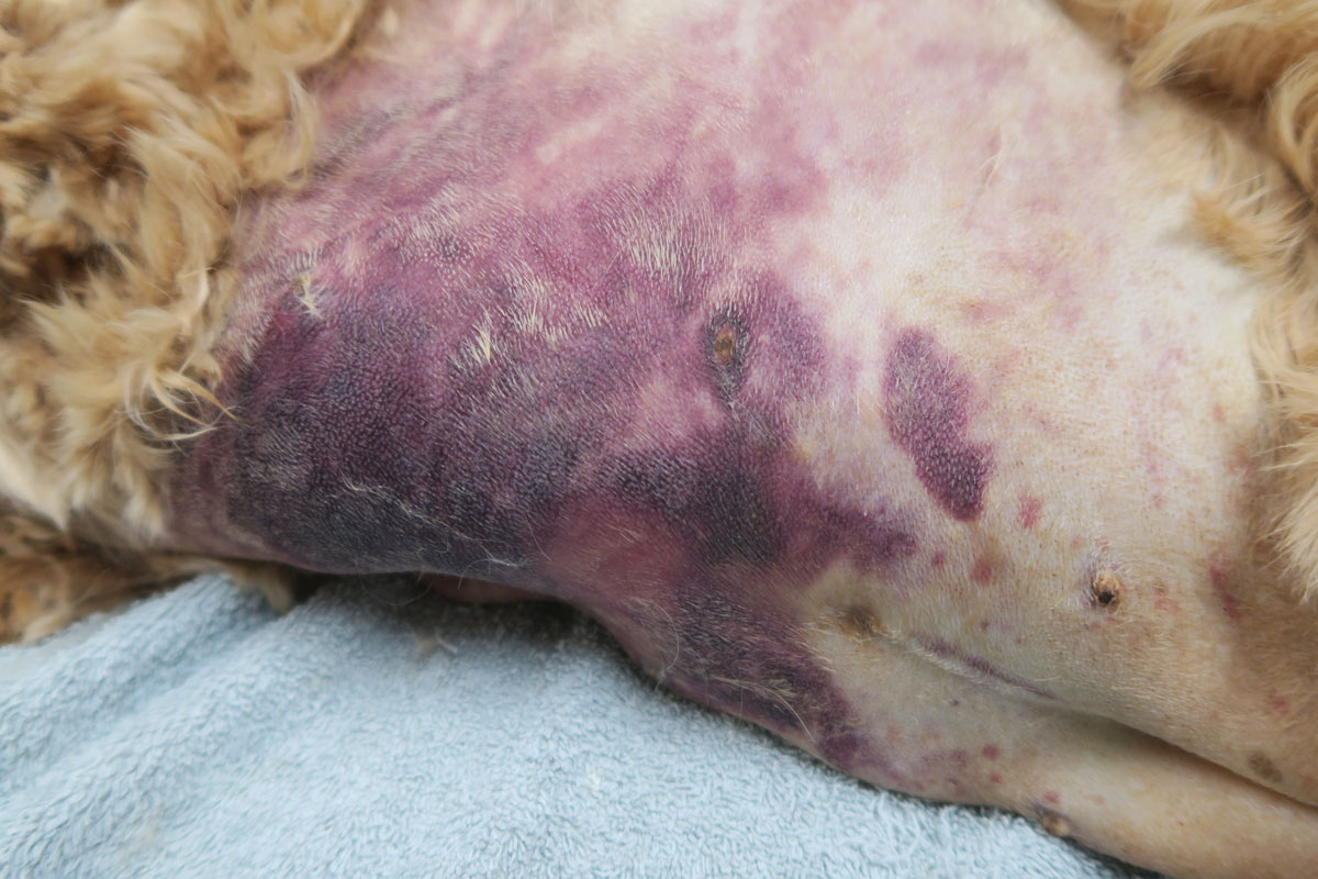
Large areas of ecchymotic haemorrhage on the skin are a quite obvious sign of thrombocytopenia.
Immunosuppression therapy is often commenced concurrently as the patient is being stabilised.
The first choice is either dexamethasone 0.5mg/kg IV every 24 hours if the patient is not stable enough for oral medications; otherwise, once stable, start prednisolone at 2mg/kg by mouth per day divided every 12 hours.
Other immunosuppressive agents include:
Azathioprine – 2mg/kg by mouth every 24 hours then 0.5mg/kg by mouth every other day. The main concerns are bone marrow suppression and hepatoxicity – also, it is very toxic in cats.
Ciclosporin – 5mg/kg to 10mg/kg by mouth divided twice a day; cats 5mg/kg by mouth every 24 hours.
Chlorambucil could also be used at a dose of 0.1mg/kg/day to 0.2mg/kg/day by mouth if the response to prednisolone is insufficient.
Management
Management of the underlying cause should be commenced if a cause is identified, but this is often not the case.
Other management options include:
Vincristine can be trialled to increase platelet number as it stimulates the release of platelets from the bone marrow.
Gastroprotectants can be considered if gastrointestinal bleeding has occurred – these include proton pump inhibitors and sucralfate.
Strict confinement, potentially sedatives and minimal blood sampling are important to minimise injury that may result in further bleeding and blood loss.
Antithrombotic therapy is not part of standard management as, unlike immune-mediated haemolytic anaemias, thrombotic events rarely occur.
When it comes to monitoring, platelet counts are performed daily until more than 40 × 109/L – this can take up to two weeks to occur.
Once above this level, take weekly counts until the numbers have normalised. Once they have, taper immunosuppressive medications over four to six months, with 20% dose reduction every couple weeks, generally with the adjunctive immunosuppressants first and prednisolone last.
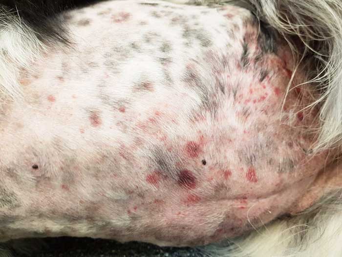
Thrombocytopenia is a condition characterised by a decrease in platelet numbers, which is often caused by increased destruction of platelets or a decrease in production.
Thrombocytopenia can manifest in many ways – the signs can be subtle and easily missed, such as small petechiae on gums, or quite obvious signs, such as large areas of ecchymotic haemorrhage on the skin.
Ecchymotic haemorrhages are often attributed to disorders of secondary haemostasis, such as rodenticide intoxication, but it can also occur with thrombocytopenia depending on the severity and chronicity.
Other common clinical signs include:
epistaxis
blood in stools, urine or vomit
pale mucus membranes
lethargy
weakness
Therefore, the first step to managing a patient with a severe thrombocytopenic episode that has resulted in significant blood loss is to manage shock, if present, with IV fluids, then administer a red blood cell transfusion.
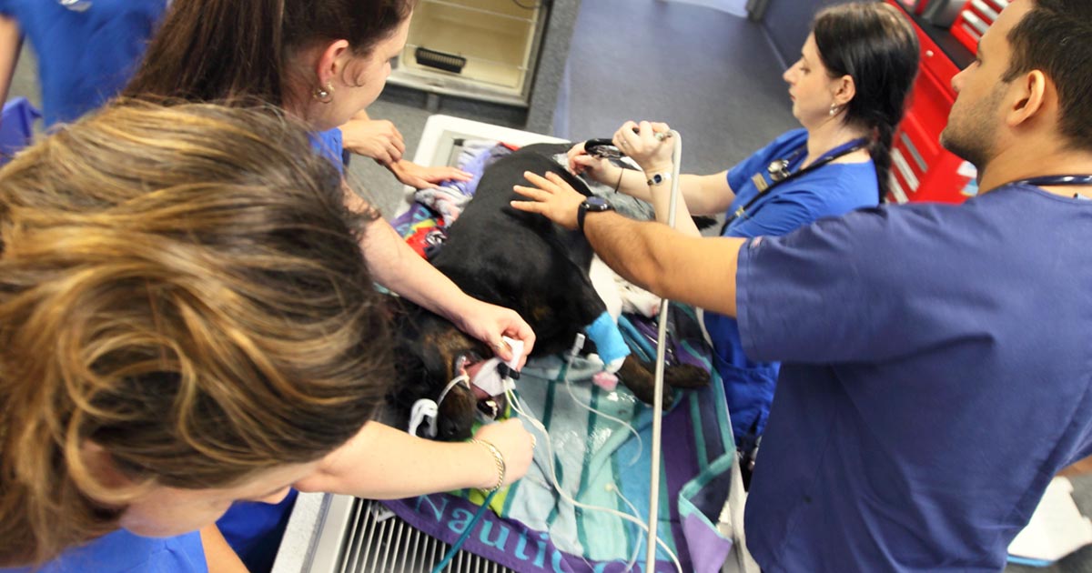
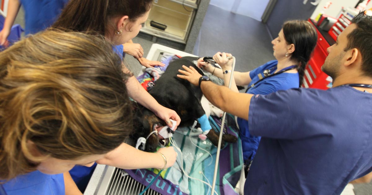
Patient handling
Careful patient handling is critical as these patients can bleed easily, leading to blown veins, large bruises that contribute to the development of anaemia and significant patient discomfort. Beyond initial patient stabilisation, the next step is to determine the underlying cause.
Diagnosing thrombocytopenia is relatively straightforward with the demonstration of low platelet counts. Generally, bleeding does not occur until the platelet count drop below 40,000 thousands per cubic milliliter (k/uL). This can be determined by either a haematology machine or manually via blood smear analysis.
When assessing blood smears, the general rule is one platelet per high-powered field on the monolayer is equal to 15,000k/uL. With either method, you must assess for platelet clumping on a blood smear as this can artefactually drop platelet numbers, leading to a false diagnosis.
The diagnostic pathway should not stop there. It needs to continue to determine the underlying cause.
The most common cause of thrombocytopenia is immune-mediated destruction. This can be either a primary (diagnosis of exclusion) or secondary cause (such as Rickettsia infection, and drugs such as sulphonamides, toxins and neoplasia). Other less common causes include:
splenomegaly, which can lead to platelet sequestration
disseminated intravascular coagulation and acute blood loss, leading to platelet consumption
bone marrow disease, which results in reduced platelet production
Signalment and history will refine such a diagnosis, as certain breeds are more prone to developing thrombocytopenia than others – for example, grey collies due to a defect in haematopoietic stem cells, and whippets and greyhounds, which traditionally have a lower platelet count than other breeds.
The generally diagnostic pathway continues to include haematology and biochemistry, thoracic radiographs, abdominal ultrasound and depending regional prevalence testing for infectious organisms with PCR and ELISA assays.
Next week I will cover the management principles of the thrombocytopenic patient.
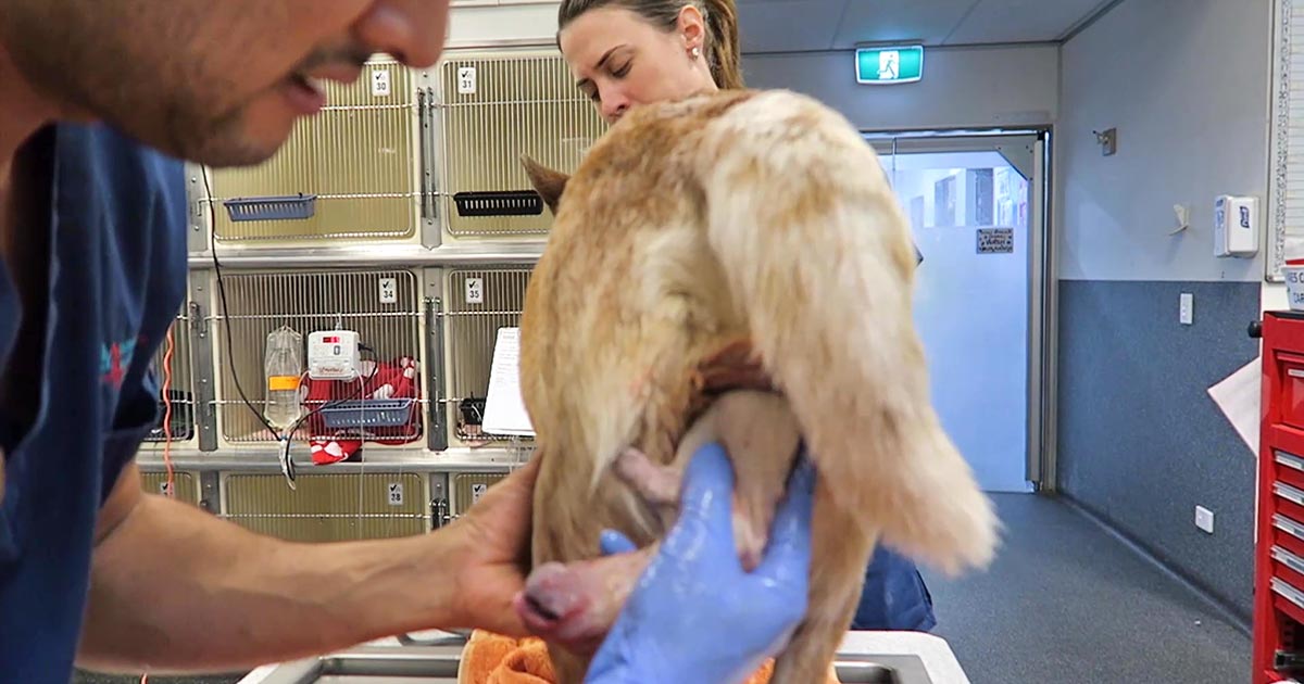
Once the bitch presents to the clinic, a few basic diagnostic checks need completing to determine the status of the bitch/queen and the fetuses.
Physical examination
The first is a thorough physical examination, starting with the bitch or queen:
Demeanour, hydration status, vital signs, mucous membrane colour, capillary refill time and temperature are important.
Pregnancy anaemia is not uncommon; however, for patients with a haemorrhagic discharge, it is important to know their cardiovascular status.
A thorough abdominal palpation should be carried out to assess comfort level and palpation for the presence of fetuses. Palpating fetuses can be difficult and cannot confirm if no fetuses are present.
A digital vaginal examination should be performed. Feathering response – also known as the Ferguson reflex in human medicine – is the neuroendocrine reflex where the self-sustained cycle of uterine contractions is initiated by firm pressure on the dorsal aspect of the vestibulovaginal wall. If this is absent, the patient is unlikely to progress with the parturition unaided.
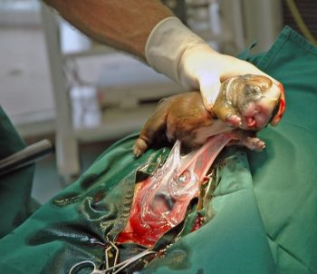
Palpation of fetuses in the canal can help decide whether surgical management is required. Obvious fetal malposition, malposture or malpresentation, or fetopelvic disparity, will be indications of caesarean. Abnormal pelvic diameter is also another reason to not proceed with medical management. To confirm these suspicions, abdominal radiography is required.
Radiographs will also help determine the number of fetuses to be expected, the signs of fetal death (presence of gas surrounding the fetus) and aforementioned fetomaternal abnormalities. I always repeat radiographs after the expected number of neonates is passed, to make sure I have not miscounted at the start.
Ultrasound
Panel 1. Heart rate ranges to help indicate stress of fetuses
Dogs:
normal – 180 to 220 beats per minute (bpm)
Stressed – 160bpm
Real concern – less than 160bpm
Cats:
normal – more than 220pbm
fetal stress – less than 180bpm
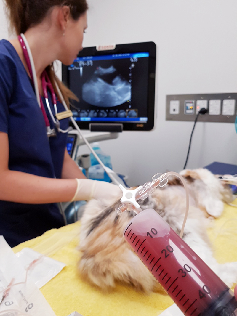
The second important diagnostic tool is ultrasound.
Fetal heart rates are good indicators of fetal stress. Some heart rate ranges that can help provide information about the status of the fetuses are detailed in Panel 1. These ranges vary between sources, but are good guidelines.
Ultrasounds can also help visualise the maturation status of the fetuses. At-term fetuses should have normal hepatic, renal and intestinal development. Intestinal peristalsis should be evident in at-term fetuses.
Other diagnostics
Other diagnostics may be indicated for patients, depending on the status of the bitch/queen:
If the patient is stable, but dystocia is present, a minimum database would include PCV/total protein, electrolytes, glucose, ionised calcium, lactate and acid-base balance.
Serum ionised calcium levels are important, as they influence the strength of contractions and how much supplementation is required.
Hypoglycaemia needs to be ruled out as a cause of dystocia, especially when large litters are involved.
If the patient is unstable or systemically unwell, include complete blood count, blood smears and biochemistry.
Physiological pregnancy anaemia can be present. The presence of regenerative response can help differentiate this from acute haemorrhage.
Abnormal leukocyte panel, especially with the presence of degenerative left shift, can indicate the presence of an infection – especially if toxic changes are present in the neutrophil.
Part three will briefly look at the medical management of dystocia and when surgical intervention is required.
Now most female canine patients are spayed, it comes as no surprise reproductive emergencies are not as common.
One confusion seems to be not knowing how to determine a true dystocia emergency – especially when given advice over the telephone – from the process of normal parturition.
Another concern is how to confidently form a diagnostic pathway to determine the cause of dystocia – especially for reasons other than obvious physical abnormalities (for example, fetopelvic disparity and fetal malposition).
Often, once we decide to go down the medical treatment pathway – whether the result of findings or owner/financial constraint – no one is confident as to what medication should be used and how often drugs can be given safely.
This series of blogs will address these issues in a step-by-step manner. Hopefully, by the end, you will be confident in the diagnosis and management of dystocia.
Labour stages
Before moving on to the signs of dystocia, let’s go through the signs of labour.
First stage labour
First stage labour is characterised by panting, tremoring, nesting behaviour, a drop in core temperature – usually a drop by almost 1°C 24 hours prior to second stage labour – and a drop of progesterone to below 2mg/ml.
Second stage labour is landmarked by the water breaking, visible abdominal contractions, and the allantoic/amniotic sac or fetal parts visible from the vulva.
If vulval discharge is present, they should be clear. Excessive amount of bright red haemorrhage, green or black discharge prior to delivery, or purulent material can indicate a pathological process requiring immediate veterinary attention.
dogs: approximately 3 to 6 hours
cats: approximately 6 to 24 hours
Third stage labour
Third stage labour this is when passage of all the placenta has occurred, generally within 15 minutes after passing a puppy or kitten.
Clues
Now we understand the normal progression of parturition, a few clues exist in the history that could suggest dystocia may be present.
Some breeders will often know the ovulation timing of the patient – especially if AI was performed. Tests such as progesterone levels, luteal hormone (LH) levels, cytology and vaginoscopy are some ways where it can help time the ovulation.
The normal gestation length should not be any longer than 66 days from the LH surge or, if the ovulation history is unknown, 72 days from the last known breeding.
History of prior dystocia is a warning, as most animals with prior parturition difficulties are more likely to develop dystocia again.
The same goes for animals that have previously required a caesarean. Their risk of requiring future caesareans is high, with further risk of uterine rupture if dystocia happens again.
Owners often telephone after the failure of normal progression of delivery. The signs that always require immediate intervention are:
more than 4 hours have passed from the rupture of the first chorioallantois
more than 2 hours between delivery
more than 30 minutes of strong abdominal contraction and no delivery
presence of green or black discharge before delivery
large amount of bright red haemorrhage
abnormal amount of pain during contractions
collapse of the bitch or distracted mothering
Any of these signs require immediate presentation to the veterinarian. Delivery of stillborn puppies is also an indication where veterinary attention is indicated.
Finally, if owners are concerned, it is best to advise veterinary assessment rather than try to convince them everything is okay based on what they describe over the telephone.
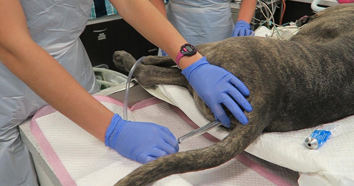
Last week we gave some hints and tips about how to perform a thoracocentesis. This week we look at what to do with the sample you collected and where to go to next.
Looking at the sample is not enough, there are several things you need to do to make sure you are getting the most information from the collected sample. This includes:
Fluid cell counts
Total protein assessment
Packed cell volume
Glucose
Lactate (if it is an exudate)
In-house cytology
Collect a sample for culture and sensitivity, and also external cytology assessment
With this information you can narrow down your list of differentials; often enough it can give you a diagnosis.
Here is the list I use. Note, it is not exhaustive and assumes you have taken three-view thoracic radiographs as part of the initial diagnosis.
Transudate
Haemorrhagic effusion.
Clear appearance – characterised by low protein and low cellularity
Transudates are caused by reduced oncotic pressure
Total nucleated cell counts = <0.5x10e9/L
Total protein = <25g/L
Differentials to consider
Liver disease
Protein-losing nephropathy
Protein-losing enteropathy
Additional diagnostics
Cytology and culture of fluid
Haematology and biochemistry
+/- dynamic liver testing
Urinalysis, urine protein/creatinine ratio, culture and sensitivity
Modified transudate
Yellow/serosanguinous/cloudy appearance
Caused by increased hydrostatic pressure leading to passive leakage of proteins and fluid into the pleural space
Total nucleated cell counts = 3.5-5x10e9/L
Total protein = variable, ~25-50g/L
Differentials to consider
Increased capillary hydrostatic pressure and pericardial disease
Diaphragmatic hernia
Neoplasia
Lymphatic obstruction, such as neoplasia, diaphragmatic hernia and abscess
Increased permeability of vessels (blood and lymphatics), such as FIP
Additional diagnostics
Cytology and culture of fluid
Haematology and biochemistry
Cardiac auscultation and ultrasound
+/- CT
Exudate
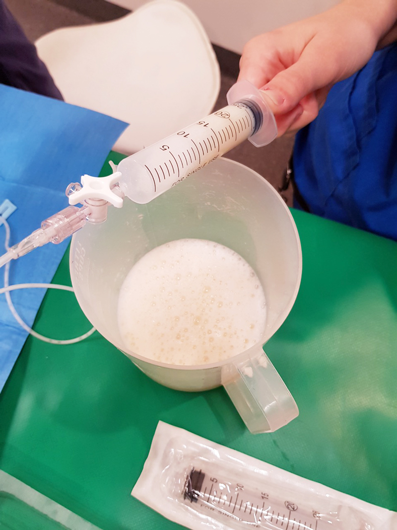
Turbid appearance – Very proteinaceous liquid, froths when shaken
Fluid is a mix of plasma and inflammatory mediators, and is caused by either septic or aseptic inflammation
Total nucleated cell counts = >3.0x10e9/L
Total protein = >30g/L
Aseptic exudate
Non-degenerate neutrophils and activated mesothelial cells predominate
Non-infectious cause
Differentials
Inflammation: FIP (can have high globulins), liver disease, lung torsion and hernia
Neoplasia
Additional diagnostics
Haematology and biochemistry
Cytology and culture of fluid
+/- ultrasound/CT
Further testing for FIP
Septic exudate
Degenerate neutrophils predominate: nuclear swelling and pale staining
Intracellular or extracelluar microorganisms
Culture and sensitivity: aerobic and anaerobic
Pleural fluid [glucose] < serum [glucose]
Pleural fluid [lactate] > serum [lactate]
Differentials to consider
Ruptured abscess
Foreign body inhalation or penetrating injury
Fungal infection
Additional diagnostics
Haematology and biochemistry
Cytology and culture of fluid
+/- ultrasound/CT
Chyle
Chyle.
Opaque (milky) to pink.
Differentials to consider
Rupture or obstruction of lymphatic flow
Neoplasia, traumatic and idiopathic
Secondary to heart failure (especially in cats)
Pseudochyle (usually formed by lymphoma)
Additional diagnostics
CBC and biochemistry
Cytology and culture of fluid
Fluid [TAG] > serum
Large number of lymphocytes and other inflammatory cells
+/- ultrasound/CT
Haemorrhage
Red blood cells
True haemorrhagic; for example, not iatrogenic: should not see platelets or erythophagocytosis on smears and sample should not clot
Time frame
Assess history
Compare fluid PCV/total protein (TP) to peripheral PCV/TP:
<1% – non-significant
1% to 20% – neoplasia, trauma, pneumonia
>50% – haemothorax
Other tips:
If PCV/TP is similar = recent bleed, if PCV is low and TP normal = chronic
If PCV is increasing or is higher than peripheral then active bleeding
Presence of erythrophagocytosis = chronic
Differentials to Consider
Trauma
Neoplasia
Coagulopathies
Ruptured granuloma
Diagnostics
Activated clotting time, activated partial thromboplastin time, prothrombin time, blood smear and other coagulation tests, see “coagulopathy”
Blood smear
CBC and biochemistry
+/- ultrasound/CT
Good luck with your next thoracocentesis. I hope this information was useful.
Clients often panic when they think their pet is having a seizure and can skip over vital information.
Often, what an owner describes as a “fit” may actually be syncope, collapse from anaphylaxis or internal haemorrhage (for example, neoplasia), a vestibular event or a behavioural condition.
True seizures
True seizures can be divided into two groups:
Generalised (grand mal) seizures, which involve both cerebral hemispheres and result in loss of consciousness, incontinence and muscle activity.
Focal/partial (petit mal) seizures, which originate from a focal region in the brain. These can also result in alterations in consciousness, but more typically only manifest in the form of repetitive twitching or limb movement.
Once you have established the owner is likely describing a true seizure, there are many important questions to ask to narrow down your differential diagnoses and treatment options.
The important questions
So, as part of a thorough history, always ask:
Was the pet conscious during the episode?
This will help to determine whether the seizure was generalised or focal.
How long did the episode last?
Status epilepticus is when a continuous seizure lasts more than five minutes or when the patient has not recovered fully before another seizure occurs. This can result in severe secondary brain injury.
How many episodes has the pet had in the past?
Epilepsy is the condition of recurrent seizures. This can be further classified as primary and symptomatic epilepsy, with symptomatic being secondary to an underlying cause (such as head trauma or a brain tumour).
How close together were the episodes?
Cluster seizures are when an animal has more than two or three episodes within a 24-hour period.
If a patient presents first time with a cluster, this carries a poorer prognosis in dogs, but has no influence in cats.
Clusters are generally an indication for commencing long-term management.
How was the pet before and after the episode?
Seizures often come with predicting (pre-ictal) and recovery (post-ictal) events.
In the pre-ictal phase, the patient may act strangely (for example, agitated or clingy) and may vomit.
Alterations in consciousness prior to a seizure usually indicate an intracranial cause.
The post-ictal phase can last anywhere between minutes and days, and animals are usually disorientated and/or lethargic. They may also appear blind.
Has the pet demonstrated any other strange activity recently?
For example, if an animal has also been circling to one side, you can start to predict the location of the lesion.
Cats more commonly present with partial seizures compared to generalised – this is seen as stereotypic behaviours and bursts of activity.
Has the pet been exposed to any toxins or chemicals?
Seizures caused by toxins (such as snail bait) generally do not stop and start, but are continuous.
In the next part of this series, we will look at differential diagnoses for seizures and highlight the differences between dogs and cats.
Great news for those who hate enemas: you may not have to do any of these ever again. This is the consensus by both the American Academy of Clinical Toxicology, and the European Association of Poisons Centres and Clinical Toxicologists.
The theory behind these procedures is legitimate – reducing systemic exposure of a toxicant by accelerating gastrointestinal tract (GIT) expulsion. But this is assuming the toxicant is absorbed very slowly, undergoes substantial enterohepatic cycling, or undergoes slow reabsorption in the lower GIT – all of which are uncommon characteristics of the types of toxicants veterinary patients are exposed to.
In fact, most toxicants of veterinary interests are generally rapidly absorbed in the upper GIT and absorption are not affected by catharsis.
Lack of evidence
No clinical evidence exists to support the use of a cathartic alone, or in combination with activated charcoal, to reduce the bioavailability of drugs or to improve the clinical outcome of poisoned patients. In fact, some evidence shows systemic exposure is increased following oral dosing of sorbitol, with activated charcoal, in canine paracetamol poisoning cases.
Similarly, no evidence exists that enemas and/or colonic irrigation improve clinical outcome in the treatment of oral poisoning.
The risks can be quite high with these procedures, with patients at risk of:
haemorrhage (in the case of anticoagulant vitamin K antagonist rodenticides, for example)
electrolyte destabilisation
bowel perforation
rectal prolapse
phosphate toxicities (cats)
The risks simply do not outweigh the benefit (or lack thereof). In fact, repeated dosing with combination preparations containing sorbitol and activated charcoal is not recommended.
This may be the most exciting news in veterinary medicine!
Until I started researching this Tip of The Week, I did not know the medical profession has abandoned the routine use of emesis in oral poisoning.
This is based on multiple medical literatures that have proven emesis induction does not influence the clinical severity of poisoning, the length of hospitalisation and the clinical outcome or mortality.
Although the rationale for inducing emesis is obvious, it is not necessarily evidence based. It is also dependent on satisfying a few large assumptions, all of which are untrue:
Emesis is a very effective way of removing gastric contents.
No separation exists of poison from its vehicle while inside the acidic environment of the stomach.
Poison is not absorbed through the stomach wall.
Ineffective method
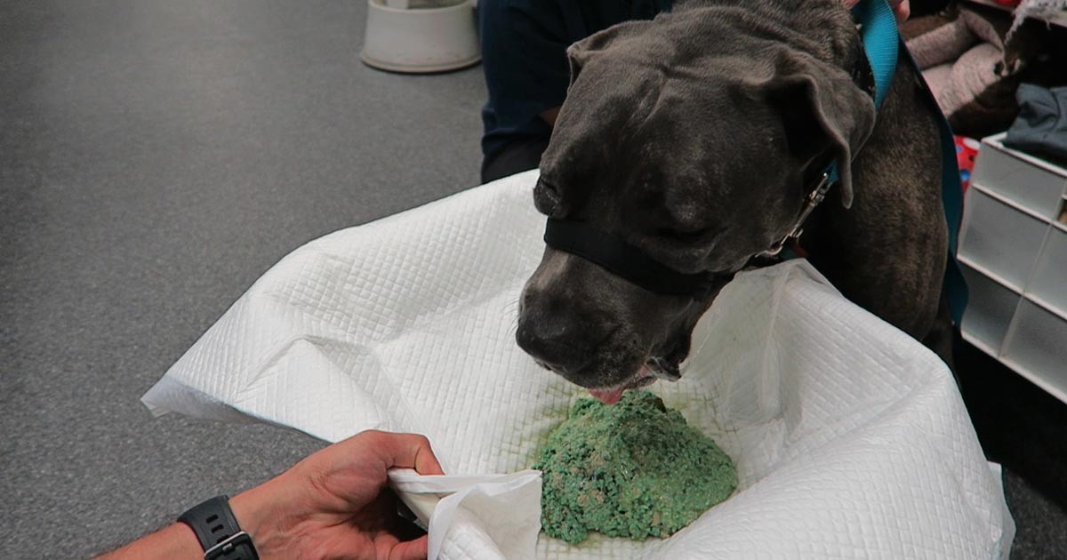
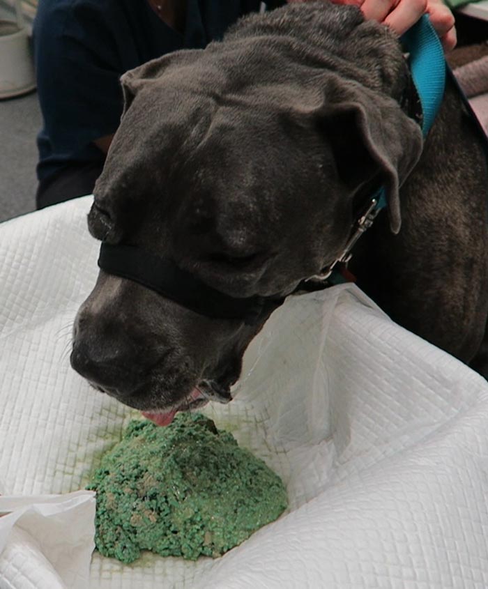
Snail bait ingestions: this patient ate 500g of snail bait containing metaldehyde.
Emesis induction is an ineffective way of clearing stomach contents. A review of the effectiveness of induced emesis, with both human and canine participants, showed at 30 minutes post-ingestion of non-absorbable markers, the recovery rate averaged between 17.5% and 52.1%, but never exceeded 62%.
In fasted puppies, this was even lower at 2% to 31%, despite inducing emesis immediately after marker administration. These have been confirmed by the presence of poisonous materials in the stomach of dead patients, despite effective emesis induction until clear fluid was brought up.
The clinical outcome only improves if the systemic exposure of a toxicant is reduced by more than half. However, considering animals rarely practice restraint, the ingested amount is unlikely to be exactly the lethal dose and no more. Therefore, even reducing the ingested toxic dose by 62% is unlikely to make a clinical difference.
Furthermore, most patients rarely present within 30 minutes of ingesting a toxicant, thus further reducing its efficiency.
The absorption conundrum
Some may argue the retrieval of metaldehyde or anticoagulant rodenticide granules from vomitus is indicative of reducing the toxicant dose. This could be true, but only if emesis was induced immediately after ingesting the poison.
The poison itself is colourless and has a different absorption characteristic to the coloured vehicle (granule); therefore, the presence of granule only serves to confirm ingestion, but is of no indication whether the poison has already been absorbed.
Contraindications
Many well-recognised absolute contraindications also exist to inducing vomiting:
Ingestion of oils, which includes waxes that melt to oil in the internal body environment, as this poses a high risk of lipoid and bacterial pneumonia. This is of significant veterinary significance, as wax is routinely used in rodenticide baits.
Ingestion of hydrocarbons and other volatile substances, or caustic or corrosive substances.
When the mental status is altered – for example, hyperexcitable or depressed mental state.
Where the patient is at risk of seizures (seizures can be induced by emesis).
Increased intracranial pressure.
Risk of intracranial or cerebral haemorrhage – for example, thrombocytopenia or abnormal clotting parameters.
Other less severe, yet important, reasons include:
delays administration of more effective treatment, such as activated charcoal, antidote or other treatments
risk of aspiration pneumonia
hypochloraemia in recurrent emetic patients
significant CNS and respiratory depression from apomorphine
rare, but reported, complications such as cerebral haemorrhage, oesophageal tear/ rupture, hiatal hernia, gastric rupture, pneumothorax and pneumomediastinum
legal implications – for example, if the product information clearly states emesis should not be induced
A place for everything
Emesis induction is not a benign procedure. It still has its place in certain circumstances, but its use in the routine management of oral poisonings may need to be reconsidered – especially if it means delaying administration of a more effective treatment, such as activated charcoal.
So, after all this, how do I tackle this information? It is a bit hard to swallow. My clinical experience is emesis is generally safe, especially in canine patients using apomorphine. So, I still feel some merit exists in reducing the amount of toxicant in the stomach if you have a chance – and in some situations, you don’t know until you try.
Emesis after ingestion of a toxic dose of chocolate can be incredibly rewarding, even six hours after ingestion, leading to patients not developing clinical signs at all.
Overall, I am biased by my personal successes with emesis, so still feel a time and place exist for emesis induction. But I now stop and question my decision to induce emesis, whereas I did not hesitate before.