Tag: Haematuria

  • How to approach the bleeding patient, part 2

    How to approach the bleeding patient, part 2

    Following on from part one, where we discussed that just by getting a good history and assessing the signalment a list of differentials can be narrowed down, we now look at how we can continue to narrow down the list based on the physical examination findings.

    Although we now understand the way coagulation occurs in the body is different from the primary and secondary haemostasis model, it is useful to use this model when it comes to diagnosing the underlying cause.

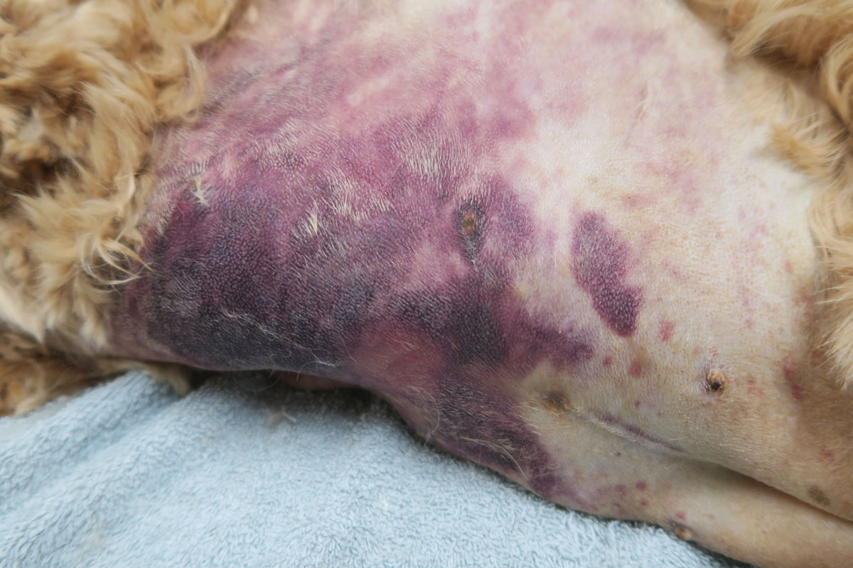
    Primary coagulopathy deficiencies – involve the platelets

    • ONSET: Usually more insidious, not usually enough to present with life-threatening blood loss (unless gastrointestinal).
    • CAUSES: Petechiae, ecchymosis, bleeding from mucosal surface.
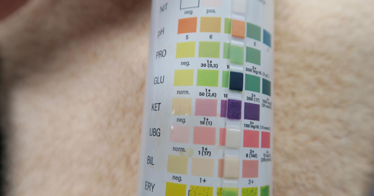
    • LEADS TO: Epistaxis, gingival bleeding, haematuria, melaena.
    • EXAMPLE: Older dog with continuous bleeding from a lump above eye.

    Pro Tip

    Think the three Ps: primary, platelets, petechiae

    gingival-bleeding
    Gingival bleeding.

    Secondary coagulopathy deficiencies – involve the clotting factors

    • ONSET: Usually more acute and present with life-threatening blood loss.
    • CAUSES: Bleeding into SC tissue, body cavities, muscles, joints.
    • LEADS TO: Haematoma, haemothorax, haemoabdomen, pulmonary haemorrhage.
    • EXAMPLE: Young dog coughing up blood, more likely secondary coagulopathy.

    Pro Tip

    Clinical signs overlap, so always rely on diagnostics to confirm your presumptive diagnosis.

    subcutaneous bleed
    Bleeding into SC tissue.

    In part three, we cover what diagnostic tests you need to perform to confirm your suspicions…

  • Urinalysis: dipstick tips

    Urinalysis: dipstick tips

    Following on from July’s post entitled Urinalysis: the neglected test, let’s have a look at the dipstick – it’s a very easy part of a urinalysis and essential to perform.

    Here are some of my tips in regards to using dipsticks:

    Poli_dipstick
    Dipstick: despite the name, DON’T DIP!
    • It may sound obvious, but you should always use veterinary-specific dipsticks. Human-specific dipsticks include panels for urobilinogen, nitrates and leukocytes, which we often do not interpret in small animal patients, as they are neither sensitive nor specific.
    • DON’T DIP! Use a syringe and drop samples on to each square, leave for 10 seconds, then flick off the excess.
    • Any amount of protein in dilute urine should raise suspicion. A reasonably large amount of protein has to be present in the urine for it to be positive on a dipstick. A urine protein to creatinine ratio may be the only way to quantify the amount of protein present, but first you must rule out evidence of inflammation or haematuria via a sediment examination.
    • The ketone panel on the dipstick test is only for acetoacetate (and not beta-hydroxybutyrate), although it is extremely rare for diabetic ketoacidosis patients to not produce any acetoacetate.
    • Trace blood can be a common artefact finding, especially during a cystocentesis where needle trauma can contaminate the sample with blood.
    • In our feline patients, any hyperbilirubinuria is abnormal, but this may be normal in a dog depending on urine concentration.