Tag: Fluid Therapy

  • Ionised hypocalcaemia, pt 2: eclampsia

    Ionised hypocalcaemia, pt 2: eclampsia

    As discussed in part one of this blog series, a myriad of disease processes can lead to ionised hypocalcaemia (iHCa).

    Despite this, only hypocalcaemia caused by eclampsia and hypoparathyroidism (primary or iatrogenic – post-surgical parathyroidectomy) are severe enough to demand immediate parenteral calcium administration.

    Hypoparathyroidism is quite rare, so this blog will not explore the detailed pathophysiology behind this syndrome. However, it is worthwhile mentioning – aside from primary hypoparathyroidism – no other disease state requires long-term calcium supplementation.

    Eclampsia, on the other hand, is the most common cause of clinical hypocalcaemia in dogs and cats. Multiple factors can predispose animals to the development of this phenomenon, so understanding the pathophysiology behind this potentially fatal disease will not only help with future diagnosis and treatment, but also help prevent this issue.

    Periparturient occurrence

    Eclampsia – also known as puerperal tetany or periparturient hypocalcaemia – occurs in the periparturient period anywhere from the final few weeks of gestation to four weeks postpartum, with the latter being the more common time frame of manifestation.

    The serum concentration of ionised calcium (iCa) is often less than 0.9mmol/L in bitches or less than 0.8mmol/L in queens. It presents as muscle fasciculation and tetany, but not usually in seizure since most patients maintain consciousness. Exceptions occur when these patients are left untreated – these patients may develop refractory seizures, cerebral oedema and death.

    The increased muscle activity generates a lot of heat and uses a significant amount of glucose; therefore, hyperthermia and hypoglycaemia are common sequelae in patients with delayed presentations.

    Reduced iCa

    Eclampsia occurs as a result of reduced iCa in the extracellular compartment. In lactation-associated hypocalcaemia, it is the result of the body’s inability to maintain serum iCa through increased osteolytic activity and gastrointestinal calcium absorption, and reduced renal calcium excretion to compensate for the loss of calcium through milk production.

    Other factors often predispose animals to developing eclampsia. These can include poor periparturient nutrition, excessive calcium supplementation and large litter size.

    Excessive calcium supplementation in the prenatal period causes parathyroid gland atrophy, preventing parathyroid hormone release – resulting in reduced gastrointestinal calcium absorption and osteoclastic activity, and increased kidney calcium loss.

    Clinical signs

    Clinical signs can progress rapidly and become fatal if left untreated.

    In the early phases, non-specific signs can present as:

    • facial pruritus
    • hyperaesthesia
    • panting
    • tremors
    • muscle fasciculations
    • paresis
    • ataxia

    Within a few hours, these clinical signs rapidly progress to rigidity, and tonic and clonic spasms with opisthotonos. By this stage, animals will develop severe tachycardia, tachypnoea and hyperthermia. Without treatment, a high mortality rate exists.

    kitten
    “Early supplementation of puppies and kittens with commercial milk formula will significantly reduce the lactation demand on the dam.” Image © Dobroslav / Adobe Stock

    Patients presenting with eclampsia require immediate medical intervention, as well as concurrent supportive therapy. The acute management of clinical iHCa is the same, regardless of the cause, and will be discussed in detail in part three.

    Supportive therapies required to manage and prevent a patient relapsing in eclampsia often include active cooling and glucose supplementation. In cases that seizure, anti-seizure medications – such as diazepam and barbiturates – and mannitol for cerebral oedema may be required.

    Prevention

    Even before getting to the stage where an animal requires treatment, all effort must be taken to prevent a dam from developing hypocalcaemia. This can be easily achieved by improving the calcium content of the food during the perinatal period, as well as reducing the milk demand by early weaning kittens or puppies. This is likely particularly helpful for those with a history of eclampsia or with large litters.

    From the second half of gestation, it is recommended a commercial formulation of puppy/kitten food (1% to 1.8% calcium and 0.8% to 1.6% phosphorus) is to be fed to the dam without any additional minerals or vitamin supplementation.

    Postpartum calcium is similar to the second half of gestation, requiring a diet containing at least 1.4% calcium with a 1:1 ratio with phosphorus (most balanced growth formula for puppies and kittens).

    Less demand

    Early supplementation of puppies and kittens with commercial milk formula will significantly reduce the lactation demand on the dam. Together with this, starting at aged three to four weeks, solids can be introduced at this time. These techniques will be particularly helpful to those with a history of previous eclampsia or those with large litter sizes.

    Aside from the parenteral calcium supplementation required, other supportive therapy – such as active cooling, IV fluid therapy and glucose supplementation – may be required.

    Long term, the dam’s nutritional content of calcium must be optimal from the second half of gestation. All additional calcium or other vitamins and mineral supplementations should not occur prior to parturition.

    In the postpartum dam with a history of eclampsia or that is at risk, changing to a nutritionally balanced commercial food aim for growing puppies and kittens is ideal. Early weaning – or abrupt weaning if hypocalcaemia is severe – may be required in severe cases or those with a high risk of relapse/development.

  • Blood transfusions, pt 3: how much?

    Blood transfusions, pt 3: how much?

    Now that you know how to spot the signs of when a blood transfusion is needed and what blood product to administer, this article will focus on the volume of blood to give.

    What PCV should I aim for?

    bloodpt3To start us off, no real benefit exists in increasing a PCV above 30%, unless you are anticipating further losses. The reason being is oxygen delivery to tissues is optimised at that level and administering more does not add significant benefit.
    How much volume to give?

    There are several formulas for determining how much red blood cells are required to be given, some more simpler and less accurate than others.  As a rough guide, if increasing PCV by 1, you need 2ml/kg of whole blood or 1ml/kg of packed red blood cells.

    This gives me a starting volume, I administer that volume and recheck. Often, I will have to give more, but it depends greatly on the underlying disease, concurrent fluid therapy and ongoing blood losses.

    How to administer?

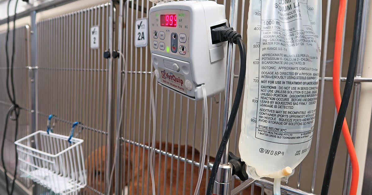
    Use a 170µm blood filter to collect any micro clots. Often, the blood is run through with a isotonic crystalloid, if so avoid administration with calcium containing crystalloid, such as Hartmann’s or lactated Ringers, as the calcium can result in activation of platelets and clots to form in the blood; so run with 0.9% saline or Plasmalyte 148.

    Packed red blood cells are usually diluted with at least 100mls of 0.9% saline. The generally rule is to administer the transfusion within 4 hours and change the fluid lines after to reduce the risk of bacterial contamination. Recheck PCV and coagulation times 30 minutes post-transfusion.

    How fast?

    Rate of administration really depends on degree of urgency. If the patient is suffering from acute haemorrhagic shock then you can administer blood as a bolus. The risk here is acute anaphylaxis if that patient is off a different blood type or if this is not their first transfusion.

    If it is not a crisis then administration should be started off slowly so monitoring for acute reactions can be performed. Start slow at 2ml/kg/hr for 15 minutes and monitor for reactions then progressively double the rate until the desired rate is reached.

  • Blood gas analysis, pt 1: why everyone needs to know about it

    Blood gas analysis, pt 1: why everyone needs to know about it

    For those of you who have received referral histories from emergency or specialists hospitals, blood gas analysis is probably no stranger to you. For those who have never heard of them before, fear not – you are in for a treat.

    In my emergency hospital, the blood gas analyser is arguably one of the most frequently used bench top lab machines, second only to centrifuge, and for good reasons…

    Acid-base disturbances are common in critically ill and emergency patients, and it can help determine the severity of their condition and sometimes provide the answer. Tracking changes in blood gas parameters can provide information about the patient’s response to your interventions.

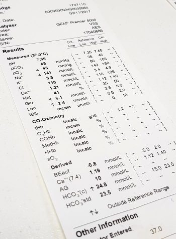
    blood-gas-analyser_output
    Blood gas analysis can help assess the severity of a patient’s condition and help guide your diagnostic plan.

    The information gained from pulse oximetry is very limited in patients with severe respiratory compromise, and the only way to accurately assess their oxygenation and/or ventilation status is by looking at their blood gas status.

    So what does the blood gas analysis actually measure?

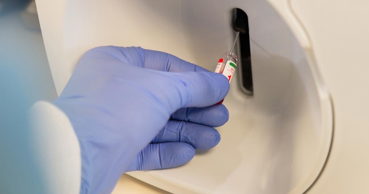
    Most blood gas panels assess the pH of the blood, partial pressure of oxygen (PO2) and partial pressure of carbon dioxide (PCO2). From these, the machine is able to derive the percentage of haemoglobin saturated with oxygen (SO2), bicarbonate (HCO3) concentration and base excess of the extracellular fluid (BEecf).

    In most machines, they are also able to measure other parameters, such as electrolytes (Na, K, Ca, Cl), glucose and lactate.

    While arterial blood gas samples are required for determining the ability of the body to oxygenate the haemoglobin, venous samples are suitable for determining the ventilation status, assessing acid base balance, electrolytes, glucose and lactate levels.

    So how can this help as a point-of-care test?

    As mentioned previously, blood gas analysis can help assess the severity of a patient’s condition and help guide your diagnostic plan. It can also provide a diagnosis (such as diabetic ketoacidosis, typical hypoadrenocorticism and high gastrointestinal obstructions).

    The changes in these parameters over time can be essential in managing critical patients in the emergency setting; it will help guide you in developing an appropriate IV fluid therapy regime and fluid choice, address the patient’s oxygenation and/or ventilation needs, correct any electrolyte and glucose abnormalities, and – although fallen out of favour – the administration of sodium-bicarbonate therapy.

    In upcoming blogs, I will teach you how to interpret the blood gas results. At the end of this, I hope everyone will incorporate blood gas analysis as their standard point-of-care test for the better assessment and management of patients.

    If given the choice between a biochemistry and a blood gas panel in a critical patient, I would hands down select blood gas every time.

  • Fluid therapy part 4: ongoing losses

    Fluid therapy part 4: ongoing losses

    This month, we will look at the final part of a fluid therapy plan – accounting for ongoing losses. This can be challenging, but some general rules can be helpful.

    Regular assessment is essential to track patients' response.
    Regular assessment is essential to track patients’ responses.

    First, let’s recap the four parts of a fluid therapy plan:

    1. Perfusion deficit
    2. Hydration deficit
    3. Maintenance requirements
    4. Ongoing losses

    When considering ongoing losses, try to not forget about patients with pre-existing polyuric diseases; chronic renal failure is a prime example. Patients with dehydrated chronic renal failure are unlikely to suddenly regain concentrating ability. Polyuria should be considered as an ongoing loss.

    Other conditions that may result in additional urinary fluid losses include post-obstructive diuresis, diabetes mellitus, hyperadrenocorticism and hyperthyroidsim.

    How much to add?

    This is the tricky part. I often add an additional half to one maintenance and frequently reassess clinical parameters, or if a urinary catheter is placed matching ins and outs.

    Gastrointestinal tract losses can be collected and weighed; 1g of vomitus or diarrhoea can be roughly equivalent to 1ml of water.

    Fluid removed from drains placed in cavities or wounds should also be measured and accounted for.

    Remember the key point is regular assessment of the patient’s hydration status, from repeat clinical exams, to track their response. Don’t forget regular retesting of electrolytes – for example, every 12 to 24 hours for patients on IV fluids and not eating.

  • Focus on GDV, part 4: the recovery

    Focus on GDV, part 4: the recovery

    Postoperatively, gastric dilatation-volvulus (GDV) patients remain in our intensive care unit for at least two to three days.

    Monitoring includes standard general physical examination parameters, invasive arterial blood pressures, ECG, urine output via urinary catheter and pain scoring.

    I repeat PCV/total protein, lactate, blood gas and activated clotting times (ACT) immediately postoperatively and then every 8-12 hours, depending on abnormalities and patient progress.

    GDV recovery
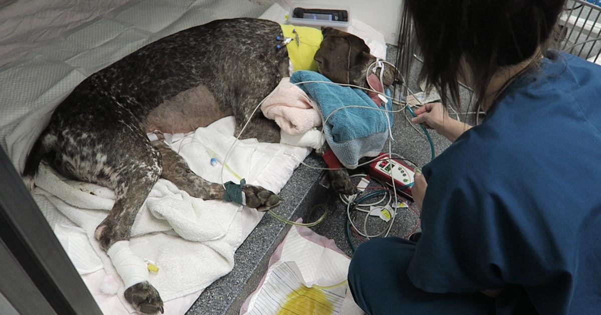
    Patient recovering in the pet intensive care unit. As well as standard monitoring parameters, GDV patients have constant ECG, arterial blood pressure and urine output monitoring to enable the early detection and correction of abnormalities.

    I always repeat these blood tests postoperatively, as IV fluids given during the resuscitation and intraoperative period often cause derangements. I use the results to guide my fluid therapy, but also take it with a grain of salt.

    IV fluids

    I generally continue a balanced and buffered crystalloid. The rate depends on blood pressures, urine output and assessment of general physical examination parameters for perfusion and hydration, but I try to avoid fluid overload and reduce the IV fluids postoperatively as soon as possible.

    Coagulopathy

    Prolonged clotting times are frequently seen as a result of consumption in a dog with GDV. However, one should note it can also occur as the result of haemodilution.

    As the underlying disease process has been corrected, and haemostasis achieved during surgery, I usually monitor ACTs, but may not necessarily treat with blood products as prolonged ACTs do not always translate to clinical bleeding. Unless clinical evidence of bleeding exists, I generally hold off treatment and monitor.

    Hypoproteinaemia

    Low total protein is also common. This is generally due to haemodilution from fluid resuscitation. However, a low total protein does not mean oedema will develop, or that it requires management. I generally track the protein levels, use conservative fluid therapy and try to correct it by instituting enteral nutrition as soon as possible.

    Electrolyte imbalances

    Hypokalaemia is a common complication of fluid therapy. This can be rectified with potassium supplementation in the IV fluids.

    Hyperlactataemia

    If present post-surgery, this is usually corrected with a fluid bolus. However, I always assess for other things that may affect oxygen delivery to the tissues, such as poor cardiac output (arrthymias), hypoxaemia (respiratory disease) and anaemia (from surgical blood loss).

    Arrhythmias

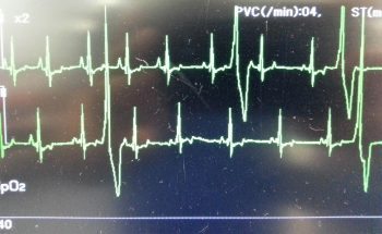
    Ventricular arrhythmias are common post-surgery. Accelerated idioventricular rhythms are the most common cause, especially if a splenectomy was performed.

    arrhythmia
    Ventricular premature contractions are common postoperative arrhythmia.

    Before reaching for anti-arrythmia medications, first check and correct:

    • electrolyte abnormalities
    • hypoxaemia
    • pain control
    • hypovolaemia or hypotension

    If they are still present, despite correction of the above, consider treating the rhythm if:

    • multifocal beats (ventricular premature contractions of various sizes)
    • overall rate greater than 190 beats per minute
    • R-on-T phenomenon
    • low blood pressure during a run of ventricular premature contractions

    I start with a bolus 2mg/kg lidocaine IV and start a constant-rate infusion of 50ug/kg/min to 75ug/kg/min.

    Anaemia

    It is common to have a mild anaemia post-surgery, due to a combination of blood loss and haemodilution. In the absence of transfusion triggers – such as increased heart rate, increased respiratory rate or hyperlactataemia – it does not require treatment.

    Vomiting

    Anti-emetics are the first line of medication. Non-prokinetic anti-emetics, such as maropitant and ondansetron, can be used immediately; otherwise, after 12 hours, metoclopramide can also be used postoperatively. If the patient remains nauseous despite these medications, the placement of a nasogastric tube can ease nausea by removing static gastric fluid.

    Excessive pain relief may also contribute to the nauseous state.

    Pain relief

    I mostly rely on potent-pure opioid agonists, such as fentanyl constant-rate infusions and patches. This is generally sufficient for most patients. Ketamine is occasionally used.

    • Some drugs listed in this article are used under the cascade.
  • Focus on GDV, part 1: resuscitation

    Focus on GDV, part 1: resuscitation

    Last month we covered a bit of pathophysiology, presenting pathophysiology, presenting clinical signs and the radiographic diagnosis of gastric dilatation-volvulus (GDV).

    Now we cover the three things you need to do as soon as a suspected case is presented:

    1. IV fluid resuscitation
    2. decompression of the stomach
    3. pain relief

    Depending on the number of staff you have, all of these can be performed simultaneously. If not, follow the above order as shock is the most imminent problem.

    Catheter placement

    Fluid resuscitation is relatively straightforward. Most GDV patients will be in some degree of shock, varying from mild to severe. Regardless of the actual degree, all patients will require IV fluids.

    The placement of IV catheters is particularly important; their numbers and diameter will influence the rate of response to treatment. Large-bore catheters allow faster flow of fluids compared to smaller ones, while multiple catheters allow concurrent delivery of two bags of fluids as opposed to one – particularly important in large dogs. Therefore, always try to place the largest catheter possible (for example, 18G or larger for large-breed dogs) into the cephalic veins.

    Once the catheters have been placed, collect 2ml to 3ml of blood for baseline measurements. These can be collected directly from the catheters and should include:

    • PCV/total protein
    • blood gas analysis
    • lactate
    • activated clotting time
    • electrolytes
    • later, full haematological and biochemical analysis

    Once the baseline bloods have been collected, fluid resuscitation should start immediately.

    How much, how fast?

    fluid
    Fluid resuscitation is relatively straightforward, says vet Gerardo Poli.

    How much fluid should you deliver, and how fast? My “go to” fluid is crystalloids and I generally start with a 20ml/kg bolus of an alkalinising crystalloid.

    I perform bolus therapy, so 10ml/kg to 20ml/kg fluid doses rather than shock rates 90ml/kg/hr, as I feel it allows me to better titrate my fluid therapy to effect. It also helps minimise excessive fluid administration and the problems with haemodilution – such as anaemia, hypoproteinaemia and prolonged coagulation times.

    As fluids are being delivered, I administer pain relief and start gastric decompression (covered next week).

    The decision to administer more fluids depends on whether I have achieved some end point resuscitation variables, such as:

    • a reduction in heart rate
    • a reduction in capillary refill time
    • an improvement of mucus membrane colour
    • improvement in pulse pressures

    Improvement in mentation is not often reliable as the sedative effect of analgesia, which I generally give during fluid resuscitation, often confounds this effect.

    Shock therapy

    If evidence of shock still exists, despite the initial fluid boluses and gastric decompression, I will consider more fluids. This can include hypertonic saline or colloids.

    In my experience, a repeat of a smaller dose of crystalloid fluid bolus is often adequate (10ml/kg). The transition on to hypertonic saline (7% solution) or colloids is influenced by the results of the aforementioned baseline diagnostics.

    A reduction in PCV/total protein suggests blood loss. In this case, I will consider either hypertonic saline (3ml/kg to 5ml/kg of 7% solution), a dose of colloids or even blood products, such as whole blood or packed red blood cells.

    If significant prolongation in activated clotting time occurs, likely from consumption, then I may incorporate fresh frozen plasma into my fluid therapy. This is in anticipation of possible surgery, where prolonged coagulation times can not only be troublesome, but life-threatening.

    Lactate

    A quick note on lactate – I don’t use the baseline reading as a prognostic indicator or an indicator of gastric necrosis. This is supported by recent findings claiming it is not the level of lactate that is predictive, but the degree of improvement in response to fluid resuscitation and gastric decompression.

    I have seen unreadable lactate levels – greater than 15mmol/L – in patients who returned to reasonably normal levels within an hour of stabilising. These patients also went on to survive surgery.

    Pain relief

    After starting IV fluid resuscitation, I generally administer pain relief while the team is preparing for gastric decompression. To keep things simple, I stick to an easily accessible pure opioid agonist at 0.2mg/kg IV. I avoid subcutaneous or even intramuscular administration as the patient is often in shock; the peripheral blood is shunted centrally to the heart and the brain and absorption can be variable.

    I find this offers a reliable and great degree of pain relief that helps reduce anxiety levels and, consequently, reduces oxygen demand. It has minimal cardiovascular effects and the mild sedative effect also helps with the process of decompression.

    >>> Read Focus on GDV, part 2: Releasing the pressure (gastric decompression)

  • Maintenance fluids

    Maintenance fluids

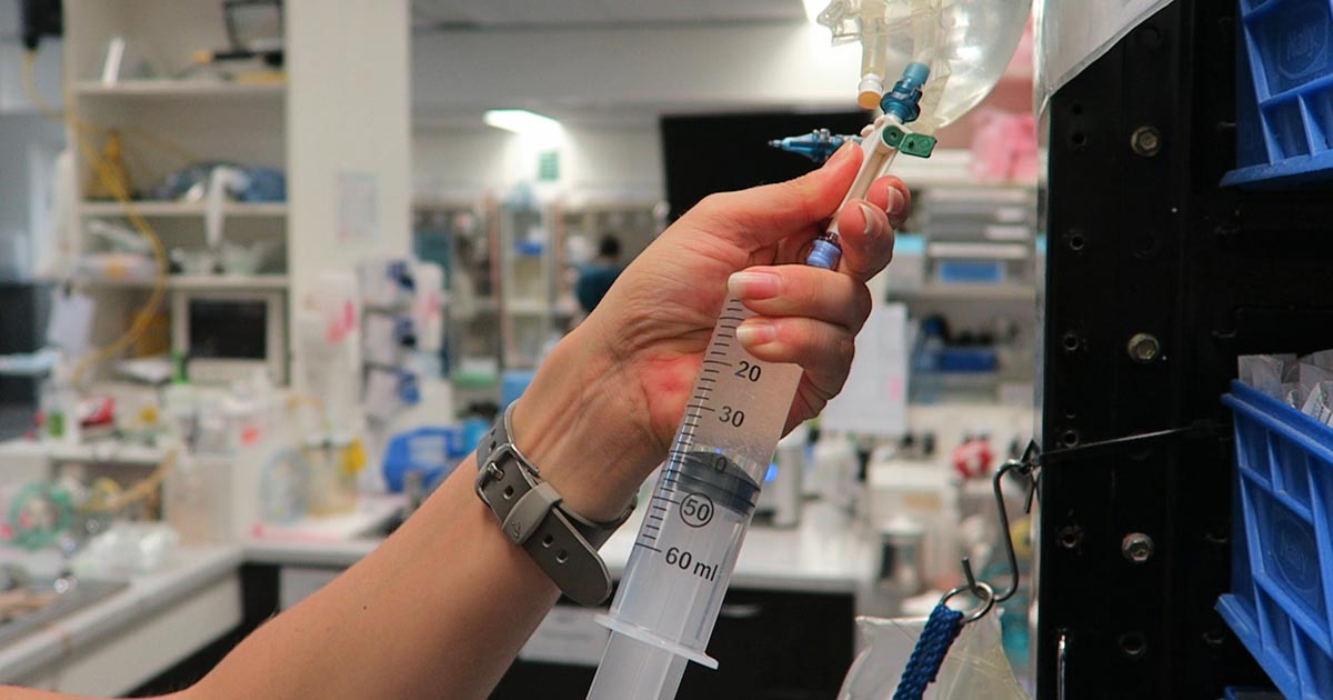
    A while ago we discussed the components of a fluid therapy plan and talked about hydration deficits. This week I want to touch on maintenance fluids.

    Gerardo_IVF
    IV fluids

    Maintenance rates are typically calculated using the following formulae:

    ml/day = 80 × bodyweight (kg)0.75 (cats)
    ml/day = 132 × bodyweight (kg)0.75 (dogs)

    or
    ml/day = 30 × bodyweight (kg) + 70

    These formulae better estimate the needs of smaller and larger patients. The flat 3ml/kg/hr underestimates for small patients and overestimates for larger patients.

    This maintenance rate is in addition to rehydration rates.

    So what sort of fluid should you use for maintenance?

    True “maintenance” crystalloids:

    • used to replace ongoing fluid and electrolyte loss from normal metabolism, not to replace perfusion and hydration deficits or ongoing losses from diarrhoea, for example
    • sodium concentration less than plasma
    • potassium concentrations higher than plasma
    • glucose sometimes added to bring solute concentrations similar to extracellular fluid

    Do you have to use maintenance crystalloids or can you use replacement crystalloids?

    Replacement crystalloids are more frequently used for maintenance fluid therapy rather than maintenance crystalloids. This is because they are more readily available, we are more familiar with their use and effect, and patients are generally continued on these after perfusion and hydration deficits have been corrected.

    In reality, most of the time it doesn’t really matter if we are using replacement crystalloids for maintenance therapy as the patient can manage the excess sodium, but some patients – especially cats – may require potassium supplementation. The key point is regular assessment of the patient’s hydration status and electrolytes – for example, every 12 to 24 hours for patients on IV fluids and not eating.

  • Perfusion deficits and fluid resuscitation: a more in-depth look

    Perfusion deficits and fluid resuscitation: a more in-depth look

    A few weeks ago in the Tip of The Week, we discussed the four basic components of a fluid therapy plan – perfusion deficit, hydration deficit, maintenance requirements and ongoing losses.

    Let’s consider perfusion deficits.

    As an emergency clinician, correcting perfusion deficits is a crucial part of stabilising a patient. So what is a perfusion deficit? It either refers to a real or relative loss of intravascular fluid volume, or low blood pressure, leading to a decrease in perfusion of tissues and, ultimately, decreased oxygen delivery – ie, shock.

    What does this look like clinically?

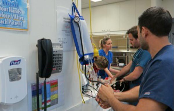
    Pressure infuser
    Operating the pressure infusor at the triage bench.

    Clinical signs of perfusion deficits include:

    • pale gums
    • reduced capillary refill time
    • tachycardia
    • reduced pulse pressure
    • dull mentation
    • cold extremities and low core body temperatures

    A low normal body temperature in a critically ill patient should alert you to the possibility the patient may be experiencing early perfusion deficits, but is not yet severe enough to result in a low body temperature.

    What are these signs caused by?

    These signs are caused by activation of the sympathetic nervous system in response to reduced blood pressure (BP).

    BP is the product of cardiac output (CO) and systemic vascular resistance (SVR):

    BP = CO × SVR

    CO is the product of the heart rate (HR) and stroke volume (SV).

    CO = HR × SV

    Reduced perfusion results in reduced BP. The body increases BP by activating the sympathetic nervous system, resulting in a compensatory increase in HR and SV (beta adrenergic), which increases CO and vasoconstriction (alpha adrenergic) of the peripheral blood vessels to increase SVR and shunt the blood to the heart and brain.

    Peripheral vasoconstriction is seen clinically as pale gums, reduced capillary refill time, cool peripheries and low body temperature as blood is being shunted away from those peripheral capillary beds. It also results in reduced pulse pressures.

    In summary, clinical signs of perfusion deficits are signs of the body trying to compensate and push blood to where it is needed most.

    Blood pressure is normal, it can’t be in shock?

    Since the body’s compensatory mechanisms are geared towards preserving blood pressure, it often remains normal in patients with shock until the body cannot compensate any longer – decompensated shock.

    So, to me, normotension does not rule out perfusion deficits and hypotension is an indicator of severe perfusion deficits.

    Are IV fluid boluses safe for all patients in shock?

    Hartmanns fluid bolus
    Preparing a Hartmann’s fluid bolus.

    The vast majority of the time I would say yes, but you must ask one question – could this patient be in cardiogenic shock? IV bolus therapy would be contraindicated in most of these patients.

    Is it a small-breed dog presenting with a murmur and dyspnoea? Or a large-breed dog with an abnormal heart rhythm? If so then diuretics and anti-arrhythmic medications may be indicated rather then IV fluid boluses.

    How much volume can you give?

    I generally give buffered crystalloid fluid boluses of 10ml/kg over 5-10 minutes. I repeat this while monitoring for resolution of clinical signs of perfusion deficits. If I get to half a blood volume – 45ml/kg in a dog and 30ml/kg in a cat – I ask myself what could be causing this ongoing perfusion deficit. Could it be:

    • Distributive or vasodilatory shock from anaphylaxis or sepsis? In which case, vasopressors – such as adrenalin, dopamine and noradrenalin – are indicated.
    • Cardiogenic shock from dilated cardiomyopathy? Are diuretics, anti-arrthymic medications or positive inotropes required?
    • Restrictive shock from a gastric dilatation-volvulus or pericardial effusion? Is gastric decompression or pericardiocentesis required?
    • Does continued hypovolaemic shock exist? If so then continued fluid therapy is indicated, but this may cause significant haemodilution. Consider colloids or blood products, such as packed red blood cells, whole blood or plasma. Does it need emergency surgery or abdominal compression to stop an internal bleed?

    Perfusion deficits need to be corrected rapidly as continued oxygen delivery to tissues increases the risk of organ dysfunction and, ultimately, organ failure.

  • Tree of life

    Tree of life

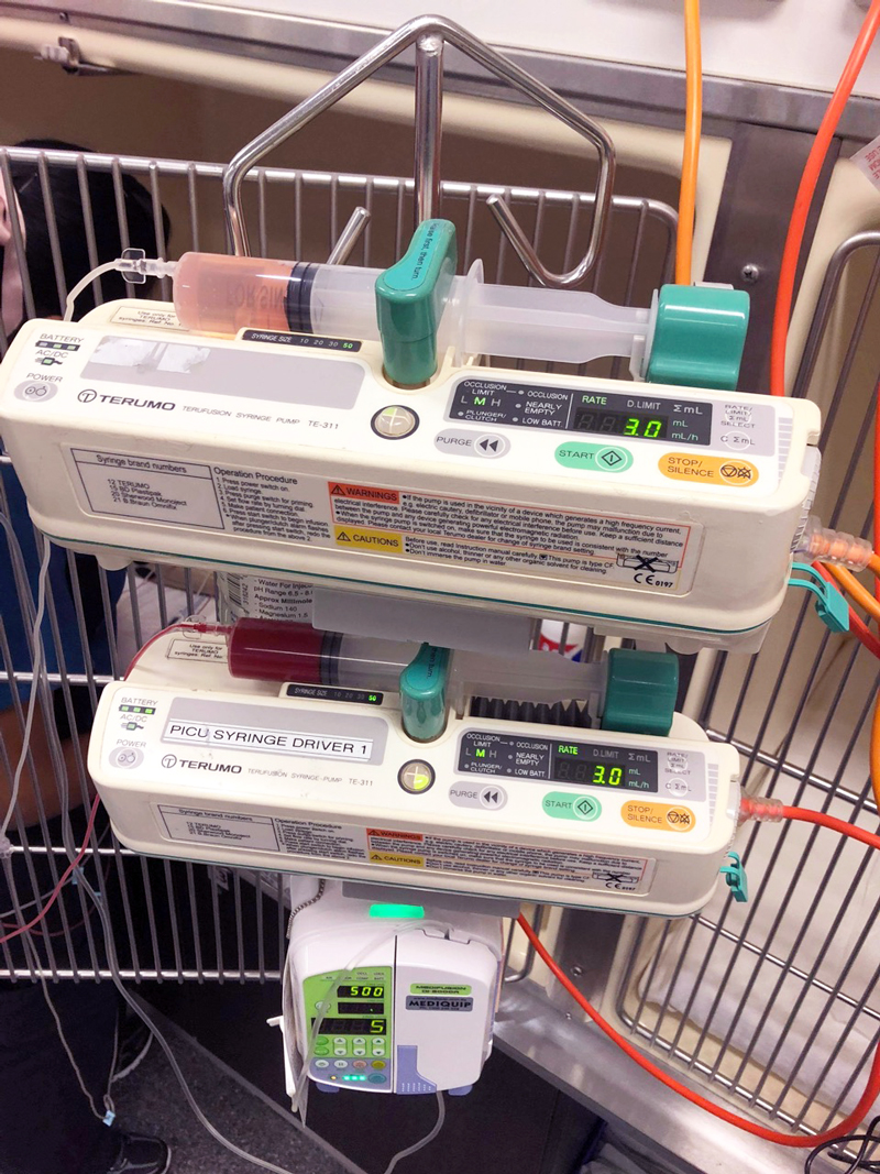
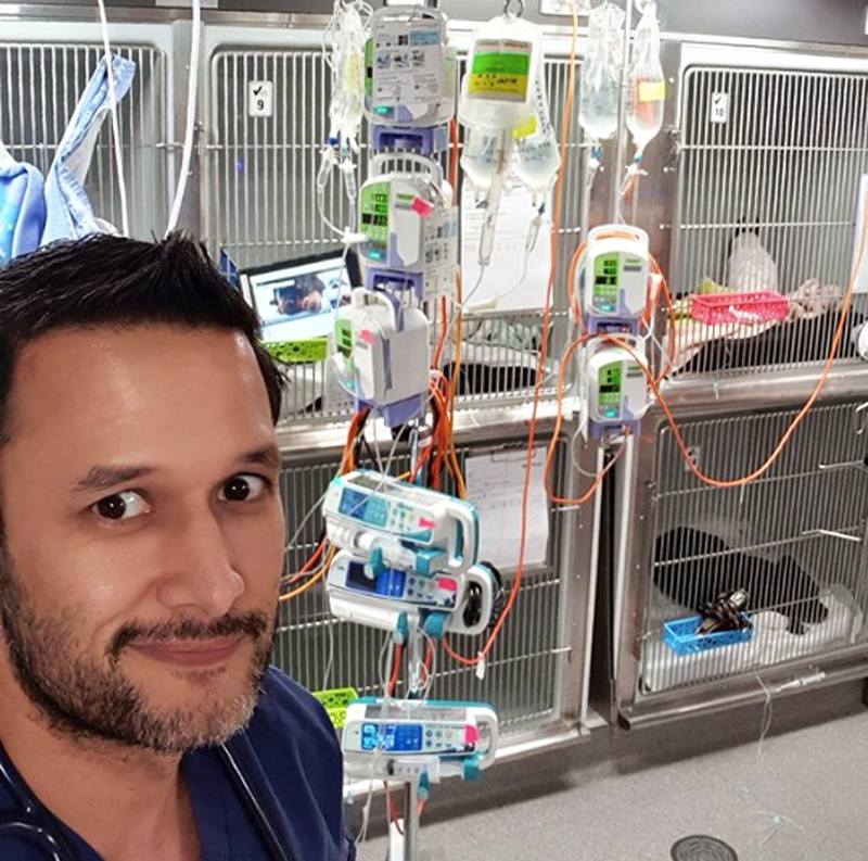
    Seeing a “tree of life” can be an intimidating sight. When you see this number of pumps for one patient, you instantly know things are pretty serious. This sight is almost a daily occurrence in our ICU.

    Now, your first reaction may be to turn around and walk quickly in the other direction. However, rather than feel overwhelmed at the whole sight, think of the saying: “How do you eat an elephant? Piece by piece.”

    Take things one step at a time.

    Step 1: stop and reset

    Gerardo
    The “tree of life” in full effect.

    Take a deep breath and reset. This is important, as these patients are complicated. If you’re not mentally ready for handover then you will miss important details critical for understanding the patient as a whole.

    Step 2: understand the patient

    It is important to know what is going on with the patient as this will provide context to the “tree of life”.

    If you are handing over, make sure it is comprehensive. If you are on the receiving end of a handover make sure you are paying attention, and ask questions or seek clarification.

    Step 3: group the meds

    Grouping the meds helps chunk them. Group them into categories such as pain relief, antibiotics, antiarrhythmics, pressor agents and supplements (such as glucose and potassium). It also puts their use into perspective.

    Step 4: know what the fluid therapy plan is

    This step is often forgotten. Always ask what the fluid status of the patient is. Also, at what phase of the fluid plan they are in – correction of perfusion, rehydration or maintenance. Also, consider if there are any on going losses being accounted for.

    The next time you walk into an ICU and are about to take handover of a patient who has a “tree of life”, it won’t be such a daunting task if you follow this process. I truly acknowledge anyone out there who has learned to see this sight as an exciting challenge, and the chance to learn and make a difference.

  • Handling an Addisonian crisis – part 2

    Handling an Addisonian crisis – part 2

    Managing an Addisonian crisis can be daunting, especially when the patient looks like it is about to check out and its baseline bloods show a sodium of 110mmol/L, a potassium of 8mmol/L and a glucose of 2.3mmol/L. That is enough to make anyone’s brain explode.

    The patient can be treated in many ways, but I find it useful to try to simplify and prioritise. I have outlined my thought process in the hope some of you will find it helpful.

    First 10 minutes: protect heart and manage hypoglycaemia

    • Protect the heart – calcium gluconate 10% 0.5mL/kg to 1.5mL/kg slow IV over 10 minutes to counter the effects of hyperkalaemia on cardiac electrical activity. This buys about 20 minutes of time.
    • Treat the hypoglycaemia – the dose depends on the severity, but 0.5ml/kg of 50% dextrose IV diluted 50:50 with Hartmann’s is a good place to start. This dose of dextrose will also help correct hyperkalaemia by stimulating endogenous insulin release.

    First 20 minutes: start addressing perfusion deficits

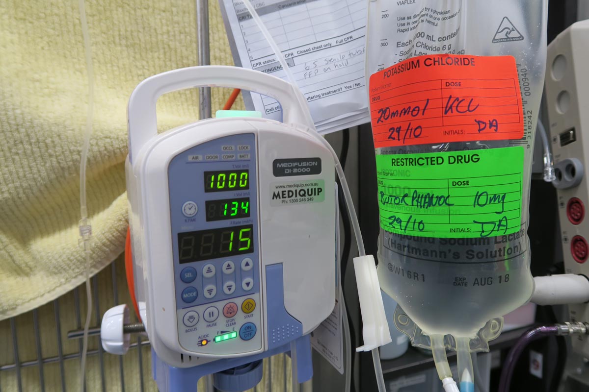
    • Create a custom IV fluid – I do not aim to increase sodium concentration at all at this stage. I am a big fan of creating custom IV fluids. I create a fluid with a same sodium concentration as the patient then use boluses of this fluid to correct signs of shock without concerns of increasing the sodium. I use Hartmann’s as my base fluid – it has the lowest sodium concentration – and add 5% dextrose to reduce the sodium concentration (you may need to remove 100ml to 200ml from the bag first). I usually run the new fluid through the electrolyte machine to check the final sodium concentration.
    • Hartmann’s contains buffers that help address metabolic acidosis (and hyperkalaemia). It also contains potassium; however, if this concentration is less than that of serum it will still help to dilute serum potassium.
    • The formula I use to create a custom sodium IV fluid bag is beyond the scope of this blog and is detailed in the fluid therapy chapter of my book, The MiniVet Guide, under hyponatraemia.

    First hour: address hyperkalaemia

    • Image © mintra / Adobe Stock
      The author warns not to rush the sodium increase in patients. Image © mintra / Adobe Stock

      If the hyperkalaemia is severe enough to warrant more aggressive management than alkalinising IV fluids, improving renal perfusion and providing a dextrose bolus (such as potassium of more than 7mmol/L to 8mmol/L) then I would use regular short acting insulin at 0.25U/kg to 0.5U/kg IV. This should always be used in combination with a bolus of dextrose at 2g of dextrose per unit of insulin or 4ml of 50% dextrose for each unit of insulin, followed by a CRI of 2.5% to 5% glucose until insulin wears off (this could be up to six hours). This should prevent hypoglycaemia.

    • I administer dexamethasone up to 0.5mg/kg IV while running the adrenocorticotropic hormone (ACTH) stimulation test. This is the only corticosteroid that can be given as it does not cross react with the ACTH stimulation test.

    Next 2 to 24 hours: correct hydration and correct hyponatraemia

    • After I have corrected perfusion deficits with my custom IV fluid, I will address hydration deficits with an appropriate fluid plan over the next 24 hours. I usually replace 50% of the hydration deficit over the first 6 hours then the remaining 50% over the following 18 hours.
    • Correction of hyponatraemia can take a couple days as sodium should only be increased by 0.5mmol/L/hr (max 12mmol/L/day). If the sodium has not increased from the initial fluids given, I would create another custom IV fluid bag with a sodium concentration 10mmol/L above that of the patient’s. I would monitor electrolytes every one to four hours, depending on response.

    Supply mineralocorticoids and glucocorticoids

    • Options for steroid supplementation include dexamethasone 0.5mg/kg IV then 0.1mg/kg IV q12hrs or IV hydrocortisone sodium succinate at 0.5mg/kg/hr. Personally I use hydrocortisone CRI, asit has equal mineralocorticoid and glucocorticoid activity. Oral steroids can be used once the patient starts eating and drinking.
    • I only use a mineralocorticoid if I see no increase in sodium after starting hydrocortisone, despite using a fluid with a higher sodium concentration than the patient.

    Addressing patients this way will generally gets them out of the crisis. One thing that I don’t do is rush the sodium increase, it can take time and I am good with that. I have seen patients develop neurological signs from sodium levels that have increased too quickly. As for the long term management; well, I will leave that to you.