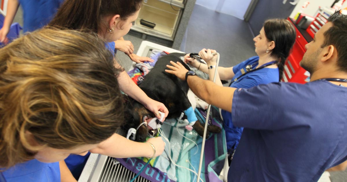
Clients often panic when they think their pet is having a seizure and can skip over vital information.
Often, what an owner describes as a “fit” may actually be syncope, collapse from anaphylaxis or internal haemorrhage (for example, neoplasia), a vestibular event or a behavioural condition.
True seizures
True seizures can be divided into two groups:
Generalised (grand mal) seizures, which involve both cerebral hemispheres and result in loss of consciousness, incontinence and muscle activity.
Focal/partial (petit mal) seizures, which originate from a focal region in the brain. These can also result in alterations in consciousness, but more typically only manifest in the form of repetitive twitching or limb movement.
Once you have established the owner is likely describing a true seizure, there are many important questions to ask to narrow down your differential diagnoses and treatment options.
The important questions
So, as part of a thorough history, always ask:
Was the pet conscious during the episode?
This will help to determine whether the seizure was generalised or focal.
How long did the episode last?
Status epilepticus is when a continuous seizure lasts more than five minutes or when the patient has not recovered fully before another seizure occurs. This can result in severe secondary brain injury.
How many episodes has the pet had in the past?
Epilepsy is the condition of recurrent seizures. This can be further classified as primary and symptomatic epilepsy, with symptomatic being secondary to an underlying cause (such as head trauma or a brain tumour).
How close together were the episodes?
Cluster seizures are when an animal has more than two or three episodes within a 24-hour period.
If a patient presents first time with a cluster, this carries a poorer prognosis in dogs, but has no influence in cats.
Clusters are generally an indication for commencing long-term management.
How was the pet before and after the episode?
Seizures often come with predicting (pre-ictal) and recovery (post-ictal) events.
In the pre-ictal phase, the patient may act strangely (for example, agitated or clingy) and may vomit.
Alterations in consciousness prior to a seizure usually indicate an intracranial cause.
The post-ictal phase can last anywhere between minutes and days, and animals are usually disorientated and/or lethargic. They may also appear blind.
Has the pet demonstrated any other strange activity recently?
For example, if an animal has also been circling to one side, you can start to predict the location of the lesion.
Cats more commonly present with partial seizures compared to generalised – this is seen as stereotypic behaviours and bursts of activity.
Has the pet been exposed to any toxins or chemicals?
Seizures caused by toxins (such as snail bait) generally do not stop and start, but are continuous.
In the next part of this series, we will look at differential diagnoses for seizures and highlight the differences between dogs and cats.
The causes of hyponatraemia can be divided into three major categories, based on serum osmolality. This is further divided based on the patient’s volume status (Table 1).
Most patients we see in clinic fall into the hypovolaemic category, except patients with diabetes mellitus.
Table 1. Causes of hyponatraemia based on osmolality and volume status (from Guillaumin and DiBartola, 2017).
Hypo-osmolar
Hyperosmolar
Normo-osmolar
Hypovolaemic
Normovolaemic
Hypervolaemic
Gastrointestinal fluid loss
Third-space fluid losses
Shock
Hypoadrenocorticism (Addison’s disease)
Renal insufficiency
Excessive diuretic administration
Salt-losing nephropathy
Cerebral salt wasting syndrome
Syndrome of inappropriate antidiuretic hormone secretion (SIADH)
Hypotonic fluid administration
Hypothyroidism
Glucocorticoid insufficiency
Psychogenic polydipsia
Reset osmostat (SIADH type B)
Congestive heart failure
Acute or chronic renal failure
Nephrotic syndrome
Hepatic cirrhosis
Accidental ingestion or injection of water (water intoxication)
Hyperglycaemia
Mannitol
Severe azotaemia
Hyperlipidaemia
Hyperproteinaemia
Common causes
In dogs, the three most common causes of hyponatraemia are:
gastrointestinal (GI) fluid loss
third-space fluid loss
fluid shift from intracellular fluid to extracellular fluid (ECF) as a result of hyperglycaemia
In cats, the three most common causes of hyponatraemia are:
urologic diseases
GI fluid loss
third-space fluid losses
In most patients, more than one pathophysiologic factor is likely to be contributing to the hyponatraemia.
Circulating volume
Hypovolaemic patients – those with, for example, GI losses, hypoadrenocorticism, renal losses and haemorrhagic shock – have a reduced effective circulating volume. ECF contraction triggers antidiuretic hormone (ADH) secretion, which leads to increases in free water absorption and thirst, and results in dilution of the serum sodium concentration. Aldosterone secretion is reduced in hypoadrenocorticism, so an overall reduction in sodium reabsorption compounds the problem.
Hypervolaemic patients are those with an increased fluid retention state, such as:
Congestive heart failure patients have a reduced cardiac output and, therefore, a decreased effective circulating volume, despite the presence of the extra fluid status. Renin-angiotensin activation leads to release of ADH and aldosterone, resulting in sodium and free water reabsorption, and increased thirst. Both lead to an excess of free water retention.
Advanced hepatic (cirrhosis) or renal failure (nephrotic syndrome) both result in hypoalbuminaemia, leading to fluid shifting into the interstitial space and third space, reducing effective circulating volumes. This leads to activation of ADH to increase free water reabsorption, to restore the circulating volume in the face of existing hypervolaemia and hyponatraemia.
Diabetic patients
Moderate to severe hyperglycaemic diabetic patients can be either hyperosmolar or normo-osmolar, depending on the serum blood glucose concentration. Hyponatraemia occurs when water shifts from the intracellular fluid to the ECF down the osmotic gradient, diluting the serum sodium content.
Despite this osmotic shift, not all diabetic patients develop hyponatraemia. Glucosuria also causes also causes a renal osmotic shift, sometimes resulting in urine water loss in excess to sodium. This offsets the hyponatraemia – in some cases, hypernatraemia results.
Treatment
Treatment of hyponatraemia hinges on how quickly it developed and the volume status of the patient. The rule of thumb is to correct hyponatraemia slowly – not exceeding 0.5meq/L/hr – especially in chronic cases, or cases where the duration of hyponatraemia is unknown. Keeping to this rate is paramount until serum sodium concentration reaches 130meq/L.
In acute patients with severe clinical signs, such as seizures, some clinicians may choose to use a higher rate of 1meq/L/hr to 2meq/L/hr until clinical signs resolved.
It should be emphasised, once again, this rate should never be used in chronic patients, patients with an unknown duration of hyponatraemia, or where frequent serum sodium concentration cannot be monitored. The rapid correction of hyponatraemia can lead to osmotic demyelination syndrome (myelinolysis).
Its effect will not be apparent until three or four days after therapy, and can result in neurological abnormalities such as:
weakness
ataxia
dysphagia
paresis
coma
For that reason, frequent electrolyte measurements are required, starting hourly then once a suitable rate of increase has been established and less frequently thereafter.
Part 3 will look at how to correct patients with hyponatraemia.
Reference
Guillaumin J and DiBartola SP (2017). A quick reference on hyponatremia, Veterinary Clinics of North America: Small Animal Practice47(2): 213-217.
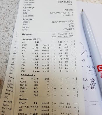
Hyponatraemia is a relatively common electrolyte disturbance encountered in critically ill patients, and the most common sodium disturbance of small animals.
In most cases, this is caused by an increased retention of free water, as opposed to the loss of sodium in excess of water.
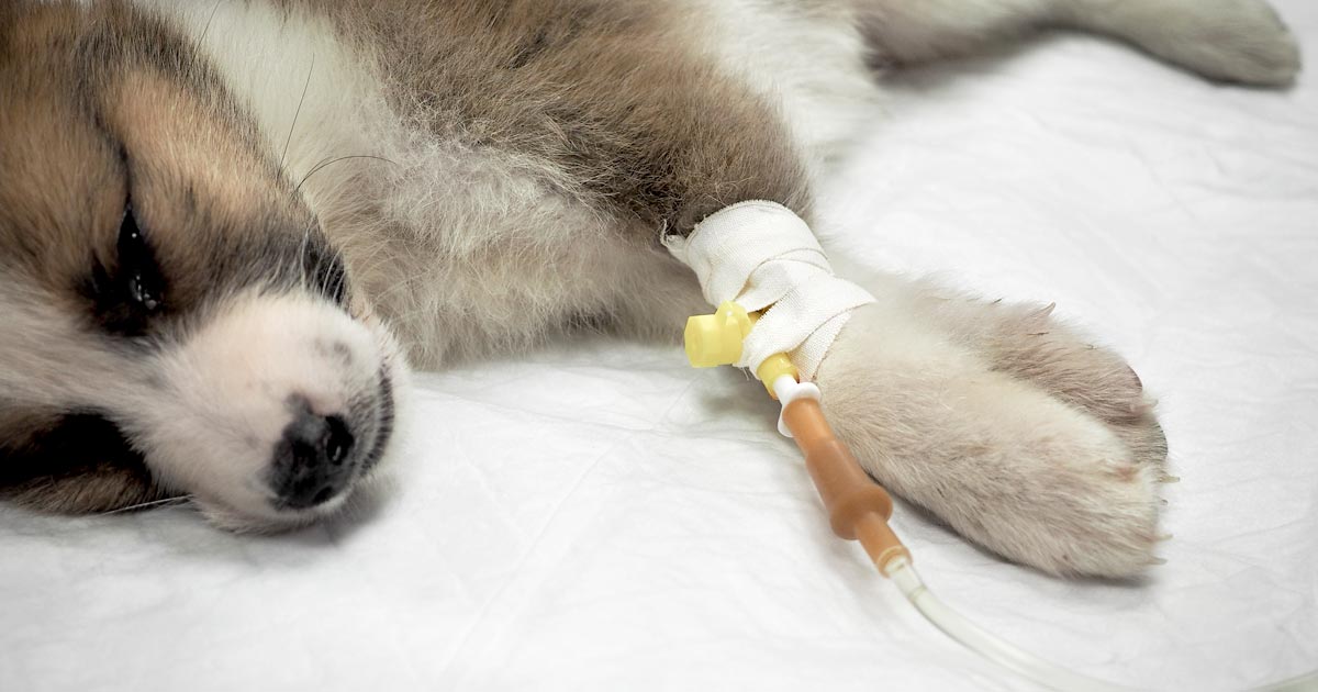
Low serum sodium concentration
Hyponatraemia is defined as serum concentration lower than 140mEq/L in dogs and lower than 149mEq/L in cats.
The serum sodium concentration measured is not the total body sodium content, but the amount of sodium relative to the volume of water in the body. For this reason, patients with hyponatraemia can actually have decreased, increased or normal total body sodium content.
This series will look briefly at the modulators of the sodium and water balance, clinical signs associated with hyponatraemia, the most common causes in small animals, the pathophysiology behind these changes, and treatment and management.
ECF volume
An example of hyponatraemia.
Sodium is the main osmotically active particle in the extracellular fluid (ECF), so is the main determining factor of the ECF volume. Any disease process that alters the patient’s ECF volume will lead to hyponatraemia, such as:
dehydration
polyuria
polydipsia
vomiting
diarrhoea
cardiac diseases
pleural or peritoneal effusion
The modulators of water and sodium balance are also different, so should be thought of as different processes.
Water balance is modulated by thirst and antidiuretic hormone, and the effect of this is to maintain normal serum osmolality and serum sodium concentration.
Modulators of sodium balance aim to maintain normal ECF volume. It adjusts this by altering the amount of renal sodium excretion; an expansion of ECF volume will lead to an increased sodium excretion, while a reduction in ECF volume will lead to increased sodium retention.
Rate and magnitude
The clinical signs of hyponatraemia are both dependent on the magnitude of the decrease and the rate at which it developed.
In mild or chronic patients, no visible clinical signs can exist. In severe (lower than 125mEq/L) and acute cases, clinical signs exhibited are typically neurological, reflecting cerebral oedema. Possibilities include:
lethargy
anorexia
weakness
incoordination
disorientation
seizures
coma
Patients with acute hyponatraemia – for example, water intoxication – are more likely to show clinical signs, compared to those with chronic hyponatraemia, because the brain takes time (at least 24 to 48 hours) to produce idiogenic osmoles, osmotically active molecules that help shift free water out of brain cells.
Therefore, any acute hyponatraemia that develops within a 24 to 48-hour period tend to show clinical signs, whereas chronic cases are less likely.
Next week’s blog will look into the different causes of hyponatraemia and how they result in sodium loss.
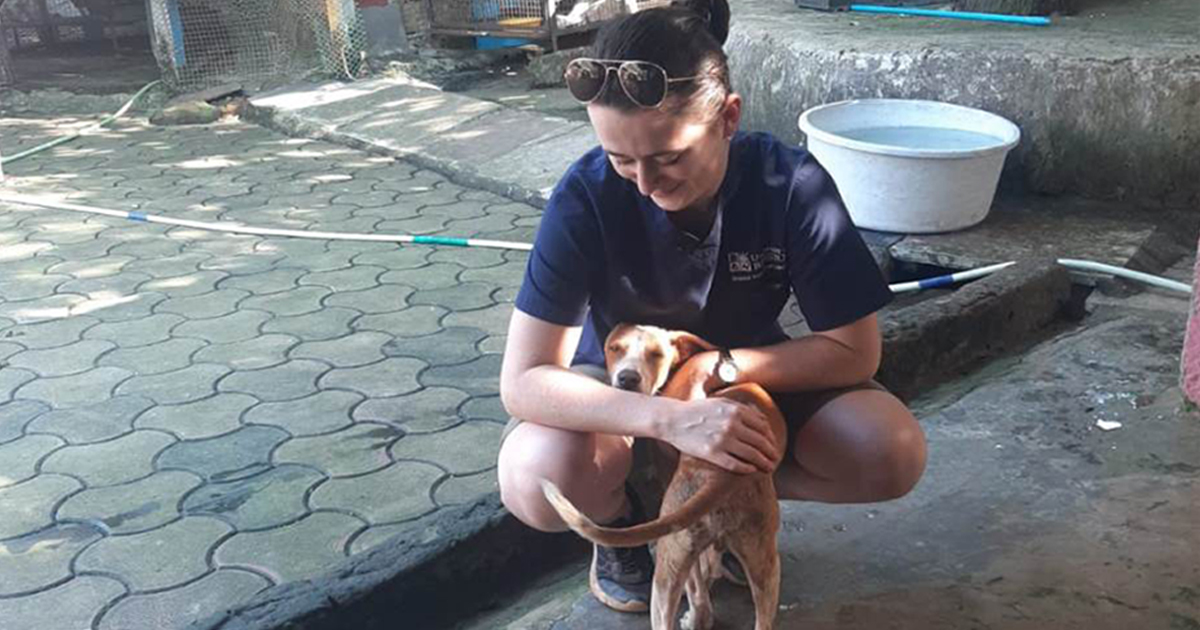
It had been an ambition of mine since the beginning of vet school to do some type of work abroad, whether it be preclinical or clinical, a paid position or volunteer work.
A big reason I undertook an intercalated MSc was for the option it presented for a three-month research period in Western Australia. Sadly, COVID-19 put a stop to that and my research never wandered further than my desk – but, if anything, the pandemic made me feel even more passionate about travelling for my EMS.
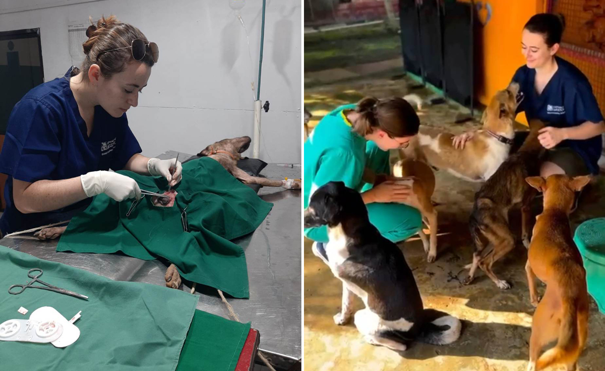
Gone to Goa
Weekends spent “lolling on the beach” were well-deserved, says Eleanor.
A friend and I both settled on a small rescue centre in Goa, India, for the placement (neither of us feeling quite brave enough to go it alone) and despite planning it almost a year in advance, the date caught up with us quite quickly. Before we knew it, we were there.
Let the record show that the motivation for this trip was not to escape from the harsh English January weather, nor to fill up on delicious curries, although the temperature did make a welcome change and I’m unsure a takeaway will ever cut it again.
The whole reason for the placement was to gain the kind of surgical experience that just isn’t readily available to students in the UK.
Understandably, vet practices can take a while to warm up to students enough to trust them to carve into somebody’s beloved animals, but this makes for generation after generation of new grads who feel completely out of depth with a scalpel in their hands.
Great(er than our) expectations
The placement’s main advertising pull had been as an opportunity to gain incredible surgical experience, but we had gone into it with some trepidation that it wasn’t going to be nearly as busy and hands-on as we’d hoped. It turned out to surpass our expectations and go right out the other side…
Weekends spent lolling on the beach were well-deserved after numerous 11-hour shifts with numb fingers and thumbs from uncooperative clamps and needle holders.
The surgical side of the trip deserves an article of its own – but suffice it to say that, between the two of us, my friend and I neutered almost 50 dogs and cats, including 15 unassisted but supervised dog spays. It was an incredible rewarding feeling when each surgery finished, knowing we were doing even just a small bit in the effort to reduce India’s stray population.
Eleanor found her EMS placement in Goa “incredible rewarding”.
Learning valuable lessons
Let it be said, I am not the most confident of travellers, and 18 hours of travel across three planes and four airports are not for the faint of heart, but neither is India – and while I have entirely fallen in love with the country, its beauty and its animals, there was a lot of disorganisation that made my poor little control-freak brain spin.
I think that learning to take each day as it comes, and constantly adapting to new situations or pressures has taught me a lot of valuable skills in a very short space of time.
In particular, the vet who taught and supervised us was invaluable in making the placement such a success. She gave us an incredible amount of patience and taught me skills in both surgery and how to face a stressful situation that I will carry with me throughout my career.
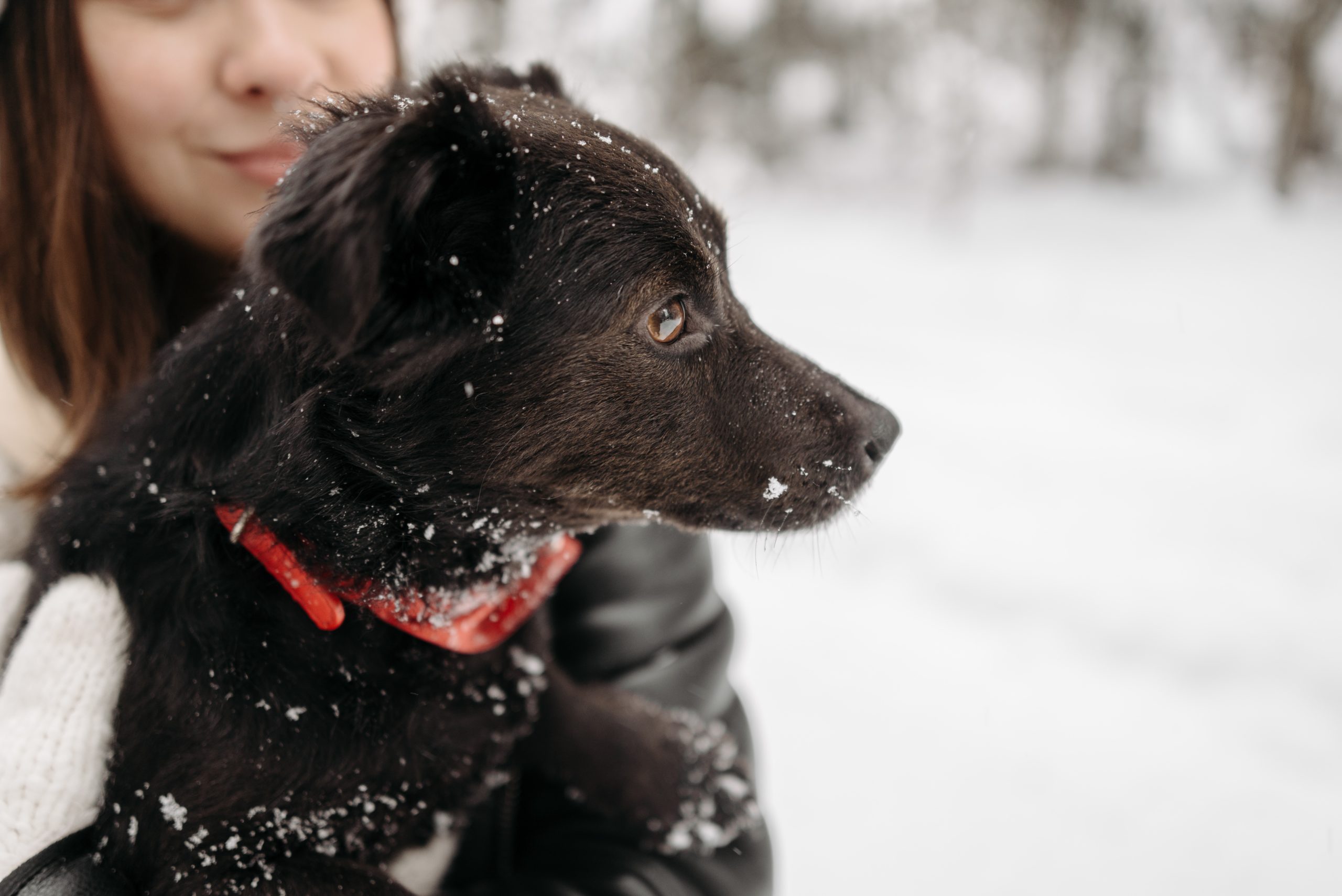
I’ve written at length about the dangers of heat and the sometimes unforeseen risks of walking your dogs during the hazy summer days, but now the cold has well and truly crept in, it is time to consider how we keep our puppies happy and healthy this winter.
Toxins
With the recent snow, we’re used to feeling grit and salt crunching beneath our feet when walking outside – but have you stopped to consider the effect that has on tiny paws?
Irritation from salt can lead to dryness, irritation and cracking of the skin on dog’s footpads, and ingestion of the salt from excessive licking of those paws can lead to toxic levels of sodium in the blood, which can lead to dehydration and even kidney damage.
Another hazardous toxin incredibly prevalent at this time of year is antifreeze. This product contains the chemical ethylene glycol, which can lead to potentially fatal kidney injury if ingested. Clinical signs range from excessive drinking to vomiting and even seizures. So, if you’re concerned your animal may have been exposed, don’t risk waiting – take them straight to your vet.
Cold snap
The cold is perhaps the most obvious danger to our beloved pets at this time of year, but it can be easy to think that their fur coats make them adapted for this kind of weather. The fact is that, aside from certain breeds like huskies, we have actually been breeding the hardiness out of our dogs for more than one hundred years.
Many breeds – especially those imported from descendants in hotter climates – are not cut out for harsh cold. According to the RSPCA, dogs shouldn’t be kept at any lower than 10°C for long periods of time, and studies have found that walking smaller, older or younger dogs (the latter of which naturally run at a higher temperature) can also be dangerous if the thermometer starts to creep towards freezing. You wouldn’t take your granny out in -1°C in just her cardigan, so maybe don’t take your geriatric pets for a walk either.
Of course, animals still need exercise, stimulation and the opportunity to visit nature’s toilet, but it’s important to be sensible and prioritise walks during the warmest times of the day, and to layer up if your pet will tolerate it.
Finally, overfeeding is something I think many of us actually aspire to in the lead up to Christmas – but while it may seem mean to leave our pets out of the festive fun (because they do have very large, cute “feed me” eyes), abstaining from feeding them human treats can actually be the kinder thing.
This isn’t just because of the dangers of chocolate and sultanas (which I’ve gotten up on my soapbox about many a time), but because even a tiny portion of human food can sometimes double the daily calories for our pets.
Depending on whether you have a chihuahua or an active collie, your animal’s dietary needs likely range from around 150 to 600 calories a day. If the average sausage is around 200 calories, you can see how things can quickly add up. Obesity is one of the leading causes of disease in our animals, and it’s so easily preventable.
Spread the love
I would love to urge everybody to spread a little extra festive love towards their furry friends this festive season and keep them healthy and safe.
Christmas is a great time for family gatherings, but this does not necessarily mean it is a great time for pets.
In fact, it can often be the opposite, with veterinary clinics seeing a major increase in patient numbers that come through the door.
One common emergency we see at the emergency hospital during the festive season is dog fight and bite wounds. As vets, we have a duty of care to educate pet owners during this time, so they – and their pet – have the best Christmas possible and do not end up in the emergency room.
Why do dogs fight and bite at Christmas?
Usually during the festive period, family or friends increasingly gather to celebrate. Whether it is people coming into their home, or them being taken to someone’s home, this can be confusing and cause anxiety levels to rise.
When a family member or friend brings a new pet into the house with an existing pet, it creates competition for food, space, affection and attention – and this can lead to dog fights. Even usually mild-mannered pets can easily feel threatened by a new pet entering their territory, and may lash out.
Increases in noise, people, decorations and general chaos during the holiday season can cause stress and anxiety. For dogs protective of their domain and the people in it, this can be a difficult and uncertain time.
Children not used to pets, and pets not used to young children, can also be a dangerous combination. Dog bites are a common injury sustained by children during the festive period and it could often be avoided.
Solutions
Although dogs are part of the family, it is important owners understand leaving their dog at home when they go to a festive gathering is not leaving them out, but protecting them and making sure they are more safe, comfortable and happy.
If hosting a party, owners can shut their dog in another room away from the chaos and noise – they will be grateful to have a peaceful space. This is a must for a dog already prone to stress.
Children and dogs should not be left alone and should be monitored at all times. If the dog starts to show signs of anxiety and stress, it should be taken somewhere it feels comfortable and calm.
Owners can take their dog to their vet for a behavior assessment. Anti-anxiety medications could be considered in extreme cases, but this would be a last resort.
Communicating messages
We can educate pet owners in the lead-up to Christmas in many ways. We can offer thoughtful, engaging and informative advice and guidance.
Some ways to communicate festive dangers to pet owners include:
infographics
videos
social media posts
posters in the hospital or clinic
blogs
email campaigns
discussing the dangers at check-ups and appointments
newsletters
flyers
special calls to clients with an anxious pet
education events, such as how to manage pets and children
Either way, as a student with a passion for both fitness and animals, I was initially intrigued. But I can’t help but have concerns for whether this practice is beneficial for all members of the class.
Five freedoms
Usually applied to the context of captive animals, the five freedoms can really be utilised to evaluate the welfare of any animal outside of its natural habitat (which, technically, every dog is).
These being freedom from pain and disease, stress, discomfort and hunger, as well as freedom to express normal behaviour.
My main concerns when it comes to puppy yoga would be stress, hunger and disease.
If classes run back to back, younger animals that require more frequent feeds may miss out on vital mealtimes, and there’s always the worry some puppies included in these classes are too young to be removed from their mothers. Ideally, no puppy should be removed from the dam or weaned before eight weeks of age. In larger breeds, puppies can appear older than they really are, and some breeders or yoga studios may be motivated by profits to use pups that are slightly shy of this age limit.
The danger here is that puppies don’t typically receive vaccinations until they’re eight weeks old, and if puppies from different litters are introduced when their mother-derived immunity is lowering, diseases can be transmitted very quickly. Most vets wouldn’t advise mixing a puppy with other dogs until at least two weeks after its second vaccines (at around 12 weeks old) to allow adequate immunity to develop.
In regard to stress, anything new or novel can be stressful to a puppy (or any animal for that matter). Loud noises, strange smells and lots of new people all at once can also be very overwhelming and scary to puppies that are yet to be properly socialised.
Socialisation
The socialisation window for puppies is from when they are roughly one to three months old. During this time, the animal’s perceptions of the outside world and its stimuli are being shaped by its experiences, and once that window closes, it can be more difficult for biases towards certain stimuli to be changed.
Since the majority of puppies used in yoga sessions are between two to four months old, on paper, the practice sounds like an excellent opportunity for animal lovers to exercise and unwind surrounded by adorable puppies, with the added benefit of those puppies being socialised to grow up more well-rounded and well-behaved pets.
Unfortunately, however, socialisation is not an exact science, and while it is incredibly beneficial to introduce puppies to lots of different things during their socialisation window, it does not mean flooding them with lots of stimulus all at once.
This is the really tricky part, because what counts as “overwhelming” to one puppy may be completely manageable to the next. Some animals may find a room full of new people and smells incredibly exciting, while others need to be introduced to new people one at a time, with plenty of opportunity to withdraw from the experience if needed.
It can also be impossible to predict what type of puppy you have until you place it in that situation. While a lot of behavioural aspects in our pets can be traced back to environment and genetics, every animal is unique, and just because a litter comes from docile, friendly and outgoing parents, doesn’t mean the offspring will share the same traits.
Ensuring every puppy’s experience of a yoga session will be adequate from a welfare perspective would take a very knowledgeable and conscientious screening process that some businesses may not know how to or be able to provide.
Yoga “pants”
I feel that puppy yoga is probably far from a black and white picture, with the level of puppy welfare and attention to their needs varying from practice to practice. For this reason, I think it’s definitely a good idea to do your research before booking a session – whether you’re a vet or not – to make sure you’re happy with where the puppies come from, if the establishment is aware of vaccine records (and so forth), and if the puppies are given adequate opportunity to rest and retreat from engaging with the customers if they wish.
In the same way that in the veterinary profession we are now seeing the outcomes of puppies raised during the pandemic lockdowns, we may soon see the influence of puppy yoga in the next generation of pets.
At the end of the day, it’s up to the individual consumer to decide if the practice is for them, or if ethical puppy yoga is a bit of a stretch…
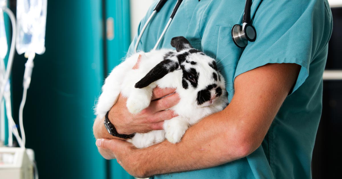
According to PDSA [PDSA Animal Wellbeing (PAW) Report 2022], rabbits are the third most popular pet in the UK behind dogs and cats. With an estimated 1.1 million pet rabbits in the country, that’s about about a tenth of the population of pet dogs and cats, which hover around the 9 to 10 million mark.
So, if the pet ratio of dogs/cats:rabbits is 10:1, why isn’t this reflected in our teaching? Despite rabbit populations being endemic to the UK for more than a thousand years, they always seem to get lumped with guinea pigs and the cold-blooded pets like lizards and corn snakes when it comes to textbooks or university curriculums.
I can confidently say my education on rabbit physiology and medicine has been dramatically less than 10% of what I’ve received for small animal medicine. Perhaps this is why many vets, especially new or recent graduates, feel more confident handing off any rabbit patients to the resident “expert” of the practice or even referring to an exotics specialist, rather than seeing it themselves.
Accessibility
It’s a sad truth that the less convenient education and health care are to access, the less people will reach for them. By extension, the less veterinary practices that advertise care for rabbits (and other exotics), the less rabbits are likely to be registered at a practice and receive regular preventive care.
For example, as a native to the land of Kent, I only know of two or three practices that would call themselves “exotic specialists” and I know that, for a lot of rabbit owners, traveling half way across the county to visit one of these few practices would not be practical or plausible. Perhaps this is why, according to PDSA reports, at least 11% of pet rabbits receive no preventive health care, including vaccinations.
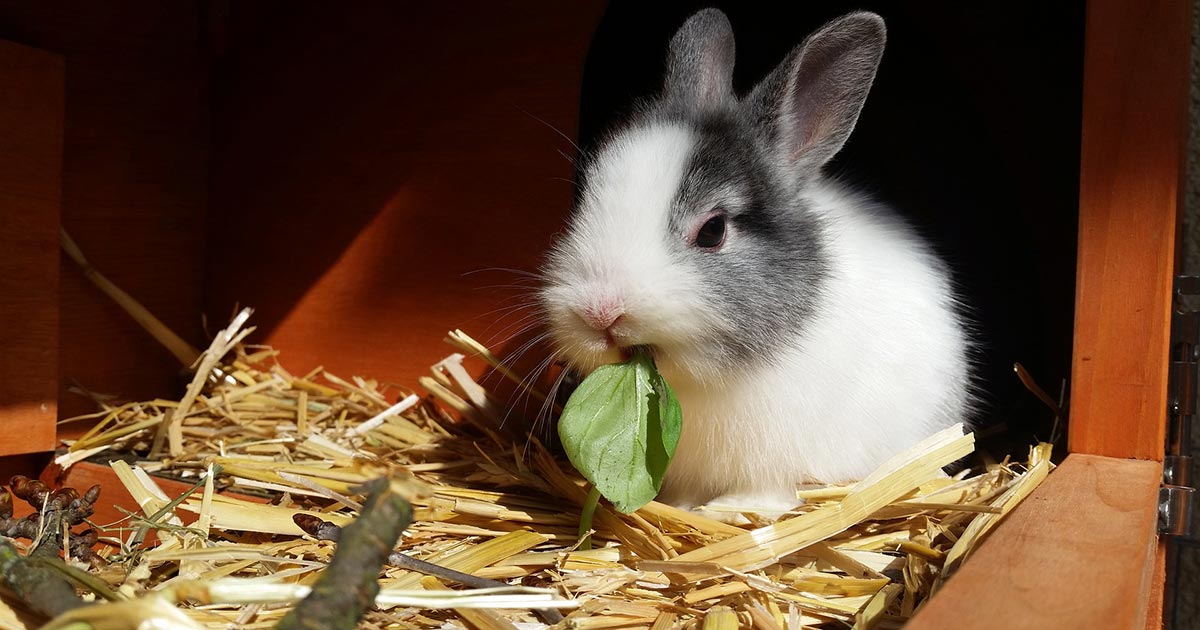
Rabbits are a social species that has evolved to live in groups, not alone.
Education
The value of a veterinary consultation is not simply to talk through clinical signs or address a flea outbreak in the home, it’s a chance for owners to discuss management issues or to ask for general advice. When rabbits aren’t brought in for routine consultations, then discussions about their diet, husbandry and behavioural needs don’t get to be had.
Some vets are already worried that the development of an annual rabbit haemorrhagic disease (RHD) booster rather than biannual is going to dramatically reduce rabbit welfare by halving the number of times these pets receive a clinical exam.
Welfare
Of course, like all “exotics”, there’s the argument to be made as to whether these animals are suitable pets in the first place. Personally, I feel that this is a moot point for the time being.
The fact that more than 50% of pet rabbits are housed by themselves with no companionship speaks volumes about the lack of knowledge the general public possesses on how to care for these animals. However, with more than a million of them currently out there, they’re not going away anytime soon.
The best we can do as professionals is educate our clients so welfare can be maximised as much as possible… and that starts with educating ourselves. I hope that in the near future the landscape of the veterinary degree can shift to better reflect the current demand for exotic vets – or at least rabbit vets.
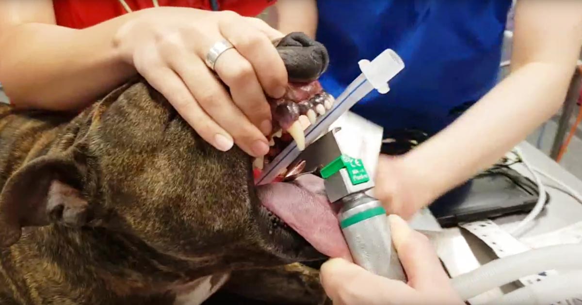
This patient was brought to us for exercise intolerance, breathing difficulty and loud airway sounds.
The patient has laryngeal paralysis. This is where the muscles controlling the arytenoids cartilages do not work and leads to failure of opening of the arytenoids during inspiration.
Most commonly seen in middle-aged large breed dogs, it can occur acutely, but more often it is a chronic problem exacerbated by heat or stress. The cause is often unknown, but it can be caused by trauma or lesion to the cervical region or some kind of neuropathy, such as myasthenia gravis or tick paralysis. Diagnosis is based on visualisation of the arytenoid cartilages failing to abduct during inspiration under light anaesthesia.
Treatment
The management of the acute presentations include oxygen and sedation (butorphanol) to improve airway dynamics – with or without active cooling triggered by heat and with or without anti-inflammatories (dexamethasone) to reduce swelling secondary to airway turbulence.
Patients in severe respiratory distress, anaesthesia and intubation may be required for a short period. Long-term management involves either surgery, such as laryngeal tieback, or conservative management strategies that involve weight loss, avoiding exercise and being kept in a cool environment.
Idiopathic acute haemorrhagic diarrhoea syndrome (AHDS) – previously known as haemorrhagic gastroenteritis – remains the one disease where constant debate exists as to whether antibiotics should be used as part of the standard treatment.
The logic behind using antibiotics to prevent bacterial translocation is sound, and if AHDS is truly initiated by Clostridium species or their toxins then the use of antibiotics can be justified.
However, no knowledge exists of the true frequency of bacterial translocation in AHDS patients and conflicting evidence supports Clostridium being the initiating cause of AHDS in dogs.
Together with new data indicating the use of antibiotic therapy in aseptic AHDS patients did not change the case outcome or time to recovery, the benefit of using antibiotics must be weighed against the very real risk of selection of antibiotic resistance and other complications associated with inappropriate antibiotic use.
In this blog, we will explore the evidence against the use of antibiotics in AHDS.
Cause unknown
AHDS is characterised by an acute onset of vomiting (of less than three days’ duration) that can quickly progress to haemetamesis, and severe and malodorous haemorrhagic diarrhoea, accompanied by marked haemoconcentration that can be fatal if left untreated.
AHDS is a diagnosis of exclusion; other diseases (such as canine parvoviral enteritis, thrombocytopenia, hypoadrenocorticism, azotaemia, hepatopathy, neoplasia, intussusception, intestinal foreign body and intestinal parasitism) must be ruled out by a combination of medical history, vaccination status, complete blood count, serum biochemistry, coagulation times, diagnostic imaging and faecal testing.
Small breed dogs – in particular, the Yorkshire terrier, miniature pinscher, miniature schnauzer and Maltese – have been found to be particularly predisposed. On average, the affected dogs were young (a median of five years old).
The cause of AHDS is still unknown. Clostridium perfringens and its toxin has been incriminated as being the initiating cause; however, conflicting studies have refuted this claim. It is also difficult to determine whether overgrowth of Clostridium speciesis primary or secondary to the intestinal injury.
Virus theory
Another theory is viruses may have a role in AHDS’ aetiology. At this stage, only single agents had been investigated. It is possible a novel agent not yet been tested is the cause of this syndrome, or possibly the syndrome is the result of a very complex interaction between multiple organisms or their toxins.
For the aforementioned reason, no indication exists for the use of antibiotics to treat for the underlying cause.
Another argument behind the use of antibiotics lies in the fact most idiopathic AHDS patients have several risk factors for bacteraemia.
Necrosis of intestinal mucosa, leading to the disruption of the gastrointestinal mucosa-blood barrier; adherence of significant numbers of bacteria to the necrotic mucosal surfaces that increases the risk of bacterial translocation; significant hypoalbuminaemia indicating substantial loss of mucosal epithelial layer; splanchnic and intestinal hypoperfusion, leading to reduced intestinal barrier function; and microbial dysbiosis all contribute to an increased risk of bacterial translocation.
Although bacterial translocation has the potential to lead to sepsis, the true incidence of bacterial translocation needs to be established in idiopathic AHDS patients, as well as their influence on the outcome of the patients.
Antibiotic requirement
Use of unnecessary antibiotics not only disrupts the protective mechanisms of a normal intestinal microflora, but also the real risk of post-antibiotic salmonellosis and Clostridium difficile-associated diarrhoea.
Multiple studies have suggested antibiotics are not required in the treatment of aseptic idiopathic AHDS patients.
In a prospective study of bacteraemia in AHDS dogs by Unterer et al (2015), the incidence of bacteraemia of patients with idiopathic AHDS was 11%, compared to those of healthy controls, where it was 14%.
Transient bacterial translocation to mesenteric lymph nodes occurred in 52% of dogs undergoing elective ovariohysterectomy (Dahlinger et al, 1997), and confirmed in studies by others (Harari et al, 1993; Howe et al, 1999; Winkler et al, 2003), where portal and systemic bacteraemia was reported in clinically normal dogs.
As long as the immune system is competent, and the functional capacity of the hepatic reticuloendothelial system is not overwhelmed, the body is usually effective at eliminating organisms from the blood.
This is reflected in the Unterer et al (2015) study result, where – regardless of the bacteraemia status – all idiopathic AHDS dogs survived to discharge.
In another prospective, placebo-controlled, blind study by Unterer et al (2011), idiopathic AHDS patients were either treated with amoxicillin/clavulanic acid for six days or a placebo, and no significant difference occurred between the treatment groups concerning mortality rate, duration of hospitalisation or severity of clinical signs.
They concluded, without the evidence of sepsis, antibiotics do not appear to change the case outcome or shorten the time to recovery.
Negative impacts
The negative impacts of inappropriate antibiotic use are undeniable – especially in a time where resistance has become a worldwide public health concern.
Use of unnecessary antibiotics not only disrupts the protective mechanisms of a normal intestinal microflora, but also the real risk of post-antibiotic salmonellosis and Clostridium difficile-associated diarrhoea.
With evidence all pointing against the use of antibiotics as routine treatment of aseptic idiopathic AHDS, next time you are about to reach for antibiotics, I urge you to reconsider. Although it has taken some time to adopt and requires clear communication with clients, all vets should feel comfortable not using antibiotics for AHDS patients.


 Depending on whether you have a chihuahua or an active collie, your animal’s dietary needs likely range from around 150 to 600 calories a day. If the average sausage is around 200 calories, you can see how things can quickly add up. Obesity is one of the leading causes of disease in our animals, and it’s so easily preventable.