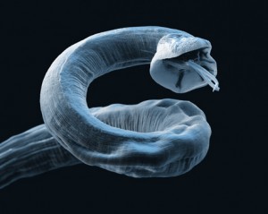
Tape cytology from dog with Malassezia dermatitis (Dif-Quik stain) – note the “peanut-shaped guys”. Image: Wikimedia Commons
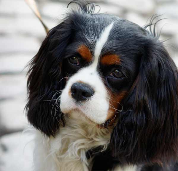
Ever had one of those cases, which seem to typically occur around this time of year, that you think must be the start of an allergic dermatitis?
These present with pruritus, erythema and sometimes a yellowish/grey, greasy feel to the skin and hair coat.
The dog is already on a regular POM-V broad-spectrum antiparasiticide.
Initial thoughts
Pyoderma immediately springs to mind – it’s 6.55pm on a Friday, you skip the cytology and start on an appropriate antibiotic; maybe even a short course of prednisolone.
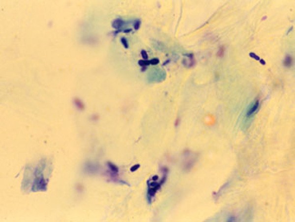
A week later and the dog has not really improved, so it’s a great time for some cytology. There is a good chance we have got a Malassezia dermatitis, and hey presto – the peanut-shaped guys are visible on microscopy. A couple of Malaseb shampoos later and we are rocking.
Then the fun really starts as we try to determine the underlying cause…
Have you ever had a puppy that just presents with lethargy, exercise intolerance and sleeps all the time?
This is normal for my teenage daughters, but not so for a young Lhasa apso that presented to my surgery. Physical exam was unremarkable, but the dog was so sleepy we administered IV fluids to perk it up.
Routine biochemistry revealed a low blood urea nitrogen (BUN), and a urine sample demonstrated the presence of urate crystals.
This triggered a request for a bile acid stimulation test, which showed markedly abnormal elevation postprandial levels.
Our diagnosis of hepatic portosystemic shunt was confirmed at Davies Veterinary Specialists and luckily this was shown to be extra hepatic. So, after some very smart surgery, the dog went on to live an energetic life.
As little as 50 grams (1.8 oz) of general chocolate can be enough to poison a small dog, but the concentration of theobromine in dark chocolates (approximately 10g/kg) is up to 10 times that of milk chocolate. (source: Wikipedia)
Chocolate is digested much more slowly by dogs than people. Therefore symptoms may not appear for many hours after the chocolate is eaten.
Do not be fooled by this into thinking that everything is okay. The earlier chocolate poisoning is treated the more likely you are to save the dog’s life.
No antidote
In addition, the very slow deactivation of theobromine by dogs means that the effects of chocolate poisoning can be very prolonged – up to three days, so the dog may need to be hospitalised throughout this time.
Theobromine has no specific antidote – cases are treated symptomatically. The prognosis depends on how much chocolate or cocoa powder the dog has eaten, and how long prior to being seen by the vet that the dog ate it.
Don’t delay
Up to 50% of dogs will die if treatment is delayed until severe, persistent vomiting has developed. If seizures have begun then an even higher proportion of dogs will die.
Treated early enough, except for dogs that have consumed very large quantities of chocolate or cocoa powder, the outlook is generally quite good. Recovered dogs show no long term ill effects from the poisoning.
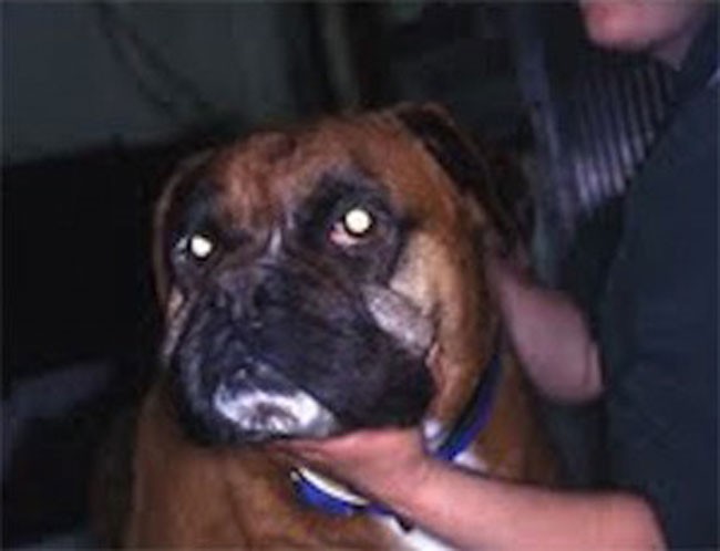
Dog with hypothyroidism presenting with myxoedema. There is facial oedema and the typical tragic facial expression. Image Jane Coatesworth / AHT.
Myxoedema most commonly occurs in moderate to severe cases of hypothyroidism in dogs.
Thickening of the skin occurs secondary to accumulation of glycosaminoglycans (mostly hyaluronic acid) in the dermis.
Myxoedema is most common on the forehead and face, causing a puffy appearance and thickened skin folds above the eyes. The puffiness, plus slight drooping of the upper eyelid, gives some dogs a “tragic” facial expression.
These changes also have been found in the GI tract, heart and skeletal muscles.
Myxoedema coma, a rare syndrome, is the extreme expression of severe hypothyroidism. The course can develop rapidly; lethargy progresses to stupor and coma. The common signs of hypothyroidism (eg hair loss) are present, but other signs, such as hypoventilation, hypotension, bradycardia and profound hypothermia, are usually seen as well.
It is advisable to always spay a bitch having a mastectomy.
Approximately 50% of malignant mammary tumours in the dog have receptors for either oestrogen or progesterone. This means the presence of these female hormones promotes the growth of these tumours.
Benign tumours also have female hormone receptors and can also be stimulated by hormonal cycling of the female dog. This means spaying is important, even if a tumour has already developed.
In one study, bitches spayed at the time of mammary tumour removal (or two years prior) lived 45% longer than those that remained unspayed.
Resolution of the hypovolaemia is the primary concern. Two large bore catheters are placed in the cephalic veins. If the cephalic veins are not available, the jugular vein is used. Fluid resuscitation through the saphenous veins is unlikely to be successful because of the caudal vena caval obstruction.
Photo of an x-ray showing gastric dilatation and volvulus in a large mixed-breed dog. The large dark area is the gas trapped in the stomach. The pylorus and duodenum are in an abnormal position cranial to the stomach and are separated by a fold in the stomach, creating a “double bubble” appearance.
Image by Joel Mills [CC-BY-SA-3.0], via Wikimedia Commons.Either isotonic crystalloids (90ml/kg in the first hour) or hypertonic saline (7% NaCl in 6% Dextran: 5 ml/kg given over five minutes) followed by crystalloid are administered.
Controversial treatments
The use of corticosteroids remains controversial. They have many theoretical benefits but have not been unequivocally demonstrated to improve survival in cases of gastric dilatation-volvulus (GDV).
Prophylactic antibiotics are also somewhat controversial, but rational arguments are made for their use. GDV dogs do have increased levels of circulating endotoxin, perhaps indicating increased GI mucosal permeability. Poor perfusion to the liver could inhibit reticuloendothelial function.
Improving tissue perfusion by fluid resuscitation and subsequent gastric decompression and de-rotation can potentially result in the production of damaging, highly reactive oxygen free radicals. These radicals can cause significant reperfusion injury that may be as damaging as the initial hypoperfusion episode.
It is possible treatment to prevent free radical generation may be beneficial in dogs with GDV. Of the drugs trialled in experimental models, deferoxamine, an iron chelator, shows the most promise for clinical application.
Gastric decompression
Ideally, a continuous electrocardiogram is connected. Once the animal has been stabilised, gastric decompression is attempted using a silicone or rubber tube. The tube is pre-measured to the level of the stomach and marked. A 2in roll of tape is placed in the dog’s mouth and the tube passed through the tape and slowly into the oesophagus and stomach.
If resistance is encountered at the level of the cranial oesophageal sphincter, the tube must not be forced, as this could cause rupture of the caudal oesophagus.
In some fractious animals, sedation and intubation is necessary for gastric decompression. If orogastric intubation is unsuccessful, the stomach is decompressed by trocarization. The abdomen is carefully palpated, and the enlarged spleen is avoided. A large gauge catheter (10-12F) is placed into the stomach percutaneously to relieve pressure.
Syringomyelia is rare in most dog breeds but has become widespread in cavalier King Charles spaniels.
Syringomyelia is a condition where fluid filled cavities develop within the spinal cord. It is sometimes known as “neck scratcher’s disease” because scratching in the air near the neck is a common sign.
Owners often report their dog is worse at night, when first getting up, during hot or cold temperature extremes, when excited, or related to posture (e.g. preferring to sleep with their head elevated).
Affected animals may seem overly sensitive to touch, or to scratch more on one side of the neck, ear, shoulder or sternum. This is typically one side only, often while the dog is moving and sometimes without making skin contact. Some dogs, especially younger patients, develop a scoliosis.
Some severe cases may have other neurological deficits such as fore and hind limb limb weakness and ataxia.
Facial nerve paralysis, deafness and seizures have also been associated with the condition, but a link has yet to be proven.
Angiostrongylus vasorum is the most common cause of haemorrhagic cerebrovascular accidents in dogs.
Bleeding in the brain or spinal cord can cause neurological symptoms. Craniotentorial bleeding can cause epileptic seizures, paresis and abnormal postural reactions.
In cases of cerebellar bleeding, hypermetria, vestibular symptoms and opisthotonus are observed, and if the brainstem is affected, abnormalities of the cranial nerves can be seen.
Inflammation, hypoxia and parasitic emboli may cause neurological signs too.
It is worth treating any dog with a suspected stroke for A vasorum with Advocate (moxidectin and imidacloprid).
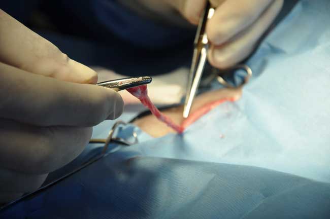
Last Saturday I had a “walk in” – a very cute spaniel that was limping slightly and had a cut pad. The owner thought she might have trodden on something.
A good palpation didn’t seem to suggest the presence of any foreign body and the dog was very stoical. My previous experiences suggest any foreign material produces a lot of pain that is exacerbated by palpation – sometimes with dramatic effect!
However, the insistence of the owner made me look closer and I could just see a glint of a firm object deeply embedded in the pad. A pair of rat toothed forceps later and I extracted a 5mm slither of glass from the pad.
The result was a happy dog and owner, but oh – I could so easily have sent that poor dog away!
The homeless come with a certain stigma – particularly those with pets at their side.
Should we be concerned for the welfare of those animals, whose owners cannot afford to feed themselves so surely cannot adequately care for a companion?
Of course we should.
However, instead of claiming these pets should be removed from their owners, Ruby Shorrock (a fourth year vet student at the University of Glasgow) took a different approach.
Being homeless can be extremely isolating and lonely. For some of these people, their dog is their only companion, and can often be the only thing keeping them going. A dog can also provide a connection to home, and so the reluctance to give them up is understandable.
Despite this, many shelters refuse to accommodate dogs and so the help available can become increasingly restricted for homeless dog owners.
In light of this, Ruby founded Trusty Paws, a non-profit organisation that hosts free clinics and provides preventative care for hounds belonging to the homeless. The clinics involve a free health check (a clinical examination performed by veterinary students, supervised by a qualified vet), microchipping, flea and worming treatment and vaccinations. Dog food packages and other supplies such as leads and dog coats are also given out at the clinics.
Trusty Paws: a vaccination clinic for dogs belonging to the homeless, run by fourth year vet students at the University of Glasgow.
There have been three Trusty Paws clinics in Glasgow so far, with several grateful clients being able to benefit from the supplies donated and the services provided by the students. Everyone involved is delighted with how the clinics have been received.
Plans for 2015 include registering as a formal charity and organising public fundraisers. The Trusty Paws team also intends to tackle the problem of local shelters and hostels not allowing dogs.
Trusty Paws relies entirely on donations and sponsorship and the response to requests for both has been exceptional. The concept has really taken off and looks to gain popularity and success in the future.
The work of Trusty Paws is a fantastic way of not only actively ensuring quality care for homeless pets, but also raising awareness within the community to tackle public perception. If these misconceptions can be eliminated, others will be willing to accept that pets are a huge part of the lives of homeless people too and, perhaps, be encouraged to help the situation instead of avoiding eye contact with that person sitting in a doorway on a rainy evening.


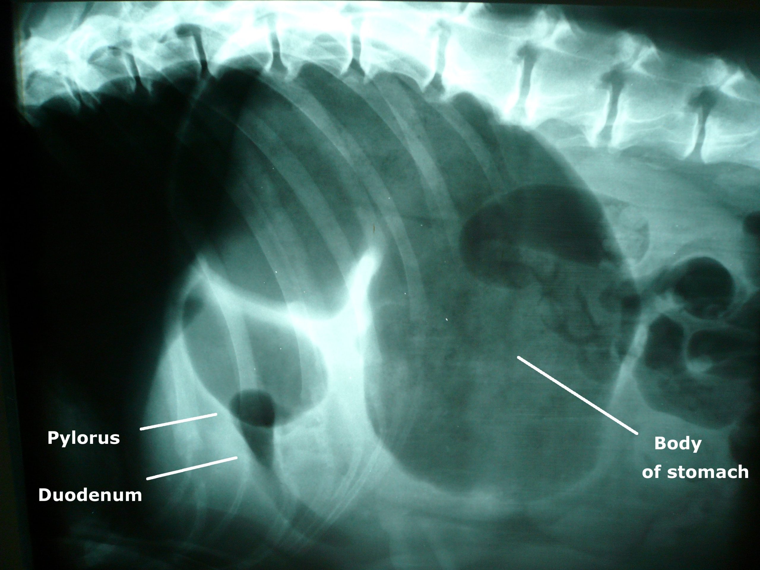
![Photo of an x-ray showing gastric dilatation and volvulus in a large mixed-breed dog. The large dark area is the gas trapped in the stomach. The pylorus and duodenum are in an abnormal position cranial to the stomach and are separated by a fold in the stomach, creating a "double bubble" appearance. By Joel Mills (Own work) [GFDL (http://www.gnu.org/copyleft/fdl.html), CC-BY-SA-3.0 (http://creativecommons.org/licenses/by-sa/3.0/) or CC-BY-SA-2.5-2.0-1.0 (http://creativecommons.org/licenses/by-sa/2.5-2.0-1.0)], via Wikimedia Commons](http://www.vettimes.co.uk/app/uploads/2013/09/GDV_x-ray.jpg)