Tag: Diarrhoea

  • Hyponatraemia, pt 1: clinical signs

    Hyponatraemia, pt 1: clinical signs

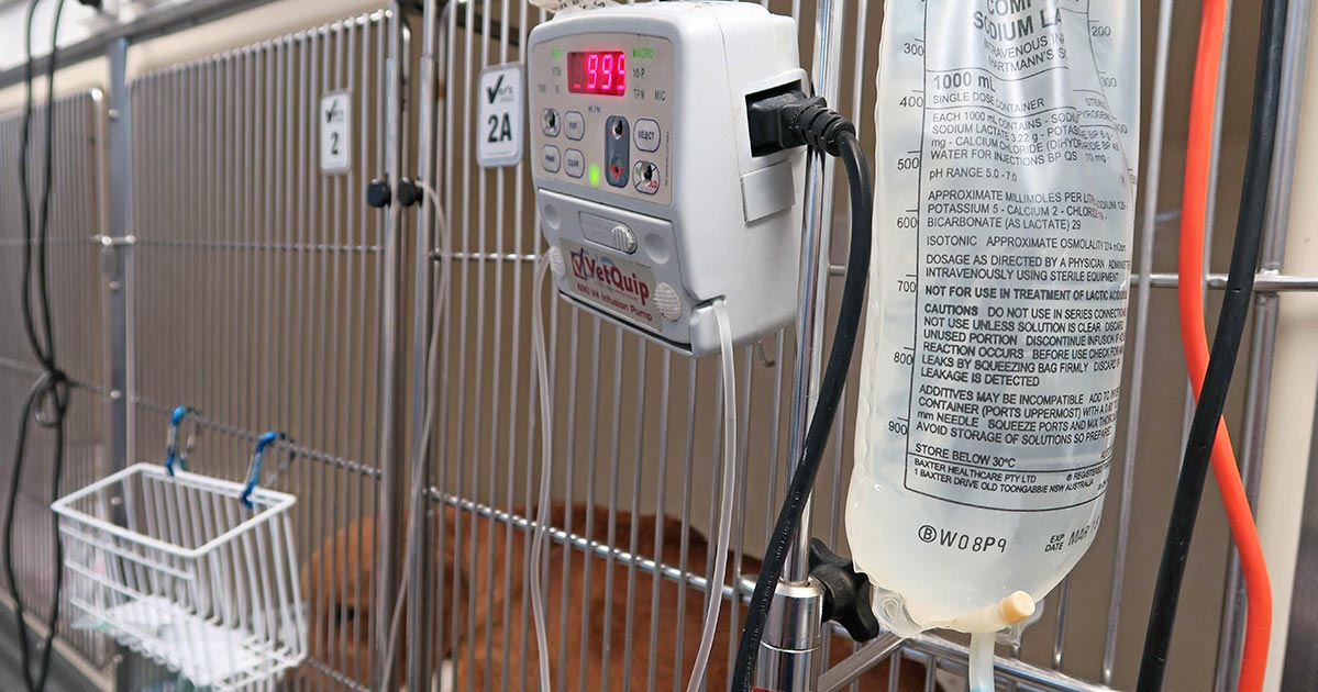
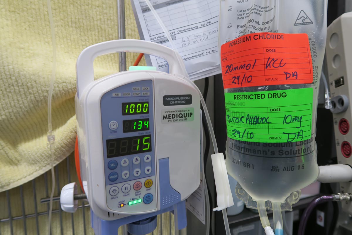
    Hyponatraemia is a relatively common electrolyte disturbance encountered in critically ill patients, and the most common sodium disturbance of small animals.

    In most cases, this is caused by an increased retention of free water, as opposed to the loss of sodium in excess of water.

    Low serum sodium concentration

    Hyponatraemia is defined as serum concentration lower than 140mEq/L in dogs and lower than 149mEq/L in cats.

    The serum sodium concentration measured is not the total body sodium content, but the amount of sodium relative to the volume of water in the body. For this reason, patients with hyponatraemia can actually have decreased, increased or normal total body sodium content.

    This series will look briefly at the modulators of the sodium and water balance, clinical signs associated with hyponatraemia, the most common causes in small animals, the pathophysiology behind these changes, and treatment and management.

    ECF volume

    hyponatraemia
    An example of hyponatraemia.

    Sodium is the main osmotically active particle in the extracellular fluid (ECF), so is the main determining factor of the ECF volume. Any disease process that alters the patient’s ECF volume will lead to hyponatraemia, such as:

    • dehydration
    • polyuria
    • polydipsia
    • vomiting
    • diarrhoea
    • cardiac diseases
    • pleural or peritoneal effusion

    The modulators of water and sodium balance are also different, so should be thought of as different processes.

    Water balance is modulated by thirst and antidiuretic hormone, and the effect of this is to maintain normal serum osmolality and serum sodium concentration.

    Modulators of sodium balance aim to maintain normal ECF volume. It adjusts this by altering the amount of renal sodium excretion; an expansion of ECF volume will lead to an increased sodium excretion, while a reduction in ECF volume will lead to increased sodium retention.

    Rate and magnitude

    The clinical signs of hyponatraemia are both dependent on the magnitude of the decrease and the rate at which it developed.

    In mild or chronic patients, no visible clinical signs can exist. In severe (lower than 125mEq/L) and acute cases, clinical signs exhibited are typically neurological, reflecting cerebral oedema. Possibilities include:

    • lethargy
    • anorexia
    • weakness
    • incoordination
    • disorientation
    • seizures
    • coma

    Patients with acute hyponatraemia – for example, water intoxication – are more likely to show clinical signs, compared to those with chronic hyponatraemia, because the brain takes time (at least 24 to 48 hours) to produce idiogenic osmoles, osmotically active molecules that help shift free water out of brain cells.

    Therefore, any acute hyponatraemia that develops within a 24 to 48-hour period tend to show clinical signs, whereas chronic cases are less likely.

    • Next week’s blog will look into the different causes of hyponatraemia and how they result in sodium loss.
  • Blood gas analysis, pt 5: metabolic acidosis and alkalosis

    Blood gas analysis, pt 5: metabolic acidosis and alkalosis

    Base excess (BE) and bicarbonate (HCO3-) represent the metabolic components of the acid base equation. In general, both components will change in the same direction.

    Decreased HCO3– and BE indicate either a primary metabolic acidosis or a metabolic compensation for a chronic respiratory alkalosis. Elevated HCO3– and BE indicate either a primary metabolic alkalosis or a metabolic compensation for a chronic respiratory acidosis.

    The exception to this rule arises when a patient hypoventilates or hyperventilates.

    Carbonic acid equation

    CO2 + H2O ↔ H2CO3 ↔ HCO3– + H+

    When a patient hypoventilates, CO2 will increase as a result of reduced expiration, so a shift to the right of the equilibrium will occur. The shift to the right will increase the bicarbonate levels proportional to the increase in CO2.

    The opposite occurs when a patient hyperventilates; the equilibrium shifts to the left, so a decrease in HCO3– is present.

    Since HCO3– is not independent to the patient’s respiratory status, it is an inaccurate way of measuring the metabolic component in patients with respiratory changes. For this reason, the BE value is the preferred.

    The BE represents the amount of acid, or base, needed to titrate 1L of the blood sample until the pH reaches exactly 7.4, with the assumption the blood sample is equilibrated to a partial pressure of CO2 of 40mmHg (the middle of the reference range) and the patient’s body temperature is normal.

    Possible causes

    The possible causes of the primary disease are:

    Metabolic acidosis

    • lactic acidosis – shock and poor perfusion
    • renal failure – reduced hydrogen ion (H+) excretion and increased loss of HCO3
    • diabetic ketoacidosis – ketone acids
    • gastrointestinal (GI) losses – loss of HCO3– through vomiting and diarrhoea

    Metabolic alkalosis

    • GI outflow obstructions – loss of H+ and chloride via vomiting
    • reduced chloride levels and resultant poor perfusion – body attempts to reabsorb water and sodium to increase intravascular volume, but inadvertently also reabsorbs HCO3– in the process, despite existing alkalosis
    • refeeding syndrome
    • severe hypokalaemia:
      • transcellular shift – potassium ions leave and H+ ions enter the cell
      • transcellular shift in cells of proximal tubules → intracellular acidosis → promotes ammonium production and excretion
      • H+ excretion in the proximal and distal tubules increases → further reabsorption of HCO3– and net acid excretion
    • renal insufficiency
    • diuretic therapy (contraction alkalosis – loss of bodily fluids that do not contain HCO3-; this causes the extracellular volume to contract around a fixed quantity of HCO3-, resulting in a rise in the concentration of HCO3– without an actual increase in HCO3– levels)

    Next step

    After ruling out the differential causes of either respiratory or metabolic acidosis/alkalosis, the next step is to determine whether a compensatory response is present and, if so, if this is adequate or whether a true mixed acid-base disorder exists.

  • Fluid therapy part 4: ongoing losses

    Fluid therapy part 4: ongoing losses

    This month, we will look at the final part of a fluid therapy plan – accounting for ongoing losses. This can be challenging, but some general rules can be helpful.

    Regular assessment is essential to track patients' response.
    Regular assessment is essential to track patients’ responses.

    First, let’s recap the four parts of a fluid therapy plan:

    1. Perfusion deficit
    2. Hydration deficit
    3. Maintenance requirements
    4. Ongoing losses

    When considering ongoing losses, try to not forget about patients with pre-existing polyuric diseases; chronic renal failure is a prime example. Patients with dehydrated chronic renal failure are unlikely to suddenly regain concentrating ability. Polyuria should be considered as an ongoing loss.

    Other conditions that may result in additional urinary fluid losses include post-obstructive diuresis, diabetes mellitus, hyperadrenocorticism and hyperthyroidsim.

    How much to add?

    This is the tricky part. I often add an additional half to one maintenance and frequently reassess clinical parameters, or if a urinary catheter is placed matching ins and outs.

    Gastrointestinal tract losses can be collected and weighed; 1g of vomitus or diarrhoea can be roughly equivalent to 1ml of water.

    Fluid removed from drains placed in cavities or wounds should also be measured and accounted for.

    Remember the key point is regular assessment of the patient’s hydration status, from repeat clinical exams, to track their response. Don’t forget regular retesting of electrolytes – for example, every 12 to 24 hours for patients on IV fluids and not eating.

  • Maintenance fluids

    Maintenance fluids

    A while ago we discussed the components of a fluid therapy plan and talked about hydration deficits. This week I want to touch on maintenance fluids.

    Gerardo_IVF
    IV fluids

    Maintenance rates are typically calculated using the following formulae:

    ml/day = 80 × bodyweight (kg)0.75 (cats)
    ml/day = 132 × bodyweight (kg)0.75 (dogs)

    or
    ml/day = 30 × bodyweight (kg) + 70

    These formulae better estimate the needs of smaller and larger patients. The flat 3ml/kg/hr underestimates for small patients and overestimates for larger patients.

    This maintenance rate is in addition to rehydration rates.

    So what sort of fluid should you use for maintenance?

    True “maintenance” crystalloids:

    • used to replace ongoing fluid and electrolyte loss from normal metabolism, not to replace perfusion and hydration deficits or ongoing losses from diarrhoea, for example
    • sodium concentration less than plasma
    • potassium concentrations higher than plasma
    • glucose sometimes added to bring solute concentrations similar to extracellular fluid

    Do you have to use maintenance crystalloids or can you use replacement crystalloids?

    Replacement crystalloids are more frequently used for maintenance fluid therapy rather than maintenance crystalloids. This is because they are more readily available, we are more familiar with their use and effect, and patients are generally continued on these after perfusion and hydration deficits have been corrected.

    In reality, most of the time it doesn’t really matter if we are using replacement crystalloids for maintenance therapy as the patient can manage the excess sodium, but some patients – especially cats – may require potassium supplementation. The key point is regular assessment of the patient’s hydration status and electrolytes – for example, every 12 to 24 hours for patients on IV fluids and not eating.

  • Giardia SNAP test

    Giardia SNAP test

    Following last week’s discussion about pancreas-specific lipase tests, this week we look at Giardia SNAP tests.

    Giardia is an important differential diagnosis in domestic species presenting with gastrointestinal disease, with a reported prevalence varying between 10% in household dogs and up to 100% in canine shelters and breeding colonies.

    Giardia
    The Giardia SNAP test.

    Younger animals – particularly younger than six months – and the presence of both acute and chronic diarrhoea have been found to have a higher likelihood to be tested positive for Giardia. However, the accurate identification of giardiasis continues to be problematic, particularly in chronic cases.

    Several reasons exist for this:

    • The shedding of cysts is often intermittent.
    • Excretion of coproantigen may continue for several weeks, despite resolution of clinical infection. This is because it is a protein expressed by the organism during cyst formation, not the whole organism.
    • Reinfection can occur after a period of clinical resolution.
    • Chronically infected animals can often be asymptomatic.

    In-house test

    The Giardia SNAP test is an in-house test that detects faecal Giardia antigens. Although this test boasts to have both a high sensitivity and high specificity – 95% and 99.3%, respectively – be cautious in interpreting the results as they are based on a population with high disease prevalence (100%), which is not characteristic in most general populations.

    In a prospective study with naturally acquired canine chronic subclinical giardiasis by Rishniw et al (2010), it was found this test has little value as a screening test because of its low positive predictive value (probability a positive result being a true positive), especially when the prevalence of disease is low (10% or less).

    This means a positive result is substantially more likely to be a false positive, supporting the complicating factor of persistent coproantigen beyond clinical resolution of disease.

    High negative predictive value

    Despite this, the test has a high negative predictive value – a negative result being truly negative – meaning it is useful in helping rule out the disease.

    In a nutshell, consider your patients’ likely risk of infection. If the risk of giardiasis is low, a negative result helps you rule out the disease, but a positive result is non-conclusive due to the high risk of false positive. However, if the risk of disease is high – for example, puppies from shelters or breeding colonies – a positive test will help confirm the diagnosis.

    With regards to tracking patients treated for Giardia, if clinical signs have resolved, due to the high chance of false positives, repeating the test does not provide valuable information.

    Giardia intestinalis
    Giardia intestinalis. Optical microscopy technique: Bright field. Magnification: 6000x (for picture width 26 cm ~ A4 format). Image by Josef Reischig / CC BY-SA 3.0
  • Being English in Scotland

    Being English in Scotland

    Coming from the centre of England, studying veterinary medicine in Scotland has its quirks. In my first week I was immersed in an entirely new language that had nothing to do with my choice of course.

    england-scotland-jigsaw-2_Fotolia_treenabeenaOne of my Scottish friends loves to remind me of the golden moment in an introductory lecture when I leaned over and whispered “who’s Ken?” (as in “I dinnae ken”, or “ken what I mean?”).

    But this week, during a lecture on bovine viral diarrhoea (BVD), I was left wondering about the geographical impact of studying in Glasgow compared to friends who stayed closer to home.

    North of the border

    The BVD virus has a pretty interesting mechanism that, while making for fascinating reading, is the reason it wreaks havoc on the UK’s cattle industries and can be pesky to both diagnose and get on top of in the herd.

    While BVD is prevalent all over the UK, Scotland is significantly further ahead than my home turf in the control of this disease, mainly due to a government-implemented eradication programme in recent years.

    In England, however, many farmers are unaware of the disease or reluctant to undertake the costly exercise of hunting persistently infected calves within the herd, which, at the moment, is not compulsory – unlike in Scotland.

    Now there was a lot of joking about England letting the side down and being a bit useless, but the reality is that until England plays catch-up and implements an eradication scheme, it’s going to be extremely difficult for the Scottish eradication to be 100% successful – short of throwing up a double fence between us and them.

    Regional issues

    If I were studying elsewhere, I wonder how the emphasis would differ depending on the prevalence in that region. Several times in my lectures I’ve heard Angiostrongylus (heartworm) brushed off as a differential if the animal has been to the south of England, with little much else said.

    Would that be given more time in an area with higher prevalence, if I were studying in London for example?

    At the end of the day, we all come out as vets, no matter where we’ve studied, and, while some topics may get more emphasis because of their regional importance, we’ll still need to pay particular attention to those conditions or diseases more commonly found in the areas we end up working in. So I don’t think geography has a dramatic impact long term (unless you never intend to leave your university city).

    And after all, Scotland knows how to ceilidh.