Tag: Congenital Tremor

  • Dystocia, pt 1: labour stages

    Dystocia, pt 1: labour stages

    Now most female canine patients are spayed, it comes as no surprise reproductive emergencies are not as common.

    One confusion seems to be not knowing how to determine a true dystocia emergency – especially when given advice over the telephone – from the process of normal parturition.

    Another concern is how to confidently form a diagnostic pathway to determine the cause of dystocia – especially for reasons other than obvious physical abnormalities (for example, fetopelvic disparity and fetal malposition).

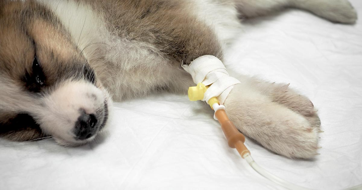
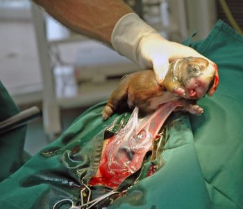
    Often, once we decide to go down the medical treatment pathway – whether the result of findings or owner/financial constraint – no one is confident as to what medication should be used and how often drugs can be given safely.

    This series of blogs will address these issues in a step-by-step manner. Hopefully, by the end, you will be confident in the diagnosis and management of dystocia.

    Labour stages

    Before moving on to the signs of dystocia, let’s go through the signs of labour.

    First stage labour

    First stage labour is characterised by panting, tremoring, nesting behaviour, a drop in core temperature – usually a drop by almost 1°C 24 hours prior to second stage labour – and a drop of progesterone to below 2mg/ml.

    Dog and puppy.
    Third stage labour occurs generally within 15 minutes after passing a puppy or kitten. Image © foto ARts / Adobe Stock
    • dogs: approximately 6 to 12 hours
    • cats: approximately 6 to 24 hours

    Second stage labour

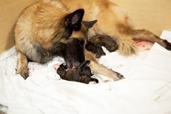
    Second stage labour is landmarked by the water breaking, visible abdominal contractions, and the allantoic/amniotic sac or fetal parts visible from the vulva.

    If vulval discharge is present, they should be clear. Excessive amount of bright red haemorrhage, green or black discharge prior to delivery, or purulent material can indicate a pathological process requiring immediate veterinary attention.

    • dogs: approximately 3 to 6 hours
    • cats: approximately 6 to 24 hours

    Third stage labour

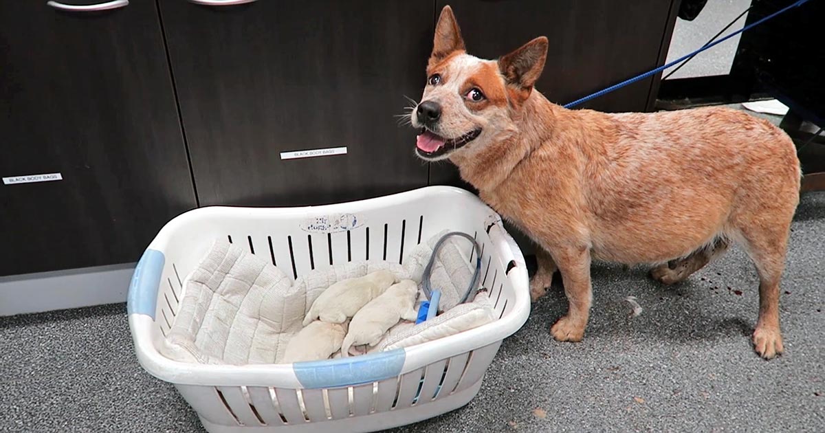
    Third stage labour this is when passage of all the placenta has occurred, generally within 15 minutes after passing a puppy or kitten.

    Clues

    Now we understand the normal progression of parturition, a few clues exist in the history that could suggest dystocia may be present.

    Some breeders will often know the ovulation timing of the patient – especially if AI was performed. Tests such as progesterone levels, luteal hormone (LH) levels, cytology and vaginoscopy are some ways where it can help time the ovulation.

    The normal gestation length should not be any longer than 66 days from the LH surge or, if the ovulation history is unknown, 72 days from the last known breeding.

    History of prior dystocia is a warning, as most animals with prior parturition difficulties are more likely to develop dystocia again.

    The same goes for animals that have previously required a caesarean. Their risk of requiring future caesareans is high, with further risk of uterine rupture if dystocia happens again.

    Image © Pilipipa / Adobe Stock
    Animals that have previously had a caesarean are at high risk of requiring future caesareans, with further risk of uterine rupture if dystocia happens again. Image © Pilipipa / Adobe Stock

    Intervention signs

    Owners often telephone after the failure of normal progression of delivery. The signs that always require immediate intervention are:

    • more than 4 hours have passed from the rupture of the first chorioallantois
    • more than 2 hours between delivery
    • more than 30 minutes of strong abdominal contraction and no delivery
    • presence of green or black discharge before delivery
    • large amount of bright red haemorrhage
    • abnormal amount of pain during contractions
    • collapse of the bitch or distracted mothering

    Any of these signs require immediate presentation to the veterinarian. Delivery of stillborn puppies is also an indication where veterinary attention is indicated.

    Finally, if owners are concerned, it is best to advise veterinary assessment rather than try to convince them everything is okay based on what they describe over the telephone.

  • Hyponatraemia, pt 2: causes

    Hyponatraemia, pt 2: causes

    The causes of hyponatraemia can be divided into three major categories, based on serum osmolality. This is further divided based on the patient’s volume status (Table 1).

    Most patients we see in clinic fall into the hypovolaemic category, except patients with diabetes mellitus.

    Table 1. Causes of hyponatraemia based on osmolality and volume status (from Guillaumin and DiBartola, 2017).
    Hypo-osmolar Hyperosmolar Normo-osmolar
    Hypovolaemic Normovolaemic Hypervolaemic
    Gastrointestinal fluid loss
    Third-space fluid losses
    Shock
    Hypoadrenocorticism (Addison’s disease)
    Renal insufficiency
    Excessive diuretic administration
    Salt-losing nephropathy
    Cerebral salt wasting syndrome
    Syndrome of inappropriate antidiuretic hormone secretion (SIADH)
    Hypotonic fluid administration
    Hypothyroidism
    Glucocorticoid insufficiency
    Psychogenic polydipsia
    Reset osmostat (SIADH type B)
    Congestive heart failure
    Acute or chronic renal failure
    Nephrotic syndrome
    Hepatic cirrhosis
    Accidental ingestion or injection of water (water intoxication)
    Hyperglycaemia
    Mannitol
    Severe azotaemia
    Hyperlipidaemia
    Hyperproteinaemia

    Common causes

    In dogs, the three most common causes of hyponatraemia are:

    • gastrointestinal (GI) fluid loss
    • third-space fluid loss
    • fluid shift from intracellular fluid to extracellular fluid (ECF) as a result of hyperglycaemia

    In cats, the three most common causes of hyponatraemia are:

    • urologic diseases
    • GI fluid loss
    • third-space fluid losses

    In most patients, more than one pathophysiologic factor is likely to be contributing to the hyponatraemia.

    Circulating volume

    Hypovolaemic patients – those with, for example, GI losses, hypoadrenocorticism, renal losses and haemorrhagic shock – have a reduced effective circulating volume. ECF contraction triggers antidiuretic hormone (ADH) secretion, which leads to increases in free water absorption and thirst, and results in dilution of the serum sodium concentration. Aldosterone secretion is reduced in hypoadrenocorticism, so an overall reduction in sodium reabsorption compounds the problem.

    Hypervolaemic patients are those with an increased fluid retention state, such as:

    • congestive heart failure (pulmonary oedema)
    • advanced hepatic failure (ascites, third-space fluid)
    • renal failure
    • free water ingestion

    Congestive heart failure patients have a reduced cardiac output and, therefore, a decreased effective circulating volume, despite the presence of the extra fluid status. Renin-angiotensin activation leads to release of ADH and aldosterone, resulting in sodium and free water reabsorption, and increased thirst. Both lead to an excess of free water retention.

    Advanced hepatic (cirrhosis) or renal failure (nephrotic syndrome) both result in hypoalbuminaemia, leading to fluid shifting into the interstitial space and third space, reducing effective circulating volumes. This leads to activation of ADH to increase free water reabsorption, to restore the circulating volume in the face of existing hypervolaemia and hyponatraemia.

    Diabetic patients

    Moderate to severe hyperglycaemic diabetic patients can be either hyperosmolar or normo-osmolar, depending on the serum blood glucose concentration. Hyponatraemia occurs when water shifts from the intracellular fluid to the ECF down the osmotic gradient, diluting the serum sodium content.

    Despite this osmotic shift, not all diabetic patients develop hyponatraemia. Glucosuria also causes also causes a renal osmotic shift, sometimes resulting in urine water loss in excess to sodium. This offsets the hyponatraemia – in some cases, hypernatraemia results.

    Treatment

    Treatment of hyponatraemia hinges on how quickly it developed and the volume status of the patient. The rule of thumb is to correct hyponatraemia slowly – not exceeding 0.5meq/L/hr – especially in chronic cases, or cases where the duration of hyponatraemia is unknown. Keeping to this rate is paramount until serum sodium concentration reaches 130meq/L.

    In acute patients with severe clinical signs, such as seizures, some clinicians may choose to use a higher rate of 1meq/L/hr to 2meq/L/hr until clinical signs resolved.

    It should be emphasised, once again, this rate should never be used in chronic patients, patients with an unknown duration of hyponatraemia, or where frequent serum sodium concentration cannot be monitored. The rapid correction of hyponatraemia can lead to osmotic demyelination syndrome (myelinolysis).

    Its effect will not be apparent until three or four days after therapy, and can result in neurological abnormalities such as:

    • weakness
    • ataxia
    • dysphagia
    • paresis
    • coma

    For that reason, frequent electrolyte measurements are required, starting hourly then once a suitable rate of increase has been established and less frequently thereafter.

    • Part 3 will look at how to correct patients with hyponatraemia.

    Reference

    Guillaumin J and DiBartola SP (2017). A quick reference on hyponatremia, Veterinary Clinics of North America: Small Animal Practice 47(2): 213-217.