Tag: Clinical signs

  • All hands on deck: GDV diagnosis

    All hands on deck: GDV diagnosis

    Gastric dilatation-volvulus (GDV) is a true veterinary emergency and while it can be daunting to be presented with a sick dog with suspected GDV, the most important thing to remember is this patient will likely succumb to this condition without your intervention.

    First, a little pathophysiology: GDV is a broad term that can refer to gastric dilation on its own, gastric dilation with volvulus, and even chronic gastric volvulus. These conditions usually present in large or giant breeds and we still know little about the underlying causes.

    GDV
    Gastric dilatation-volvulus (click to zoom).

    Once dilation and volvulus occurs, perfusion to the stomach and other abdominal organs is compromised. Along with general shock – which can be fatal in its own right – decreased stomach wall perfusion can result in stomach wall necrosis, rupture and peritonitis.

    Clinical signs

    Quite often, a GDV case starts with a telephone call from a panicking owner. He or she usually reports an acute onset of retching, regurgitation or vomiting in their large or giant breed dog after feeding.

    Other common signs include:

    • hypersalivation
    • agitation
    • palpable abdominal distension

    When presented, many of these cases will be obvious and the animal already in some degree of shock. You need to institute fluid resuscitation and gastric decompression immediately to restore perfusion as soon as possible.

    Confirmation

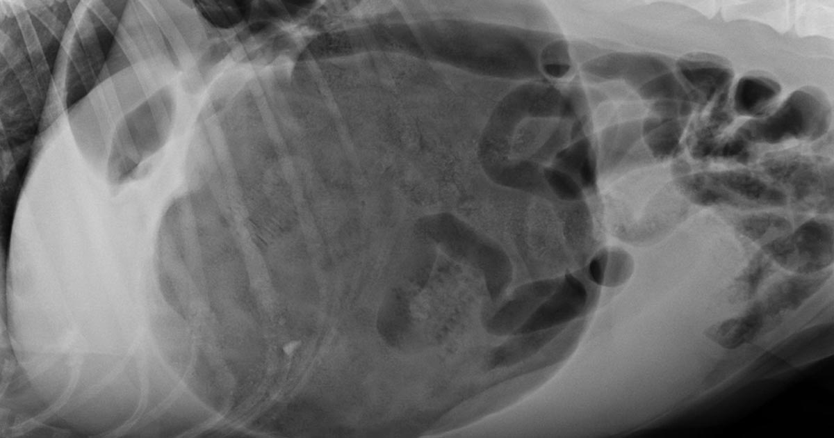
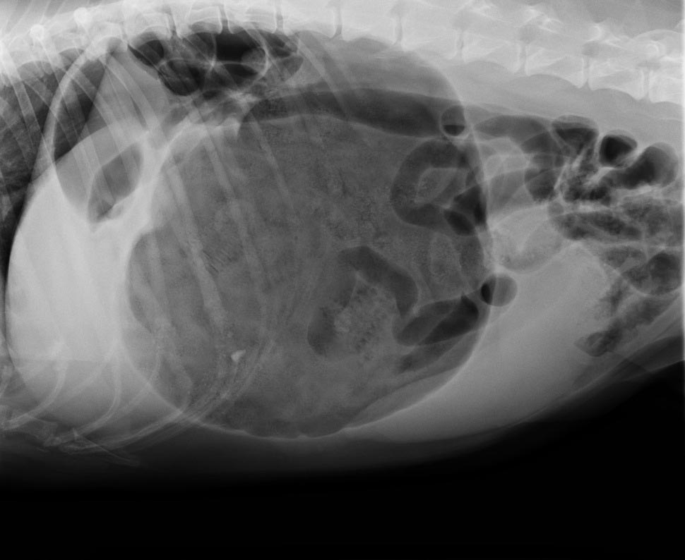
    To confirm the patient truly has GDV, as some patients may present with simple gastric dilation from over-engorgement, you need to perform an abdominal radiograph.

    Always keep an eye out for the large, deep-chested dog that presents with vomiting or retching, but doesn’t appear bloated. Don’t be fooled into ruling out GDV in these patients based on physical examination alone – often, no visible or palpable gastric distension exists as the ribs cover the stomach. That is where the abdominal radiographs play an especially important role.

    It is common practice at our hospital to perform abdominal radiographs as soon as possible, so as to not miss a hidden or subtle GDV in these large breed dogs.

    Which view is best?

    The classic approach is to lie the patient in right-lateral recumbency, in this view, you would see the classic “Smurf’s hat”, “boxing glove”, “Popeye’s arm”, “double bubble”, etc. This is compartmentalisation of the stomach, indicating not only gastric dilatation, but volvulus as well.

    You should also look for evidence of pneumoperitoneum, as it may suggest gastric wall rupture.

    At this stage, it is also important to collect blood for biochemistry, haematology, electrolytes and, if available to you, blood gas analysis. ECG readings should also be taken to determine if the patient has any life-threatening arrhythmias, such as ventricular tachycardia.

    Next month, we will talk about stabilising and treating these patients.

  • Perfusion deficits and fluid resuscitation: a more in-depth look

    Perfusion deficits and fluid resuscitation: a more in-depth look

    A few weeks ago in the Tip of The Week, we discussed the four basic components of a fluid therapy plan – perfusion deficit, hydration deficit, maintenance requirements and ongoing losses.

    Let’s consider perfusion deficits.

    As an emergency clinician, correcting perfusion deficits is a crucial part of stabilising a patient. So what is a perfusion deficit? It either refers to a real or relative loss of intravascular fluid volume, or low blood pressure, leading to a decrease in perfusion of tissues and, ultimately, decreased oxygen delivery – ie, shock.

    What does this look like clinically?

    Pressure infuser
    Operating the pressure infusor at the triage bench.

    Clinical signs of perfusion deficits include:

    • pale gums
    • reduced capillary refill time
    • tachycardia
    • reduced pulse pressure
    • dull mentation
    • cold extremities and low core body temperatures

    A low normal body temperature in a critically ill patient should alert you to the possibility the patient may be experiencing early perfusion deficits, but is not yet severe enough to result in a low body temperature.

    What are these signs caused by?

    These signs are caused by activation of the sympathetic nervous system in response to reduced blood pressure (BP).

    BP is the product of cardiac output (CO) and systemic vascular resistance (SVR):

    BP = CO × SVR

    CO is the product of the heart rate (HR) and stroke volume (SV).

    CO = HR × SV

    Reduced perfusion results in reduced BP. The body increases BP by activating the sympathetic nervous system, resulting in a compensatory increase in HR and SV (beta adrenergic), which increases CO and vasoconstriction (alpha adrenergic) of the peripheral blood vessels to increase SVR and shunt the blood to the heart and brain.

    Peripheral vasoconstriction is seen clinically as pale gums, reduced capillary refill time, cool peripheries and low body temperature as blood is being shunted away from those peripheral capillary beds. It also results in reduced pulse pressures.

    In summary, clinical signs of perfusion deficits are signs of the body trying to compensate and push blood to where it is needed most.

    Blood pressure is normal, it can’t be in shock?

    Since the body’s compensatory mechanisms are geared towards preserving blood pressure, it often remains normal in patients with shock until the body cannot compensate any longer – decompensated shock.

    So, to me, normotension does not rule out perfusion deficits and hypotension is an indicator of severe perfusion deficits.

    Are IV fluid boluses safe for all patients in shock?

    Hartmanns fluid bolus
    Preparing a Hartmann’s fluid bolus.

    The vast majority of the time I would say yes, but you must ask one question – could this patient be in cardiogenic shock? IV bolus therapy would be contraindicated in most of these patients.

    Is it a small-breed dog presenting with a murmur and dyspnoea? Or a large-breed dog with an abnormal heart rhythm? If so then diuretics and anti-arrhythmic medications may be indicated rather then IV fluid boluses.

    How much volume can you give?

    I generally give buffered crystalloid fluid boluses of 10ml/kg over 5-10 minutes. I repeat this while monitoring for resolution of clinical signs of perfusion deficits. If I get to half a blood volume – 45ml/kg in a dog and 30ml/kg in a cat – I ask myself what could be causing this ongoing perfusion deficit. Could it be:

    • Distributive or vasodilatory shock from anaphylaxis or sepsis? In which case, vasopressors – such as adrenalin, dopamine and noradrenalin – are indicated.
    • Cardiogenic shock from dilated cardiomyopathy? Are diuretics, anti-arrthymic medications or positive inotropes required?
    • Restrictive shock from a gastric dilatation-volvulus or pericardial effusion? Is gastric decompression or pericardiocentesis required?
    • Does continued hypovolaemic shock exist? If so then continued fluid therapy is indicated, but this may cause significant haemodilution. Consider colloids or blood products, such as packed red blood cells, whole blood or plasma. Does it need emergency surgery or abdominal compression to stop an internal bleed?

    Perfusion deficits need to be corrected rapidly as continued oxygen delivery to tissues increases the risk of organ dysfunction and, ultimately, organ failure.

  • SNAP cortisol test

    SNAP cortisol test

    While hyperadrenocorticism is not an uncommon incidental finding in patients presenting to our emergency clinic, hypoadrenocorticism is a lot less common. Or, possibly, more frequently underdiagnosed.

    Textbook clinical presentations combined with haematology and biochemicial changes can make diagnosis straightforward, but not all patients will present with all the classic signs.

    SNAP cortisol test
    The SNAP cortisol test is a quantitative ELISA test that measures the level of serum cortisol in dogs.

    To complicate things further, hypoadrenocorticism is the great mimicker of diseases; it is often impossible to arrive at a definitive diagnosis without knowing the cortisol levels.

    The SNAP cortisol test allows clinicians to determine cortisol levels in-house – a blessing to those of us who work out-of-hours – but is not without its limitations.

    Suspicious signs

    Patients with hypoadrenocorticism often present with vague and non-specific clinical signs, but certain clinicopathological changes help raise the suspicion:

    • a decrease in sodium-to-potassium ratio (below 1:27)
    • azotaemia
    • an inappropriately low urinary specific gravity, despite evidence of dehydration or hypovolaemia
    • a leukogram unfitting to the degree of illness of the patient (a “reverse stress leukogram”- neutropenia, lymphocytosis, eosinophilia)
    • anaemia
    • hypoglycaemia
    • hypercalcaemia

    Although most Addisonian patients will not present with all these signs – especially those in the early stages of disease or those with atypical Addisonian disease (glucocorticoid insufficiency only) – any patients showing any of these haematology and biochemicial changes should have hypoadrenocorticism ruled out as part of the diagnostic plan.

    Imperfect ELISA

    The SNAP cortisol test has been advertised as an in-house assay to aid the diagnosis, treatment and management of both hyperadrenocorticism and hypoadrenocorticism, although the quality of the result is not perfect. This quantitative ELISA test measures the level of serum cortisol in dogs.

    In one study1, the SNAP cortisol test appears to have a good correlation with an external laboratory chemiluminescent assay test; however, in 12.8% of cases (5 of 39 patients), the SNAP test result could have led to a different clinical decision regarding the management of the patient.

    Since long-term Cushing’s management relies on reliable, repeatable cortisol level detection, this high level of discrepancy is unacceptable, especially when more accurate alternatives are available at external laboratories.

    Still useful

    Despite this, it is still very useful helping to assess for the presence or absence of hypoadrenocorticism, especially in an emergency setting.

    I use the SNAP cortisol to measure the resting cortisol level. If it is below 2ug/dL or in inconclusive range (between 2ug/L and 6ug/L), but the clinical picture suggests hypoadrenocorticism, I would perform an adrenocorticotropic hormone (ACTH) stimulation test and send samples to an external laboratory. If it is well above the inconclusive range, I would not perform an ACTH stimulation test.

    In summary, I think the SNAP cortisol test can be useful in helping assess for hypoadrenocorticism, but would still recommend performing an ACTH stimulation test and running the samples externally.

    However, use it with caution for hyperadrenocorticism diagnosis and its long-term management – especially when more accurate and economical alternatives are available.

  • Giardia SNAP test

    Giardia SNAP test

    Following last week’s discussion about pancreas-specific lipase tests, this week we look at Giardia SNAP tests.

    Giardia is an important differential diagnosis in domestic species presenting with gastrointestinal disease, with a reported prevalence varying between 10% in household dogs and up to 100% in canine shelters and breeding colonies.

    Giardia
    The Giardia SNAP test.

    Younger animals – particularly younger than six months – and the presence of both acute and chronic diarrhoea have been found to have a higher likelihood to be tested positive for Giardia. However, the accurate identification of giardiasis continues to be problematic, particularly in chronic cases.

    Several reasons exist for this:

    • The shedding of cysts is often intermittent.
    • Excretion of coproantigen may continue for several weeks, despite resolution of clinical infection. This is because it is a protein expressed by the organism during cyst formation, not the whole organism.
    • Reinfection can occur after a period of clinical resolution.
    • Chronically infected animals can often be asymptomatic.

    In-house test

    The Giardia SNAP test is an in-house test that detects faecal Giardia antigens. Although this test boasts to have both a high sensitivity and high specificity – 95% and 99.3%, respectively – be cautious in interpreting the results as they are based on a population with high disease prevalence (100%), which is not characteristic in most general populations.

    In a prospective study with naturally acquired canine chronic subclinical giardiasis by Rishniw et al (2010), it was found this test has little value as a screening test because of its low positive predictive value (probability a positive result being a true positive), especially when the prevalence of disease is low (10% or less).

    This means a positive result is substantially more likely to be a false positive, supporting the complicating factor of persistent coproantigen beyond clinical resolution of disease.

    High negative predictive value

    Despite this, the test has a high negative predictive value – a negative result being truly negative – meaning it is useful in helping rule out the disease.

    In a nutshell, consider your patients’ likely risk of infection. If the risk of giardiasis is low, a negative result helps you rule out the disease, but a positive result is non-conclusive due to the high risk of false positive. However, if the risk of disease is high – for example, puppies from shelters or breeding colonies – a positive test will help confirm the diagnosis.

    With regards to tracking patients treated for Giardia, if clinical signs have resolved, due to the high chance of false positives, repeating the test does not provide valuable information.

    Giardia intestinalis
    Giardia intestinalis. Optical microscopy technique: Bright field. Magnification: 6000x (for picture width 26 cm ~ A4 format). Image by Josef Reischig / CC BY-SA 3.0
  • Eating disorders and the veterinary profession

    Eating disorders and the veterinary profession

    The general public associates the word “anorexia” with the eating disorder characterised by refusing to eat to lose weight, which, in human medicine, has the more specific name of “anorexia nervosa”. As vets, we use the word the term “anorexic” in the slightly different sense of being a clinical sign our patients exhibit – defined as “a lack or loss of appetite”.

    Kid eating noodles.
    Is the veterinary profession practising what it preaches when it comes to nutrition?

    When referring to vets themselves, however, these definitions blur together a little, but I believe many vets frequently exhibit clinical signs that may or may not be part of an eating disorder.

    While there is a lack of hard evidence or figures for eating disorders within the veterinary profession, it is estimated 10% of UK veterinary students suffer from eating disorders (not limited to anorexia nervosa), which is higher than the figure for the general UK population, which sits at 6.4%. (vetlife.org.uk).

    Another branch

    We are all made very aware of the mental health statistics and suicide risk of vets, and eating disorders are another branch of that tree.

    Despite the lack of evidence to back up the theory, based on anecdotes alone, I’m willing to bet eating disorders, or even intermittent “anorexia” (the clinical sign), are more prevalent in qualified vets than the general population.

    I’ve lost count of the number of times I’ve been on EMS and spent the entire day in the car with the same vet, going from call to call and not seeing them eat once.

    I’m probably on the other end of the scale; I tend to get indigestion and heartburn from excess stomach acid production if I don’t eat regularly enough, so I tend to stress about the next time I’m going to eat (which turns into a vicious cycle because those symptoms also develop as a result of that stress). Therefore, I always try to ensure I have a packed lunch so I never get caught out.

    On the road

    Burger in a car.
    Veterinary professionals on the road can often find themselves picking up fast food, which will be of long-term detriment to their health.

    The number of ambulatory equine or farm vets who don’t appear to carry food in their cars is astounding.

    They often rely on getting time to stop for food – which, inevitably, results in them not eating for the whole working day or picking up unhealthy fast food or snacks, which isn’t really much better.

    When discussing my observations with fellow students, many of them have similar tales, and the problem is not limited to ambulatory practice.

    While many vets and nurses in small animal practice will have a slightly more routine structure to their working lives, there’s always the odd mad day, week or month when they just can’t seem to grab a minute to supply their digestive system between consults, surgeries and emergencies.

    Despite my own claim of always being prepared to avert such situations, sometimes they’re unavoidable.

    I’ve found myself a few select times during rotations when I’ve been so rushed off my feet, trying to get everything done or see clients, that I haven’t eaten lunch until well after 4pm. Then, it’s likely I’ve got to that stage of being “past hungry”, but, as it is at that point I normally get headachey and feel a bit weak, I force something down despite not really wanting it any more.

    Can’t or won’t?

    Now, there’s a subtle difference between anorexia (a “lack of appetite”) and physically not having a chance to eat despite the grumbling in your stomach and the agonising knowledge there’s a pretty decent lunch waiting in the fridge in the next room – but it’s a fine line.

    If a client has been waiting 20 minutes for you already, surely another two won’t hurt while you inhale a sandwich? Are vet staff just too busy to eat sometimes or are they not finding the time themselves? No matter how stressed and busy you are, you should still be able to satisfy the basic human right of being able to eat.

    I believe the problem of the profession not eating properly is a combination of possible eating disorders, stress related anorexia and the working environment.

    Take responsibility

    Peanut butter.
    Finding time to eat is crucial, says Jordan, for the health of both the profession and its patients.

    I have previously expressed my opinion of the poor work-life balance within the UK veterinary profession, and ignoring rest breaks that are a legal requirement in almost any other field – while not entirely to blame – certainly doesn’t make it easy for vets to look after themselves.

    On the other hand, vets need to take personal responsibility for their own health and find time to eat during the working day – if you don’t look after number one, you won’t be providing optimal care to your patients.

    As much as skipping one meal might seem like a short-term solution to helping a patient that little bit sooner, it will be at the detriment of your clinical ability in the long term.

    Evidently, this is not a clear cut problem and, as such, there’s no fix-all solution.

    However, I think vocalising these issues is a good starting point if we wish to become a more healthy, sustainable profession in the future.