Tag: Clinical signs

  • Ionised hypocalcaemia, pt 3: acute treatment and management

    Ionised hypocalcaemia, pt 3: acute treatment and management

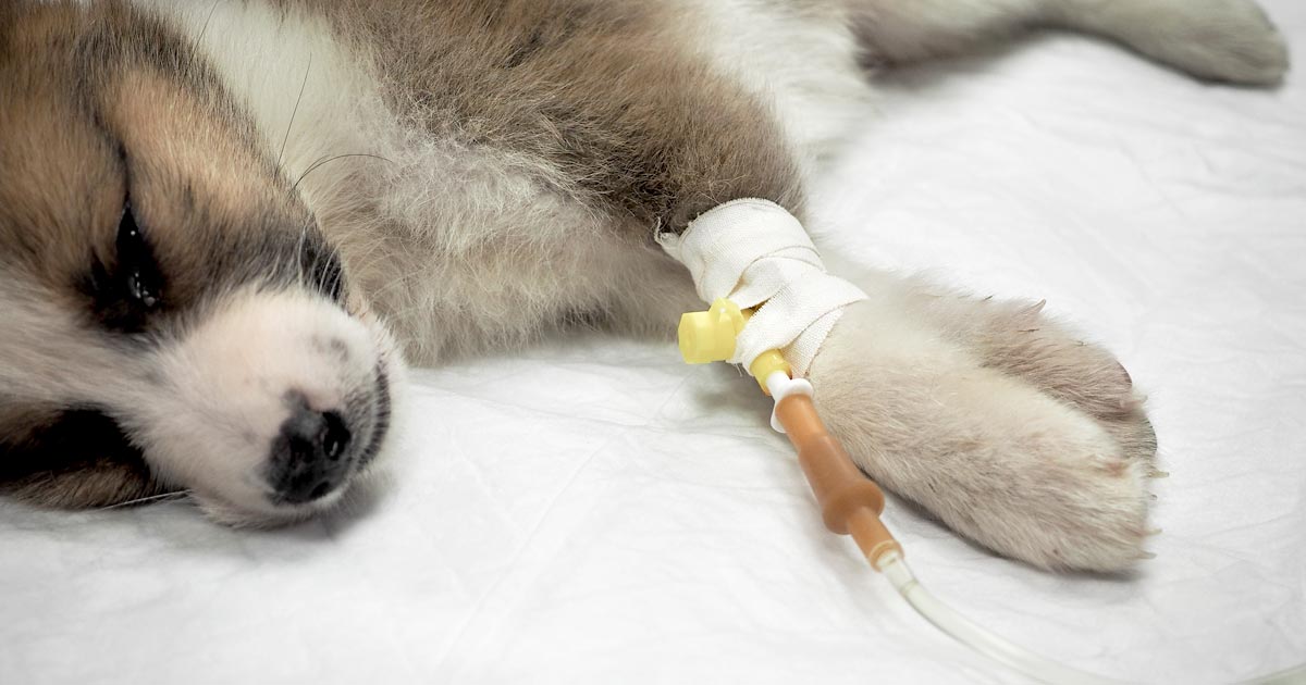
    Treatment of ionised hypocalcaemia (iHCa) is reserved for patients with supportive clinical signs, then divided into acute and chronic management.

    Since the most common cases of clinical hypocalcaemia in canine and feline patients are acute to peracute cases, this blog will focus on the acute treatment and management of hypocalcaemia.

    Clinical signs

    The severity of clinical signs of iHCa is proportional to the magnitude, as well as the rate of decline in ionised calcium (iCa) concentration.

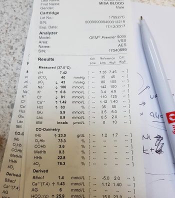
    The normal reference range for iCa is 1.2mmol/L to 1.5mmol/L in dogs and 1.1mmol/L to 1.4mmol/L in cats. Serum iCa concentrations in younger dogs and cats are, on average, 0.025mmol/L to  0.1mmol/L higher than adults.

    Mild iHCa (0.9mmol/L to 1.1mmol/L) – as seen in critically ill dogs and cats with diabetic ketoacidosis, acute pancreatitis, protein-losing enteropathies, sepsis, trauma, tumour lysis syndrome or urethral obstructions – often has no observable clinical signs.

    Moderately (0.8mmol/L to 0.9mmol/L) to severely (lower than 0.8mmol/L) affected animals – in the case of eclampsia and those with parathyroid disease – often display severe signs.

    Early signs of iHCa are often non-specific, and include:

    • anorexia
    • rubbing of the face
    • agitation
    • restlessness
    • hypersensitivity
    • stiff and stilted gait

    As the serum iCa concentration further decreases, patients often progress to:

    • paresthesia
    • tachypnoea
    • generalised muscle fasciculations
    • cramping
    • tetany
    • seizures

    In cats, the gastrointestinal system can also be affected, presenting as anorexia and vomiting.

    Treatment

    The need for treatment of hypocalcaemia is dependent on the presence of clinical signs, rather than a specific cut-off of serum concentration of iCa itself.

    Moderate to severe iHCa should always be treated. Mild hypocalcaemia, on the other hand, may not be necessary, especially if it is well tolerated. It should be remembered the threshold for development of clinical signs is variable, and treatment may benefit critical cases with an iCa concentration of less than 1.0mmol/L.

    Treatment is divided into the acute treatment phase and chronic management.

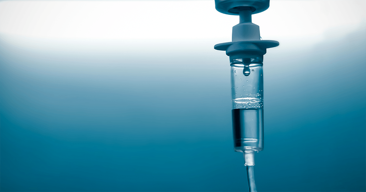
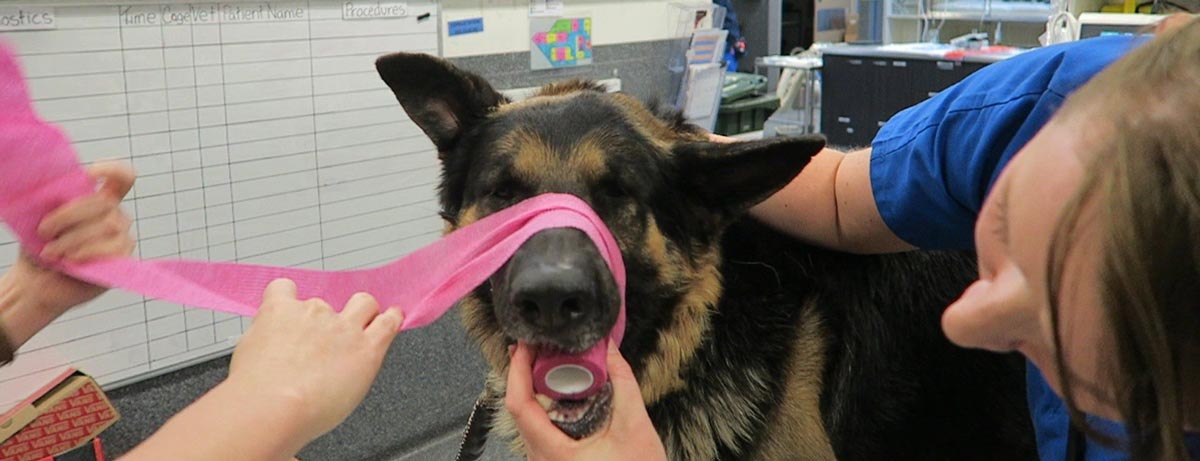
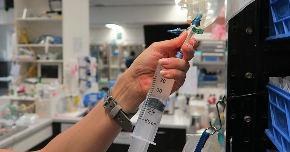
    In the tetanic phase, IV calcium is required – 10% calcium gluconate (equivalent to 9.3mg/ml) administered at 0.5ml/kg to 1.5ml/kg dosing to effect. This should be administered slowly with concurrent ECG monitoring. Infusion of calcium needs to be stopped if bradycardia develops or if shortening of the QT interval occurs.

    Some suggest calcium gluconate (diluted 1:1 with 0.9% sodium chloride) of half or the full IV dose can be given SC and repeated every six to eight hours until the patient is stable enough to receive oral supplementation. However, be aware calcium salts SC can cause severe necrosis or skin mineralisation.

    Calcium chloride should never be given SC, as it is a severe perivascular irritant.

    Correcting iCa

    Irrespective of the chronicity of the treatment, the rule of thumb is correction of calcium should not exceed 1.1mmol/L.

    Correction of iCa to normal or hypercalcaemic concentration should always be avoided, as this will result in the desensitisation of the parathyroid response, predisposing renal mineralisation and formation of urinary calculi.

    Some of the more common calcium supplementation medications – both parenteral and oral formulas – are detailed in Table 1. Supplementation of magnesium may also benefit some patients, as it is a common concurrent finding in critically ill patients with iHCa.

    Table 1. Common calcium supplementation medications
    Drug Calcium Content Dose Comment
    Parenteral calcium
    Calcium gluconate
    (10% solution)
    9.3mg/ml
    i) slow IV dosing to effect (0.5ml/kg to 1.5ml/kg); acute crisis, 50mg/kg to 150mg/kg over 20 to 30 minutes
    ii) 5mg/kg/hr to 15mg/kg/hr IV or 1,000mg/kg/day to 1,500mg/kg/day (or 42mg/kg/hr to 63mg/kg/hr)
    Stop if bradycardia or shortened QT interval occurs.
    Infusion to maintain normal Ca level
    SC calcium salts can cause severe skin necrosis/mineralisation.
    Calcium chloride
    (10% solution)
    27.2mg/ml 5mg/kg/hr to 15mg/kg/hr IV Do not give SC as severe perivascular irritant
    Oral calcium
    Calcium carbonate
    (many sizes)
    40% tablet 5mg/kg/day to 15mg/kg/day
    Calcium lactate
    (325mg, 650mg)
    13% tablet 25mg/kg/day to 50mg/kg/day
    Calcium chloride
    (powder)
    27.2% 25mg/kg/day to 50mg/kg/day May cause gastric irritation
    Calcium gluconate (many sizes) 10% 25mg/kg/day to 50mg/kg/day

    Next time…

    The next blog will look at the pathophysiology behind iHCa among critically ill animals. It will also look at the controversy regarding treatment of non-clinical iHCa cases and the prognostic indications of iCa concentrations.

  • Ionised hypocalcaemia, pt 2: eclampsia

    Ionised hypocalcaemia, pt 2: eclampsia

    As discussed in part one of this blog series, a myriad of disease processes can lead to ionised hypocalcaemia (iHCa).

    Despite this, only hypocalcaemia caused by eclampsia and hypoparathyroidism (primary or iatrogenic – post-surgical parathyroidectomy) are severe enough to demand immediate parenteral calcium administration.

    Hypoparathyroidism is quite rare, so this blog will not explore the detailed pathophysiology behind this syndrome. However, it is worthwhile mentioning – aside from primary hypoparathyroidism – no other disease state requires long-term calcium supplementation.

    Eclampsia, on the other hand, is the most common cause of clinical hypocalcaemia in dogs and cats. Multiple factors can predispose animals to the development of this phenomenon, so understanding the pathophysiology behind this potentially fatal disease will not only help with future diagnosis and treatment, but also help prevent this issue.

    Periparturient occurrence

    Eclampsia – also known as puerperal tetany or periparturient hypocalcaemia – occurs in the periparturient period anywhere from the final few weeks of gestation to four weeks postpartum, with the latter being the more common time frame of manifestation.

    The serum concentration of ionised calcium (iCa) is often less than 0.9mmol/L in bitches or less than 0.8mmol/L in queens. It presents as muscle fasciculation and tetany, but not usually in seizure since most patients maintain consciousness. Exceptions occur when these patients are left untreated – these patients may develop refractory seizures, cerebral oedema and death.

    The increased muscle activity generates a lot of heat and uses a significant amount of glucose; therefore, hyperthermia and hypoglycaemia are common sequelae in patients with delayed presentations.

    Reduced iCa

    Eclampsia occurs as a result of reduced iCa in the extracellular compartment. In lactation-associated hypocalcaemia, it is the result of the body’s inability to maintain serum iCa through increased osteolytic activity and gastrointestinal calcium absorption, and reduced renal calcium excretion to compensate for the loss of calcium through milk production.

    Other factors often predispose animals to developing eclampsia. These can include poor periparturient nutrition, excessive calcium supplementation and large litter size.

    Excessive calcium supplementation in the prenatal period causes parathyroid gland atrophy, preventing parathyroid hormone release – resulting in reduced gastrointestinal calcium absorption and osteoclastic activity, and increased kidney calcium loss.

    Clinical signs

    Clinical signs can progress rapidly and become fatal if left untreated.

    In the early phases, non-specific signs can present as:

    • facial pruritus
    • hyperaesthesia
    • panting
    • tremors
    • muscle fasciculations
    • paresis
    • ataxia

    Within a few hours, these clinical signs rapidly progress to rigidity, and tonic and clonic spasms with opisthotonos. By this stage, animals will develop severe tachycardia, tachypnoea and hyperthermia. Without treatment, a high mortality rate exists.

    kitten
    “Early supplementation of puppies and kittens with commercial milk formula will significantly reduce the lactation demand on the dam.” Image © Dobroslav / Adobe Stock

    Patients presenting with eclampsia require immediate medical intervention, as well as concurrent supportive therapy. The acute management of clinical iHCa is the same, regardless of the cause, and will be discussed in detail in part three.

    Supportive therapies required to manage and prevent a patient relapsing in eclampsia often include active cooling and glucose supplementation. In cases that seizure, anti-seizure medications – such as diazepam and barbiturates – and mannitol for cerebral oedema may be required.

    Prevention

    Even before getting to the stage where an animal requires treatment, all effort must be taken to prevent a dam from developing hypocalcaemia. This can be easily achieved by improving the calcium content of the food during the perinatal period, as well as reducing the milk demand by early weaning kittens or puppies. This is likely particularly helpful for those with a history of eclampsia or with large litters.

    From the second half of gestation, it is recommended a commercial formulation of puppy/kitten food (1% to 1.8% calcium and 0.8% to 1.6% phosphorus) is to be fed to the dam without any additional minerals or vitamin supplementation.

    Postpartum calcium is similar to the second half of gestation, requiring a diet containing at least 1.4% calcium with a 1:1 ratio with phosphorus (most balanced growth formula for puppies and kittens).

    Less demand

    Early supplementation of puppies and kittens with commercial milk formula will significantly reduce the lactation demand on the dam. Together with this, starting at aged three to four weeks, solids can be introduced at this time. These techniques will be particularly helpful to those with a history of previous eclampsia or those with large litter sizes.

    Aside from the parenteral calcium supplementation required, other supportive therapy – such as active cooling, IV fluid therapy and glucose supplementation – may be required.

    Long term, the dam’s nutritional content of calcium must be optimal from the second half of gestation. All additional calcium or other vitamins and mineral supplementations should not occur prior to parturition.

    In the postpartum dam with a history of eclampsia or that is at risk, changing to a nutritionally balanced commercial food aim for growing puppies and kittens is ideal. Early weaning – or abrupt weaning if hypocalcaemia is severe – may be required in severe cases or those with a high risk of relapse/development.

  • Hyponatraemia, pt 2: causes

    Hyponatraemia, pt 2: causes

    The causes of hyponatraemia can be divided into three major categories, based on serum osmolality. This is further divided based on the patient’s volume status (Table 1).

    Most patients we see in clinic fall into the hypovolaemic category, except patients with diabetes mellitus.

    Table 1. Causes of hyponatraemia based on osmolality and volume status (from Guillaumin and DiBartola, 2017).
    Hypo-osmolar Hyperosmolar Normo-osmolar
    Hypovolaemic Normovolaemic Hypervolaemic
    Gastrointestinal fluid loss
    Third-space fluid losses
    Shock
    Hypoadrenocorticism (Addison’s disease)
    Renal insufficiency
    Excessive diuretic administration
    Salt-losing nephropathy
    Cerebral salt wasting syndrome
    Syndrome of inappropriate antidiuretic hormone secretion (SIADH)
    Hypotonic fluid administration
    Hypothyroidism
    Glucocorticoid insufficiency
    Psychogenic polydipsia
    Reset osmostat (SIADH type B)
    Congestive heart failure
    Acute or chronic renal failure
    Nephrotic syndrome
    Hepatic cirrhosis
    Accidental ingestion or injection of water (water intoxication)
    Hyperglycaemia
    Mannitol
    Severe azotaemia
    Hyperlipidaemia
    Hyperproteinaemia

    Common causes

    In dogs, the three most common causes of hyponatraemia are:

    • gastrointestinal (GI) fluid loss
    • third-space fluid loss
    • fluid shift from intracellular fluid to extracellular fluid (ECF) as a result of hyperglycaemia

    In cats, the three most common causes of hyponatraemia are:

    • urologic diseases
    • GI fluid loss
    • third-space fluid losses

    In most patients, more than one pathophysiologic factor is likely to be contributing to the hyponatraemia.

    Circulating volume

    Hypovolaemic patients – those with, for example, GI losses, hypoadrenocorticism, renal losses and haemorrhagic shock – have a reduced effective circulating volume. ECF contraction triggers antidiuretic hormone (ADH) secretion, which leads to increases in free water absorption and thirst, and results in dilution of the serum sodium concentration. Aldosterone secretion is reduced in hypoadrenocorticism, so an overall reduction in sodium reabsorption compounds the problem.

    Hypervolaemic patients are those with an increased fluid retention state, such as:

    • congestive heart failure (pulmonary oedema)
    • advanced hepatic failure (ascites, third-space fluid)
    • renal failure
    • free water ingestion

    Congestive heart failure patients have a reduced cardiac output and, therefore, a decreased effective circulating volume, despite the presence of the extra fluid status. Renin-angiotensin activation leads to release of ADH and aldosterone, resulting in sodium and free water reabsorption, and increased thirst. Both lead to an excess of free water retention.

    Advanced hepatic (cirrhosis) or renal failure (nephrotic syndrome) both result in hypoalbuminaemia, leading to fluid shifting into the interstitial space and third space, reducing effective circulating volumes. This leads to activation of ADH to increase free water reabsorption, to restore the circulating volume in the face of existing hypervolaemia and hyponatraemia.

    Diabetic patients

    Moderate to severe hyperglycaemic diabetic patients can be either hyperosmolar or normo-osmolar, depending on the serum blood glucose concentration. Hyponatraemia occurs when water shifts from the intracellular fluid to the ECF down the osmotic gradient, diluting the serum sodium content.

    Despite this osmotic shift, not all diabetic patients develop hyponatraemia. Glucosuria also causes also causes a renal osmotic shift, sometimes resulting in urine water loss in excess to sodium. This offsets the hyponatraemia – in some cases, hypernatraemia results.

    Treatment

    Treatment of hyponatraemia hinges on how quickly it developed and the volume status of the patient. The rule of thumb is to correct hyponatraemia slowly – not exceeding 0.5meq/L/hr – especially in chronic cases, or cases where the duration of hyponatraemia is unknown. Keeping to this rate is paramount until serum sodium concentration reaches 130meq/L.

    In acute patients with severe clinical signs, such as seizures, some clinicians may choose to use a higher rate of 1meq/L/hr to 2meq/L/hr until clinical signs resolved.

    It should be emphasised, once again, this rate should never be used in chronic patients, patients with an unknown duration of hyponatraemia, or where frequent serum sodium concentration cannot be monitored. The rapid correction of hyponatraemia can lead to osmotic demyelination syndrome (myelinolysis).

    Its effect will not be apparent until three or four days after therapy, and can result in neurological abnormalities such as:

    • weakness
    • ataxia
    • dysphagia
    • paresis
    • coma

    For that reason, frequent electrolyte measurements are required, starting hourly then once a suitable rate of increase has been established and less frequently thereafter.

    • Part 3 will look at how to correct patients with hyponatraemia.

    Reference

    Guillaumin J and DiBartola SP (2017). A quick reference on hyponatremia, Veterinary Clinics of North America: Small Animal Practice 47(2): 213-217.

  • Hyponatraemia, pt 1: clinical signs

    Hyponatraemia, pt 1: clinical signs

    Hyponatraemia is a relatively common electrolyte disturbance encountered in critically ill patients, and the most common sodium disturbance of small animals.

    In most cases, this is caused by an increased retention of free water, as opposed to the loss of sodium in excess of water.

    Low serum sodium concentration

    Hyponatraemia is defined as serum concentration lower than 140mEq/L in dogs and lower than 149mEq/L in cats.

    The serum sodium concentration measured is not the total body sodium content, but the amount of sodium relative to the volume of water in the body. For this reason, patients with hyponatraemia can actually have decreased, increased or normal total body sodium content.

    This series will look briefly at the modulators of the sodium and water balance, clinical signs associated with hyponatraemia, the most common causes in small animals, the pathophysiology behind these changes, and treatment and management.

    ECF volume

    hyponatraemia
    An example of hyponatraemia.

    Sodium is the main osmotically active particle in the extracellular fluid (ECF), so is the main determining factor of the ECF volume. Any disease process that alters the patient’s ECF volume will lead to hyponatraemia, such as:

    • dehydration
    • polyuria
    • polydipsia
    • vomiting
    • diarrhoea
    • cardiac diseases
    • pleural or peritoneal effusion

    The modulators of water and sodium balance are also different, so should be thought of as different processes.

    Water balance is modulated by thirst and antidiuretic hormone, and the effect of this is to maintain normal serum osmolality and serum sodium concentration.

    Modulators of sodium balance aim to maintain normal ECF volume. It adjusts this by altering the amount of renal sodium excretion; an expansion of ECF volume will lead to an increased sodium excretion, while a reduction in ECF volume will lead to increased sodium retention.

    Rate and magnitude

    The clinical signs of hyponatraemia are both dependent on the magnitude of the decrease and the rate at which it developed.

    In mild or chronic patients, no visible clinical signs can exist. In severe (lower than 125mEq/L) and acute cases, clinical signs exhibited are typically neurological, reflecting cerebral oedema. Possibilities include:

    • lethargy
    • anorexia
    • weakness
    • incoordination
    • disorientation
    • seizures
    • coma

    Patients with acute hyponatraemia – for example, water intoxication – are more likely to show clinical signs, compared to those with chronic hyponatraemia, because the brain takes time (at least 24 to 48 hours) to produce idiogenic osmoles, osmotically active molecules that help shift free water out of brain cells.

    Therefore, any acute hyponatraemia that develops within a 24 to 48-hour period tend to show clinical signs, whereas chronic cases are less likely.

    • Next week’s blog will look into the different causes of hyponatraemia and how they result in sodium loss.
  • Intoxication: working out possible ingested dose

    Intoxication: working out possible ingested dose

    We frequently field telephone calls from owners concerned about their pet being intoxicated or having access to a toxic compound.

    These are the list of questions I always ask owners:

    What is your pet doing?

    The main reason I ask this question first is to determine if the pet’s life is in danger. If the pet is seizing, collapsed, neurological, bleeding or having difficulty breathing then they need to come into practice immediately.

    What led to the suspicion of toxic exposure?

    This can help provide useful background information.

    What is the product?

    In some situations, owners can tell you accurately over the telephone what they think they have been exposed to.

    Asking them to bring the packaging, and whatever is remaining of the toxin, with them can help determine a possible dose they have been exposed to.

    When did this occur?

    A timeline, and when they think the pet could have been exposed to the toxin, is critical as it can help put presenting clinical signs into perspective.

    I always ask if they could have had prior exposure to the toxin. An example where this may be important is with rodenticides.

    What have you done in response to this?

    Owners may have tried to address the situation themselves, using information gained from the internet.

    Attempts to induce emesis can also make pets incredibly ill and result in neurological signs.

    Have they passed faeces or vomitus with the toxin?

    If the answer is yes, ask them to bring the pet into the practice. This can help identify the toxin; some baits are coloured and can easily be seen. These samples may even be able to be sent away for further testing.

    Do you have any other pets that may have also had access to the toxin?

    Other pets that may have had access will need to be seen in practice as well. A classic example is a multi-dog household where one pet is the scavenger. Owners may neglect to inform you their other pets may have been the culprits, but did not because they assumed it was the one with the history of being a scavenger.

    Next week, we will cover the decontamination steps owners can carry out at home.

  • PCV/total solids: getting the most from simple test

    PCV/total solids: getting the most from simple test

    The PCV and total solids (TS) test is simple, yet informative – but is often misinterpreted or underused.

    Table 1. Changes that can be found on a PCV/TS and possible causes (click to view).
    Table 1. Changes that can be found on a PCV/TS and possible causes (click to zoom).

    It is important to remember all test results need to be interpreted in light of the patient’s history, presenting clinical signs and general physical examination findings.

    The various changes that can be found on a PCV/TS, and the possible causes, are detailed in Table 1. Many of the differentials can be included or excluded based on the history, clinical signs and examination findings.

    Misconceptions

    I would like to highlight some common misconceptions I find with PCV/TS interpretation.

    A normal PCV/TS means the patient cannot be dehydrated

    The concept all dehydrated patients will have an elevated PCV/TS is inaccurate. Patients will have to be severely dehydrated to see an elevation in both PCV/TS.

    Dehydration should be based primarily on physical examination findings, not based primarily on PCV/TS results.

    A patient with a normal PCV could not have lost blood as the PCV should be low

    Patients can have acute whole blood loss, which is not reflected in the PCV at presentation. This could have been caused by a number of reasons – for example:

    • the extravascular fluid has not yet shifted down the hydrostatic pressure gradients
    • the patient has not ingested water since the time of blood loss
    • IV fluid has not been given to correct the hypovolaemia

    Once the fluid shifts, the patient drinks water or IV fluids are administered, the PCV/TS will drop due to haemodilution.

    An elevated PCV means the patient is dehydrated

    PCV tubes
    PCV tubes.

    This is probably the most common change I see in my patients – and it is not because they are all dehydrated.

    The most common cause of this change is stress-induced splenic contraction. The spleen stores red blood cells. Under the influence of adrenalin, the smooth muscle in the spleen contracts and the stored red blood cells are pushed into circulation.

    The next most common cause would be haemorrhagic gastroenteritis, where a fluid shift into the gastrointestinal tract has occurred.

    Assessment of the serum colour can also provide valuable information. White or lipaemic serum can cause artifactually high TS. Haemolysed serum with a low PCV can indicate a haemolytic anaemia.

    Next time you perform a PCV/TS, look at Table 1 and consider the other differentials for your results.

  • Focus on GDV, part 2: releasing the pressure

    Focus on GDV, part 2: releasing the pressure

    Last week we covered IV fluid resuscitation and pain relief. This week we will go into more detail about gastric decompression.

    stomach tube
    Passing the stomach tube inside the roll down into the oesophagus (click to zoom).

    Gastric decompression can be achieved in two ways:

    1. trocarisation
    2. stomach tube (orogastric tube) placement

    The decision on which method to use depends on many factors – personal preferences, past experiences and clinical protocols, to name a few.

    So, which one is best? A retrospective analysis of 116 gastric dilatation-volvulus (GDV) patients (Goodrich et al, 2013) found both methods of gastric decompression had low complication and high success rates, and either technique is acceptable.

    If one method fails to achieve gastric decompression, the other can be tried.

    How to decide

    Personally, I use either or sometimes both. Which one I choose first depends on the situation. My decision-making process goes something like this:

    Not clinically obvious or mild GDV

    These are often diagnosed based on supportive radiographic findings as history and presenting clinical signs making me suspicious of a GDV.

    I would always try to pass a stomach tube in these patients first, as the tube is passes easier when the gastric distention is milder. Although this procedure generally requires prior opioid analgesia administration to help reduce the stress, it can achieve rapid and lasting decompression of the stomach.

    I often leave the tube in throughout stabilisation, just prior to induction of anaesthesia for surgical correction of the torsion. The tube allows continual release of gastric gases that can accumulate again rapidly if the tube is removed prior to surgery.

    Obvious or severe GDV

    The abdomen in these animals is often distended and tympanic. I will perform trocarisation in these cases first, as passing a stomach tube in these patients is often unsuccessful. It allows rapid gastric decompression, which is particularly important in cases with evidence of respiratory compromise.

    After the trocar is no longer releasing gas, I will then pass a stomach tube. At this stage, it is often easier to pass the stomach tube once the gastric pressure has been reduced. Once again, I often leave the tube in during stabilisation.

    How to perform

    Stomach tube

    • The main risk is rupture of the oesophagus or gastric wall.
    • Pre-measure and mark the tube from the mouth to the level of the last rib.
    • Use a roll of adhesive bandaging material as the mouth gag. I prefer to use Elastoplast as it has an incompressible plastic core and the diameter is just large enough to fit our largest diameter stomach tube.
    • Unwrap approximately 30cm of Elastoplast before placing the roll of tape inside the mouth.
    • Wrap the tape snugly around the muzzle to prevent the dog from opening its mouth and dislodging the roll.
    • Lubricate the tube to reduce frictional trauma to the oesophagus.
    • Pass the stomach tube through the core of roll and into the mouth. You will feel a dead end at the level of the lower oesophageal sphincter, where the volvulus has torsed the oesophagus.
    • Apply gentle constant pressure and, most times, the tube will pass through into the stomach. Sometimes a puff of gas can be heard and felt from the aboral end of the tube when it enters the stomach. The tube can also be palpated when the stomach is decompressed.
    • The tube is taped to the muzzle to prevent dislodgement and the aboral end placed in a bucket to allow fluid to exit via gravity and siphon.
    • If it does not pass, reassess to see if trocarisation is required to relieve some pressure in the stomach

    As mentioned above, I generally leave the stomach tube in while continuing to stabilise the patient and prepare for surgery. Gas can rapidly accumulate in the stomach and cause rapid deterioration if the tube is not left in. The tube is removed just prior to induction of anaesthesia.

    tape
    Placing a roll in the mouth to prevent biting down on the stomach tube.

    Trocarisation

    • The main risk is hitting the spleen while trying trocarisation. To avoid this, identify the most tympanic site by palpation, or use the ultrasound to confirm the absence of the spleen.
    • A 3in, 14g catheter is usually sufficient.
    • Clip and surgically prep a 10cm by 10cm area where you intend to place the catheter.
    • Insert the catheter to the hub and remove the stylet.
    • Although local anaesthetic in the area is ideal, you will not have time to do this in most cases – especially the very unstable ones. Also, since I administer pure opioid agonist intravenously to most confirmed GDV cases on presentation, local anaesthetic is not required.
    • Remove the stylet and gas should come blowing out under pressure.
    • Once the gas flow starts to slow down, gently apply inward pressure or pressure on the dilated stomach, which helps ensure the stylet does not fall out of the stomach and as much of the gas is removed as possible.

    >>> Read Focus on GDV, part 3: surgery tips

  • Focus on GDV, part 1: resuscitation

    Focus on GDV, part 1: resuscitation

    Last month we covered a bit of pathophysiology, presenting pathophysiology, presenting clinical signs and the radiographic diagnosis of gastric dilatation-volvulus (GDV).

    Now we cover the three things you need to do as soon as a suspected case is presented:

    1. IV fluid resuscitation
    2. decompression of the stomach
    3. pain relief

    Depending on the number of staff you have, all of these can be performed simultaneously. If not, follow the above order as shock is the most imminent problem.

    Catheter placement

    Fluid resuscitation is relatively straightforward. Most GDV patients will be in some degree of shock, varying from mild to severe. Regardless of the actual degree, all patients will require IV fluids.

    The placement of IV catheters is particularly important; their numbers and diameter will influence the rate of response to treatment. Large-bore catheters allow faster flow of fluids compared to smaller ones, while multiple catheters allow concurrent delivery of two bags of fluids as opposed to one – particularly important in large dogs. Therefore, always try to place the largest catheter possible (for example, 18G or larger for large-breed dogs) into the cephalic veins.

    Once the catheters have been placed, collect 2ml to 3ml of blood for baseline measurements. These can be collected directly from the catheters and should include:

    • PCV/total protein
    • blood gas analysis
    • lactate
    • activated clotting time
    • electrolytes
    • later, full haematological and biochemical analysis

    Once the baseline bloods have been collected, fluid resuscitation should start immediately.

    How much, how fast?

    fluid
    Fluid resuscitation is relatively straightforward, says vet Gerardo Poli.

    How much fluid should you deliver, and how fast? My “go to” fluid is crystalloids and I generally start with a 20ml/kg bolus of an alkalinising crystalloid.

    I perform bolus therapy, so 10ml/kg to 20ml/kg fluid doses rather than shock rates 90ml/kg/hr, as I feel it allows me to better titrate my fluid therapy to effect. It also helps minimise excessive fluid administration and the problems with haemodilution – such as anaemia, hypoproteinaemia and prolonged coagulation times.

    As fluids are being delivered, I administer pain relief and start gastric decompression (covered next week).

    The decision to administer more fluids depends on whether I have achieved some end point resuscitation variables, such as:

    • a reduction in heart rate
    • a reduction in capillary refill time
    • an improvement of mucus membrane colour
    • improvement in pulse pressures

    Improvement in mentation is not often reliable as the sedative effect of analgesia, which I generally give during fluid resuscitation, often confounds this effect.

    Shock therapy

    If evidence of shock still exists, despite the initial fluid boluses and gastric decompression, I will consider more fluids. This can include hypertonic saline or colloids.

    In my experience, a repeat of a smaller dose of crystalloid fluid bolus is often adequate (10ml/kg). The transition on to hypertonic saline (7% solution) or colloids is influenced by the results of the aforementioned baseline diagnostics.

    A reduction in PCV/total protein suggests blood loss. In this case, I will consider either hypertonic saline (3ml/kg to 5ml/kg of 7% solution), a dose of colloids or even blood products, such as whole blood or packed red blood cells.

    If significant prolongation in activated clotting time occurs, likely from consumption, then I may incorporate fresh frozen plasma into my fluid therapy. This is in anticipation of possible surgery, where prolonged coagulation times can not only be troublesome, but life-threatening.

    Lactate

    A quick note on lactate – I don’t use the baseline reading as a prognostic indicator or an indicator of gastric necrosis. This is supported by recent findings claiming it is not the level of lactate that is predictive, but the degree of improvement in response to fluid resuscitation and gastric decompression.

    I have seen unreadable lactate levels – greater than 15mmol/L – in patients who returned to reasonably normal levels within an hour of stabilising. These patients also went on to survive surgery.

    Pain relief

    After starting IV fluid resuscitation, I generally administer pain relief while the team is preparing for gastric decompression. To keep things simple, I stick to an easily accessible pure opioid agonist at 0.2mg/kg IV. I avoid subcutaneous or even intramuscular administration as the patient is often in shock; the peripheral blood is shunted centrally to the heart and the brain and absorption can be variable.

    I find this offers a reliable and great degree of pain relief that helps reduce anxiety levels and, consequently, reduces oxygen demand. It has minimal cardiovascular effects and the mild sedative effect also helps with the process of decompression.

    >>> Read Focus on GDV, part 2: Releasing the pressure (gastric decompression)

  • Christmas dangers

    Christmas dangers

    Christmas can be a busy time for vet clinics, so here is a list of common intoxications and conditions to keep an eye out on during the festive period.

    Chocolate

    • Michael Pettigrew / fotoliaNumerous online calculators can determine whether a toxic dose has been consumed and they are a great place to start.
    • I always perform emesis in patients that have ingested chocolate, even hours after ingestion as often large amounts can reside in the stomach.
    • Remember that cardiac arrhythmias can also occur in clinically normal looking patients, so perform an ECG.
    • The toxic components can be reabsorbed through the bladder wall; therefore, urinary catheterisation is a part of management of this intoxication.

    Onions

    • Onions used in roasts and on BBQ’s can cause Heinz body formation, haemolytic anaemias and pigmenturia.
    • This is not a common intoxication, but should be considered in anaemia patients and those with discoloured urine.

    Raisins

    • Commonly used in Christmas cakes and puddings. They can cause acute kidney failure, the exact mechanism of action is unknown, and there does not appear to be a dose-dependent relationship.
    • It should always be a differential for azotemic patients this time of year.
    • IV fluid induced diuresis for 48 hours is the safest way to manage raisin exposure.

    Mistletoe

    • The berries can be fatal, even if only a couple are ingested.

    Ethylene glycol

    • In colder climates, ethylene glycol can be a very common toxicity.
    • This sweet liquid is very attractive to pets and can cause acute renal failure, with the first signs being acute onset ataxia.

    Macadamia nuts

    • Macadamia nuts are common in some parts of the world. They result in joint pain in the hocks and carpus leading to weakness and ataxia.
    • Often confused with trauma and soft tissue injuries. Hyperextension of the hocks and sometimes flexion of the carpus are the clinical features.

    Xylitol

    • Xylitol is a sugar-free product used in lollies and baking.
    • In dogs, it triggers endogenous insulin to be released and a subsequent hypoglycemia develops. It can also cause hepatic failure.
    • As a general rule, I approach all intoxications as if they could be fatal as it is rare to know exactly how much of the toxic agent they have been exposed to. I consider if a patient I am treating for intoxication never develops clinical signs and wonder whether it was going to or not is the best outcome.

    Strings

    • Look under the tongue.
    • Linear foreign bodies can be difficult to diagnose. Some features on abdominal radiographs to look out for include abnormal bunching of the small intestines, and “c” and “comma” shaped gas patterns.

    Christmas meals

    • Gastroenteritis is the most common presenting condition over the Christmas period, with dietary change and indiscretion often being the culprit.
    • Bones can lead to obstructions from oral cavity to the intestines and can also cause constipation.
    • Leftover meat trimmings, often fat laden, are a common cause of pancreatitis.

    BBQ skewers

    • In some parts of the world (Australia especially) BBQs are common around Christmas time.
    • BBQ skewers can cause gastrointestinal tract perforation and septic peritonitis.
    • Because they are not radiopaque they are often difficult to diagnose.
  • Emesis: a thing of the past?

    Emesis: a thing of the past?

    Until I started researching this Tip of The Week, I did not know the medical profession has abandoned the routine use of emesis in oral poisoning.

    This is based on multiple medical literatures that have proven emesis induction does not influence the clinical severity of poisoning, the length of hospitalisation and the clinical outcome or mortality.

    Although the rationale for inducing emesis is obvious, it is not necessarily evidence based. It is also dependent on satisfying a few large assumptions, all of which are untrue:

    • Emesis is a very effective way of removing gastric contents.
    • No separation exists of poison from its vehicle while inside the acidic environment of the stomach.
    • Poison is not absorbed through the stomach wall.

    Ineffective method

    snail bait
    Snail bait ingestions: this patient ate 500g of snail bait containing metaldehyde.

    Emesis induction is an ineffective way of clearing stomach contents. A review of the effectiveness of induced emesis, with both human and canine participants, showed at 30 minutes post-ingestion of non-absorbable markers, the recovery rate averaged between 17.5% and 52.1%, but never exceeded 62%.

    In fasted puppies, this was even lower at 2% to 31%, despite inducing emesis immediately after marker administration. These have been confirmed by the presence of poisonous materials in the stomach of dead patients, despite effective emesis induction until clear fluid was brought up.

    The clinical outcome only improves if the systemic exposure of a toxicant is reduced by more than half. However, considering animals rarely practice restraint, the ingested amount is unlikely to be exactly the lethal dose and no more. Therefore, even reducing the ingested toxic dose by 62% is unlikely to make a clinical difference.

    Furthermore, most patients rarely present within 30 minutes of ingesting a toxicant, thus further reducing its efficiency.

    The absorption conundrum

    Some may argue the retrieval of metaldehyde or anticoagulant rodenticide granules from vomitus is indicative of reducing the toxicant dose. This could be true, but only if emesis was induced immediately after ingesting the poison.

    The poison itself is colourless and has a different absorption characteristic to the coloured vehicle (granule); therefore, the presence of granule only serves to confirm ingestion, but is of no indication whether the poison has already been absorbed.

    Contraindications

    Many well-recognised absolute contraindications also exist to inducing vomiting:

    • Ingestion of oils, which includes waxes that melt to oil in the internal body environment, as this poses a high risk of lipoid and bacterial pneumonia. This is of significant veterinary significance, as wax is routinely used in rodenticide baits.
    • Ingestion of hydrocarbons and other volatile substances, or caustic or corrosive substances.
    • When the mental status is altered – for example, hyperexcitable or depressed mental state.
    • Where the patient is at risk of seizures (seizures can be induced by emesis).
    • Increased intracranial pressure.
    • Risk of intracranial or cerebral haemorrhage – for example, thrombocytopenia or abnormal clotting parameters.

    Other less severe, yet important, reasons include:

    • delays administration of more effective treatment, such as activated charcoal, antidote or other treatments
    • risk of aspiration pneumonia
    • hypochloraemia in recurrent emetic patients
    • significant CNS and respiratory depression from apomorphine
    • rare, but reported, complications such as cerebral haemorrhage, oesophageal tear/ rupture, hiatal hernia, gastric rupture, pneumothorax and pneumomediastinum
    • legal implications – for example, if the product information clearly states emesis should not be induced

    A place for everything

    Emesis induction is not a benign procedure. It still has its place in certain circumstances, but its use in the routine management of oral poisonings may need to be reconsidered – especially if it means delaying administration of a more effective treatment, such as activated charcoal.

    So, after all this, how do I tackle this information? It is a bit hard to swallow. My clinical experience is emesis is generally safe, especially in canine patients using apomorphine. So, I still feel some merit exists in reducing the amount of toxicant in the stomach if you have a chance – and in some situations, you don’t know until you try.

    Emesis after ingestion of a toxic dose of chocolate can be incredibly rewarding, even six hours after ingestion, leading to patients not developing clinical signs at all.

    Overall, I am biased by my personal successes with emesis, so still feel a time and place exist for emesis induction. But I now stop and question my decision to induce emesis, whereas I did not hesitate before.

    • Some drugs listed are used under the cascade.