Nasotracheal intubation can be used as an easy, less traumatic method of rabbit intubation when compared with orotracheal intubation.
Nasotracheal intubation takes advantage of the fact the rabbit is an obligate nasal breather.
Rabbits normally have their epiglottis entrapped on the dorsal surface of the soft palate, thus allowing direct passage of air from the nasopharynx into the larynx and trachea. A tube placed nasally will naturally traverse this pathway from the nasopharynx into the larynx and trachea.
Disadvantages
Potential complications include the possibility of introducing pathogens into the lungs and need for high oxygen flow rates. However, rabbits that received nasotracheal intubation in one study were observed over two months, and no clinical signs of respiratory disease were noted. In addition, high oxygen flow rates were unnecessary.
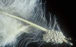
Lice eggs at the base of the feather shaft. Credit: Ohio State University Extension.
Several species of lice live on chickens and cement their eggs on the base of feather shafts.
Identification of the species is based on examining an adult microscopically and the area of the chicken’s body they are found on.
The eggs of Menopon gallinae, the chicken shaft louse, appear as clusters of tiny cream-coloured balls. These lice are generally not regarded as pathogenic, and most birds have small populations of several species at the same time.
If the bird is immunosuppressed by another condition, then the number of lice increases and the feathers appear moth-eaten. The lice are large enough to be visible to the naked eye.
Abnormally large numbers of lice on a chicken should initiate a full clinical examination in addition to a husbandry and diet review, as usually there is an underlying problem.
Some quite loud murmurs may occur with relatively small defects.
It is sensible to assess the patient for clinical signs that would suggest an underlying problem (e.g. lethargy, abnormal breathing pattern or effort, pale gums).
The presence of such signs indicate further diagnostic work up such as echocardiography.
However, if the cat appears very well, is showing no other clinical signs of a problem, and exercises normally, then it is fair to suggest a repeat examination in a few months to reassess the heart murmur and see if it has changed, or to see if the cat has developed any other clinical signs.
Four parameters can be measured to objectively assess dehydration.
PCV and Total Protein should be interpreted together. An increase in both suggests fluid loss except that resulting from acute haemorrhage.
Lactate. Hypoperfusion of tissue increases the concentration of lactate due to anaerobic metabolism of cells. A portable lactate meter quickly and cheaply measures blood lactate in the clinical setting and functions similarly to a glucometer. Lactate <2 mmol/l is normal. Increased lactate correlates with outcome and is a poor prognostic indicator in dogs. Serial lactate measurements can be used to monitor and guide fluid therapy.
Blood gas analysis. The equipment for measuring blood gas analysis is expensive and not usually available in general practice.
Tyzzer’s disease is caused by Clostridium piliforme and can cause severe disease in many animal species.
Transmission is mainly through the fecal-oral route.
It is mostly seen in mice, but infected mice often do not exhibit clinical symptoms. Mice become carriers of the disease and spread the pathogen to other mice and other animal species.
Different mouse strains differ in their susceptibility to the pathogen.
Tyzzer’s disease can occasionally infect rats and should be considered as a possible cause in cases of diarrhoea and sudden death in this species.
The prognosis for affected individuals is very poor. Infection causes necrotic lesions in the liver, digestive organs and heart.
Diagnosing CKD before the appearance of clinical signs is difficult, as no symptoms will be seen until approximately 70% of renal function is lost. However, common signs include increased thirst (polydipsia) and excessive urination (polyuria).
The overall aim is to maintain the phosphate concentration in the lower end of normal range: <1.45mmol/l.
Cats with hyperthyroidism are more vulnerable to bacterial UTIs. One study reported bacterial lower UTIs were diagnosed in 12% of hyperthyroid cats.
Bacterial UTIs are clinically “silent” in a high proportion of older cats, with no haematuria, dysuria, or other signs to indicate their presence.
Where possible, bacterial culture of cystocentesis-obtained urine samples (right) is recommended in hyperthyroid cats at time of diagnosis and periodically thereafter, especially if indicated by clinical signs or previous history.
Resolution of the hypovolaemia is the primary concern. Two large bore catheters are placed in the cephalic veins. If the cephalic veins are not available, the jugular vein is used. Fluid resuscitation through the saphenous veins is unlikely to be successful because of the caudal vena caval obstruction.
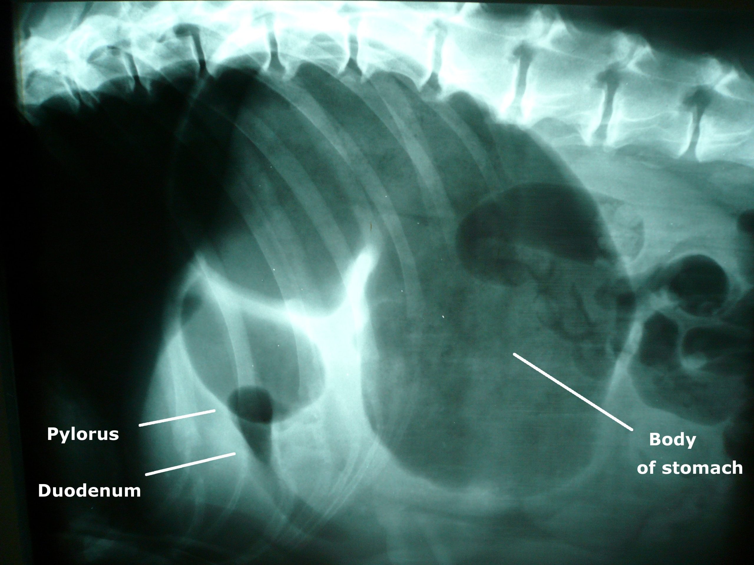
Photo of an x-ray showing gastric dilatation and volvulus in a large mixed-breed dog. The large dark area is the gas trapped in the stomach. The pylorus and duodenum are in an abnormal position cranial to the stomach and are separated by a fold in the stomach, creating a “double bubble” appearance.
Image by Joel Mills [CC-BY-SA-3.0], via Wikimedia Commons.Either isotonic crystalloids (90ml/kg in the first hour) or hypertonic saline (7% NaCl in 6% Dextran: 5 ml/kg given over five minutes) followed by crystalloid are administered.
Controversial treatments
The use of corticosteroids remains controversial. They have many theoretical benefits but have not been unequivocally demonstrated to improve survival in cases of gastric dilatation-volvulus (GDV).
Prophylactic antibiotics are also somewhat controversial, but rational arguments are made for their use. GDV dogs do have increased levels of circulating endotoxin, perhaps indicating increased GI mucosal permeability. Poor perfusion to the liver could inhibit reticuloendothelial function.
Improving tissue perfusion by fluid resuscitation and subsequent gastric decompression and de-rotation can potentially result in the production of damaging, highly reactive oxygen free radicals. These radicals can cause significant reperfusion injury that may be as damaging as the initial hypoperfusion episode.
It is possible treatment to prevent free radical generation may be beneficial in dogs with GDV. Of the drugs trialled in experimental models, deferoxamine, an iron chelator, shows the most promise for clinical application.
Gastric decompression
Ideally, a continuous electrocardiogram is connected. Once the animal has been stabilised, gastric decompression is attempted using a silicone or rubber tube. The tube is pre-measured to the level of the stomach and marked. A 2in roll of tape is placed in the dog’s mouth and the tube passed through the tape and slowly into the oesophagus and stomach.
If resistance is encountered at the level of the cranial oesophageal sphincter, the tube must not be forced, as this could cause rupture of the caudal oesophagus.
In some fractious animals, sedation and intubation is necessary for gastric decompression. If orogastric intubation is unsuccessful, the stomach is decompressed by trocarization. The abdomen is carefully palpated, and the enlarged spleen is avoided. A large gauge catheter (10-12F) is placed into the stomach percutaneously to relieve pressure.
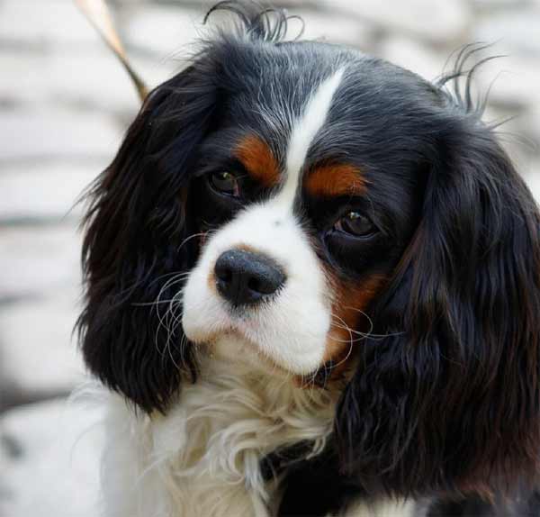
Syringomyelia is rare in most dog breeds but has become widespread in cavalier King Charles spaniels.
Syringomyelia is a condition where fluid filled cavities develop within the spinal cord. It is sometimes known as “neck scratcher’s disease” because scratching in the air near the neck is a common sign.
Owners often report their dog is worse at night, when first getting up, during hot or cold temperature extremes, when excited, or related to posture (e.g. preferring to sleep with their head elevated).
Affected animals may seem overly sensitive to touch, or to scratch more on one side of the neck, ear, shoulder or sternum. This is typically one side only, often while the dog is moving and sometimes without making skin contact. Some dogs, especially younger patients, develop a scoliosis.
Some severe cases may have other neurological deficits such as fore and hind limb limb weakness and ataxia.
Facial nerve paralysis, deafness and seizures have also been associated with the condition, but a link has yet to be proven.


![Photo of an x-ray showing gastric dilatation and volvulus in a large mixed-breed dog. The large dark area is the gas trapped in the stomach. The pylorus and duodenum are in an abnormal position cranial to the stomach and are separated by a fold in the stomach, creating a "double bubble" appearance. By Joel Mills (Own work) [GFDL (http://www.gnu.org/copyleft/fdl.html), CC-BY-SA-3.0 (http://creativecommons.org/licenses/by-sa/3.0/) or CC-BY-SA-2.5-2.0-1.0 (http://creativecommons.org/licenses/by-sa/2.5-2.0-1.0)], via Wikimedia Commons](http://www.vettimes.co.uk/app/uploads/2013/09/GDV_x-ray.jpg)