Tag: Cats

  • Hyponatraemia, pt 2: causes

    Hyponatraemia, pt 2: causes

    The causes of hyponatraemia can be divided into three major categories, based on serum osmolality. This is further divided based on the patient’s volume status (Table 1).

    Most patients we see in clinic fall into the hypovolaemic category, except patients with diabetes mellitus.

    Table 1. Causes of hyponatraemia based on osmolality and volume status (from Guillaumin and DiBartola, 2017).
    Hypo-osmolar Hyperosmolar Normo-osmolar
    Hypovolaemic Normovolaemic Hypervolaemic
    Gastrointestinal fluid loss
    Third-space fluid losses
    Shock
    Hypoadrenocorticism (Addison’s disease)
    Renal insufficiency
    Excessive diuretic administration
    Salt-losing nephropathy
    Cerebral salt wasting syndrome
    Syndrome of inappropriate antidiuretic hormone secretion (SIADH)
    Hypotonic fluid administration
    Hypothyroidism
    Glucocorticoid insufficiency
    Psychogenic polydipsia
    Reset osmostat (SIADH type B)
    Congestive heart failure
    Acute or chronic renal failure
    Nephrotic syndrome
    Hepatic cirrhosis
    Accidental ingestion or injection of water (water intoxication)
    Hyperglycaemia
    Mannitol
    Severe azotaemia
    Hyperlipidaemia
    Hyperproteinaemia

    Common causes

    In dogs, the three most common causes of hyponatraemia are:

    • gastrointestinal (GI) fluid loss
    • third-space fluid loss
    • fluid shift from intracellular fluid to extracellular fluid (ECF) as a result of hyperglycaemia

    In cats, the three most common causes of hyponatraemia are:

    • urologic diseases
    • GI fluid loss
    • third-space fluid losses

    In most patients, more than one pathophysiologic factor is likely to be contributing to the hyponatraemia.

    Circulating volume

    Hypovolaemic patients – those with, for example, GI losses, hypoadrenocorticism, renal losses and haemorrhagic shock – have a reduced effective circulating volume. ECF contraction triggers antidiuretic hormone (ADH) secretion, which leads to increases in free water absorption and thirst, and results in dilution of the serum sodium concentration. Aldosterone secretion is reduced in hypoadrenocorticism, so an overall reduction in sodium reabsorption compounds the problem.

    Hypervolaemic patients are those with an increased fluid retention state, such as:

    • congestive heart failure (pulmonary oedema)
    • advanced hepatic failure (ascites, third-space fluid)
    • renal failure
    • free water ingestion

    Congestive heart failure patients have a reduced cardiac output and, therefore, a decreased effective circulating volume, despite the presence of the extra fluid status. Renin-angiotensin activation leads to release of ADH and aldosterone, resulting in sodium and free water reabsorption, and increased thirst. Both lead to an excess of free water retention.

    Advanced hepatic (cirrhosis) or renal failure (nephrotic syndrome) both result in hypoalbuminaemia, leading to fluid shifting into the interstitial space and third space, reducing effective circulating volumes. This leads to activation of ADH to increase free water reabsorption, to restore the circulating volume in the face of existing hypervolaemia and hyponatraemia.

    Diabetic patients

    Moderate to severe hyperglycaemic diabetic patients can be either hyperosmolar or normo-osmolar, depending on the serum blood glucose concentration. Hyponatraemia occurs when water shifts from the intracellular fluid to the ECF down the osmotic gradient, diluting the serum sodium content.

    Despite this osmotic shift, not all diabetic patients develop hyponatraemia. Glucosuria also causes also causes a renal osmotic shift, sometimes resulting in urine water loss in excess to sodium. This offsets the hyponatraemia – in some cases, hypernatraemia results.

    Treatment

    Treatment of hyponatraemia hinges on how quickly it developed and the volume status of the patient. The rule of thumb is to correct hyponatraemia slowly – not exceeding 0.5meq/L/hr – especially in chronic cases, or cases where the duration of hyponatraemia is unknown. Keeping to this rate is paramount until serum sodium concentration reaches 130meq/L.

    In acute patients with severe clinical signs, such as seizures, some clinicians may choose to use a higher rate of 1meq/L/hr to 2meq/L/hr until clinical signs resolved.

    It should be emphasised, once again, this rate should never be used in chronic patients, patients with an unknown duration of hyponatraemia, or where frequent serum sodium concentration cannot be monitored. The rapid correction of hyponatraemia can lead to osmotic demyelination syndrome (myelinolysis).

    Its effect will not be apparent until three or four days after therapy, and can result in neurological abnormalities such as:

    • weakness
    • ataxia
    • dysphagia
    • paresis
    • coma

    For that reason, frequent electrolyte measurements are required, starting hourly then once a suitable rate of increase has been established and less frequently thereafter.

    • Part 3 will look at how to correct patients with hyponatraemia.

    Reference

    Guillaumin J and DiBartola SP (2017). A quick reference on hyponatremia, Veterinary Clinics of North America: Small Animal Practice 47(2): 213-217.

  • Hyponatraemia, pt 1: clinical signs

    Hyponatraemia, pt 1: clinical signs

    Hyponatraemia is a relatively common electrolyte disturbance encountered in critically ill patients, and the most common sodium disturbance of small animals.

    In most cases, this is caused by an increased retention of free water, as opposed to the loss of sodium in excess of water.

    Low serum sodium concentration

    Hyponatraemia is defined as serum concentration lower than 140mEq/L in dogs and lower than 149mEq/L in cats.

    The serum sodium concentration measured is not the total body sodium content, but the amount of sodium relative to the volume of water in the body. For this reason, patients with hyponatraemia can actually have decreased, increased or normal total body sodium content.

    This series will look briefly at the modulators of the sodium and water balance, clinical signs associated with hyponatraemia, the most common causes in small animals, the pathophysiology behind these changes, and treatment and management.

    ECF volume

    hyponatraemia
    An example of hyponatraemia.

    Sodium is the main osmotically active particle in the extracellular fluid (ECF), so is the main determining factor of the ECF volume. Any disease process that alters the patient’s ECF volume will lead to hyponatraemia, such as:

    • dehydration
    • polyuria
    • polydipsia
    • vomiting
    • diarrhoea
    • cardiac diseases
    • pleural or peritoneal effusion

    The modulators of water and sodium balance are also different, so should be thought of as different processes.

    Water balance is modulated by thirst and antidiuretic hormone, and the effect of this is to maintain normal serum osmolality and serum sodium concentration.

    Modulators of sodium balance aim to maintain normal ECF volume. It adjusts this by altering the amount of renal sodium excretion; an expansion of ECF volume will lead to an increased sodium excretion, while a reduction in ECF volume will lead to increased sodium retention.

    Rate and magnitude

    The clinical signs of hyponatraemia are both dependent on the magnitude of the decrease and the rate at which it developed.

    In mild or chronic patients, no visible clinical signs can exist. In severe (lower than 125mEq/L) and acute cases, clinical signs exhibited are typically neurological, reflecting cerebral oedema. Possibilities include:

    • lethargy
    • anorexia
    • weakness
    • incoordination
    • disorientation
    • seizures
    • coma

    Patients with acute hyponatraemia – for example, water intoxication – are more likely to show clinical signs, compared to those with chronic hyponatraemia, because the brain takes time (at least 24 to 48 hours) to produce idiogenic osmoles, osmotically active molecules that help shift free water out of brain cells.

    Therefore, any acute hyponatraemia that develops within a 24 to 48-hour period tend to show clinical signs, whereas chronic cases are less likely.

    • Next week’s blog will look into the different causes of hyponatraemia and how they result in sodium loss.
  • Tales of an Indian winter

    Tales of an Indian winter

    It had been an ambition of mine since the beginning of vet school to do some type of work abroad, whether it be preclinical or clinical, a paid position or volunteer work.

    A big reason I undertook an intercalated MSc was for the option it presented for a three-month research period in Western Australia. Sadly, COVID-19 put a stop to that and my research never wandered further than my desk – but, if anything, the pandemic made me feel even more passionate about travelling for my EMS.

    Gone to Goa

    Weekends spent “lolling on the beach” were well-deserved, says Eleanor.

    A friend and I both settled on a small rescue centre in Goa, India, for the placement (neither of us feeling quite brave enough to go it alone) and despite planning it almost a year in advance, the date caught up with us quite quickly. Before we knew it, we were there.

    Let the record show that the motivation for this trip was not to escape from the harsh English January weather, nor to fill up on delicious curries, although the temperature did make a welcome change and I’m unsure a takeaway will ever cut it again.

    The whole reason for the placement was to gain the kind of surgical experience that just isn’t readily available to students in the UK.

    Understandably, vet practices can take a while to warm up to students enough to trust them to carve into somebody’s beloved animals, but this makes for generation after generation of new grads who feel completely out of depth with a scalpel in their hands.

    Great(er than our) expectations

    The placement’s main advertising pull had been as an opportunity to gain incredible surgical experience, but we had gone into it with some trepidation that it wasn’t going to be nearly as busy and hands-on as we’d hoped. It turned out to surpass our expectations and go right out the other side…

    Weekends spent lolling on the beach were well-deserved after numerous 11-hour shifts with numb fingers and thumbs from uncooperative clamps and needle holders.

    The surgical side of the trip deserves an article of its own – but suffice it to say that, between the two of us, my friend and I neutered almost 50 dogs and cats, including 15 unassisted but supervised dog spays. It was an incredible rewarding feeling when each surgery finished, knowing we were doing even just a small bit in the effort to reduce India’s stray population.

    Eleanor found her EMS placement in Goa “incredible rewarding”.

    Learning valuable lessons

    Let it be said, I am not the most confident of travellers, and 18 hours of travel across three planes and four airports are not for the faint of heart, but neither is India – and while I have entirely fallen in love with the country, its beauty and its animals, there was a lot of disorganisation that made my poor little control-freak brain spin.

    I think that learning to take each day as it comes, and constantly adapting to new situations or pressures has taught me a lot of valuable skills in a very short space of time.

    In particular, the vet who taught and supervised us was invaluable in making the placement such a success. She gave us an incredible amount of patience and taught me skills in both surgery and how to face a stressful situation that I will carry with me throughout my career.

  • Feline aortic thromboembolism

    Feline aortic thromboembolism

    If a cat comes in unable to walk, consider the three Ps:

    • pain
    • paralysis
    • pulselessness

    gerardo_paws
    Figure 1. Colour change in the paws of a cat.

    Feline aortic thromboembolism (FATE) should be on top of your differentials.

    Figure 1 demonstrates the colour change in the paws of an affected cat outlining blood flow: the pink pad is the unaffected cat’s front paw, while the pale pad is on the affected hind limb that will be cold to the touch.

    Cardiological problems

    Often FATE is a secondary condition in cats with heart disease.

    The heart forms clots in the distal aorta that occlude flow to the femoral arteries. With the femoral arteries being the main arteries providing blood flow to the hind limbs, symptoms become apparent.

    Symptoms can include:

    • sudden hind limb paralysis
    • cold hind limbs
    • vocalising
    • pain
  • Nutritional healing power

    Nutritional healing power

    Nutrition is a key factor in a patients recovery; in fact, numerous studies show getting patients to eat as soon as possible or providing nutritional support early has several benefits:

    • Patients start to eat on their own earlier.
    • They are less nauseous once they start.
    • Reduced mortality.
    • Improved wound healing.

    All of these contribute to overall improved outcomes for the patient.

    Encouraging patients

    At Animal Emergency Service we treat the sickest of the sick so we work hard towards encouraging patients – just like the kitten pictured above – to eat as soon as possible. So, after they have recovered from their anaesthesia we make sure there are no contraindications, we address their nausea and pain, then offer food.

    It is best for the patient and feeding to use as much of the gastrointestinal tract as possible, meaning it’s better if they eat on their own, otherwise the next best thing is an oesophageal tube, followed by a tube into the stomach, such as a nasogastric tube.

    Focused approach

    So, in combination with the management of pain, nausea and the underlying illness, we first encourage them to eat on their own. We begin with offering an assortment of different foods, warmed up to increase aromas, or ask owners to try to feed their pet.

    If they are critically ill we will take a more focused approach with feeding tubes, as they are unlikely to eat on their own by themselves for several days.

    We feel a proactive approach to early nutrition helps get our patients home to their families earlier.

  • Are rabbits really ‘exotic’?

    Are rabbits really ‘exotic’?

    According to PDSA [PDSA Animal Wellbeing (PAW) Report 2022], rabbits are the third most popular pet in the UK behind dogs and cats. With an estimated 1.1 million pet rabbits in the country, that’s about about a tenth of the population of pet dogs and cats, which hover around the 9 to 10 million mark.

    So, if the pet ratio of dogs/cats:rabbits is 10:1, why isn’t this reflected in our teaching? Despite rabbit populations being endemic to the UK for more than a thousand years, they always seem to get lumped with guinea pigs and the cold-blooded pets like lizards and corn snakes when it comes to textbooks or university curriculums.

    I can confidently say my education on rabbit physiology and medicine has been dramatically less than 10% of what I’ve received for small animal medicine. Perhaps this is why many vets, especially new or recent graduates, feel more confident handing off any rabbit patients to the resident “expert” of the practice or even referring to an exotics specialist, rather than seeing it themselves.

    Accessibility

    It’s a sad truth that the less convenient education and health care are to access, the less people will reach for them. By extension, the less veterinary practices that advertise care for rabbits (and other exotics), the less rabbits are likely to be registered at a practice and receive regular preventive care.

    For example, as a native to the land of Kent, I only know of two or three practices that would call themselves “exotic specialists” and I know that, for a lot of rabbit owners, traveling half way across the county to visit one of these few practices would not be practical or plausible. Perhaps this is why, according to PDSA reports, at least 11% of pet rabbits receive no preventive health care, including vaccinations.

    rabbit
    Rabbits are a social species that has evolved to live in groups, not alone.

    Education

    The value of a veterinary consultation is not simply to talk through clinical signs or address a flea outbreak in the home, it’s a chance for owners to discuss management issues or to ask for general advice. When rabbits aren’t brought in for routine consultations, then discussions about their diet, husbandry and behavioural needs don’t get to be had.

    Some vets are already worried that the development of an annual rabbit haemorrhagic disease (RHD) booster rather than biannual is going to dramatically reduce rabbit welfare by halving the number of times these pets receive a clinical exam.

    Welfare

    Of course, like all “exotics”, there’s the argument to be made as to whether these animals are suitable pets in the first place. Personally, I feel that this is a moot point for the time being.

    The fact that more than 50% of pet rabbits are housed by themselves with no companionship speaks volumes about the lack of knowledge the general public possesses on how to care for these animals. However, with more than a million of them currently out there, they’re not going away anytime soon.

    The best we can do as professionals is educate our clients so welfare can be maximised as much as possible… and that starts with educating ourselves. I hope that in the near future the landscape of the veterinary degree can shift to better reflect the current demand for exotic vets – or at least rabbit vets.

  • Blood gas analysis, pt 3: interpreting pH

    Blood gas analysis, pt 3: interpreting pH

    After taking note of the direction of the pH shift – acidaemia or alkalaemia – it is important to determine the primary and secondary causes.

    If an acidaemia is present (pH less than 7.35), an underlying respiratory or metabolic acidosis, or both, must exist. Similarly, if an alkalaemia is present (pH more than 7.45), an underlying respiratory or metabolic alkalosis, or both, must be present.

    This is usually very simple, with the exception of cases presenting with a normal pH (between 7.35 and 7.45, slightly higher for cats).

    Cases with normal pH

    uparrow
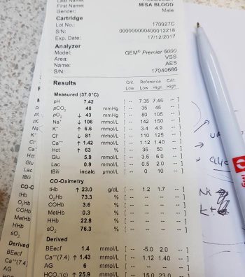
For cases with a normal pH, we need to determine which category it falls into:

    1. No acid-base disturbance
      • Both respiratory and metabolic components are within the normal reference range.
    2. Complete compensation for the acid-base disturbance
      • This requires specific calculations that will be discussed in a later blog.
      • This cannot be determined by glancing at the figures alone.
    3. Two opposing acid-base disturbances (a mixed disorder), which are cancelling the effect of each other out in terms of pH.
      • Both the respiratory and metabolic components will be outside of their reference range, going in the opposite direction to each other.

    Determining primary disorder

    Since these animals are within the normal pH range – particularly those with complete compensation – how can you tell which is the primary disease process?

    A golden rule of thumb is: even with maximal compensation, the pH will still usually move in the same direction as the primary problem.

    Therefore, if the pH lies towards the acidaemic side of the mid-point of the pH range (less than 7.4), the primary disease process is an acidosis. By the same token, if the pH lies towards the alkalaemic side of the midpoint of the pH range (more than 7.4), it will have a primary alkalosis disorder.

    The reason behind this is the body does not usually overcompensate for an acid-base disturbance.

    Secondary disorder

    downarrowOnce the primary disorder has been identified, we need to look at whether a secondary disorder is also present and, if so, whether this is the result of compensation or a true mixed process.

    To determine whether compensation occurred, you need to understand the timeline for when compensation usually occurs.

    With respiratory compensation, this typically starts immediately, but may take up to 8 to 12 hours to occur. This is because adjusting levels of CO2 is relatively easy, with the change of respiratory rate and patterns.

    On the other hand, metabolic compensations take approximately one to three days to occur, since renal excretion of hydrogen ions or retention of bicarbonate takes longer.

    Variation magnitude

    If the magnitude of the observed variation is compatible with compensation alone (this requires calculation), a compensatory mechanism is likely. A mixed process (mixed acid-base disorder) is present if the magnitude:

    • does not correspond to the clinical status of the patient
    • falls outside of the compensation time frame
    • is outside of the expected magnitude of compensation

    All other causes for why the acid base is moving in the opposite direction must be ruled out before determining a secondary process is present.

    Once the primary and, if present, secondary disorder are determined, the next step is to determine the cause of the respiratory and metabolic acidosis and alkalosis.

  • Catharsis, enemas and colonic irrigation for acute oral poisoning

    Catharsis, enemas and colonic irrigation for acute oral poisoning

    Great news for those who hate enemas: you may not have to do any of these ever again. This is the consensus by both the American Academy of Clinical Toxicology, and the European Association of Poisons Centres and Clinical Toxicologists.

    The theory behind these procedures is legitimate – reducing systemic exposure of a toxicant by accelerating gastrointestinal tract (GIT) expulsion. But this is assuming the toxicant is absorbed very slowly, undergoes substantial enterohepatic cycling, or undergoes slow reabsorption in the lower GIT – all of which are uncommon characteristics of the types of toxicants veterinary patients are exposed to.

    In fact, most toxicants of veterinary interests are generally rapidly absorbed in the upper GIT and absorption are not affected by catharsis.

    Lack of evidence

    No clinical evidence exists to support the use of a cathartic alone, or in combination with activated charcoal, to reduce the bioavailability of drugs or to improve the clinical outcome of poisoned patients. In fact, some evidence shows systemic exposure is increased following oral dosing of sorbitol, with activated charcoal, in canine paracetamol poisoning cases.

    Similarly, no evidence exists that enemas and/or colonic irrigation improve clinical outcome in the treatment of oral poisoning.

    The risks can be quite high with these procedures, with patients at risk of:

    • haemorrhage (in the case of anticoagulant vitamin K antagonist rodenticides, for example)
    • electrolyte destabilisation
    • bowel perforation
    • rectal prolapse
    • phosphate toxicities (cats)

    The risks simply do not outweigh the benefit (or lack thereof). In fact, repeated dosing with combination preparations containing sorbitol and activated charcoal is not recommended.

    This may be the most exciting news in veterinary medicine!

  • Temporary catheters in obstructed FLUTDs: buying time with a blocked cat

    Temporary catheters in obstructed FLUTDs: buying time with a blocked cat

    Obstructive feline lower urinary tract disease (FLUTD) is a common presentation in both general practice and emergency settings.

    Every clinician has his or her own approach to treating and managing a cat with obstructive FLUTD signs. Working in an emergency setting, once I have confirmed an obstructed bladder via palpation, I focus on trying to relieve the obstruction as quickly as possible.

    The first step is obtaining consent from the client to administer pain relief (an opioid IV or IM), place an IV catheter, collect blood for biochemistry, electrolyte and blood gas analysis, and temporarily relieve the obstruction.

    At our hospital, we achieve temporary relief of the obstruction generally within 15 minutes of patient arrival.

    Process

    blocked cat
    Obstructive feline lower urinary tract disease is a common presentation in general practice.

    We do this in three steps:

    1. Assess the tip of the penis, occasionally a crystal/mucus plug is all that is blocking the penis.
    2. If this is not the case, I pass a pre-lubricated 22g IV catheter tip (without the stylet) into the penis with a 10ml syringe, containing 0.9% NaCl, connected for hydropropulsion. In the vast majority of cases, this helps to dislodge the urethral blockage enough to enable some urine to pass (urination suggests active urination by the cat).
    3. Once urine is flowing, I pass a 12cm or 14cm rigid catheter, tape it to the tail and leave it in place to allow constant drainage.

    If the 22g IV catheter does not relieve the obstruction, I would use a rigid catheter and progressively advance it up the urethra while hydropropulsing with the saline the entire time. Once unblocked, then I will tape it to the tail as aforementioned.

    Quick Tip: Once you have the catheter in the tip of the penis, pull the prepuce straight out to straighten the penis and thus the penile urethra. Otherwise, the bend in the penile urethra may hinder the passage of the catheter.

    Benefits

    The benefits I see of placing a temporary urinary catheter include:

    • immediate relief to the patient and reduces their stress levels
    • provides a sample for urinalysis
    • allows you time to run through the diagnostic and treatment plan in more detail with clients
    • buys you time to stabilise the patient for their anaesthetic later to place a closed system indwelling urinary catheter and then bladder lavage

    Quite often, your patient would present unwell enough that you should have no issues (resistance to) passing this temporary urinary catheter, provided you have given pain relief on presentation.

    In fractious patients, I usually forgo the temporary catheter and focus on stabilising the patient. The aim is to have them stable as soon as possible for sedation or a general anaesthesia to place a longer indwelling urinary catheter.

  • Maintenance fluids

    Maintenance fluids

    A while ago we discussed the components of a fluid therapy plan and talked about hydration deficits. This week I want to touch on maintenance fluids.

    Gerardo_IVF
    IV fluids

    Maintenance rates are typically calculated using the following formulae:

    ml/day = 80 × bodyweight (kg)0.75 (cats)
    ml/day = 132 × bodyweight (kg)0.75 (dogs)

    or
    ml/day = 30 × bodyweight (kg) + 70

    These formulae better estimate the needs of smaller and larger patients. The flat 3ml/kg/hr underestimates for small patients and overestimates for larger patients.

    This maintenance rate is in addition to rehydration rates.

    So what sort of fluid should you use for maintenance?

    True “maintenance” crystalloids:

    • used to replace ongoing fluid and electrolyte loss from normal metabolism, not to replace perfusion and hydration deficits or ongoing losses from diarrhoea, for example
    • sodium concentration less than plasma
    • potassium concentrations higher than plasma
    • glucose sometimes added to bring solute concentrations similar to extracellular fluid

    Do you have to use maintenance crystalloids or can you use replacement crystalloids?

    Replacement crystalloids are more frequently used for maintenance fluid therapy rather than maintenance crystalloids. This is because they are more readily available, we are more familiar with their use and effect, and patients are generally continued on these after perfusion and hydration deficits have been corrected.

    In reality, most of the time it doesn’t really matter if we are using replacement crystalloids for maintenance therapy as the patient can manage the excess sodium, but some patients – especially cats – may require potassium supplementation. The key point is regular assessment of the patient’s hydration status and electrolytes – for example, every 12 to 24 hours for patients on IV fluids and not eating.