Tag: Cats

  • Dystocia, pt 4: caesarean tips

    Dystocia, pt 4: caesarean tips

    Prolonged hypoxaemia, hypotension and hypoventilation are the top three causes of periparturient fetal mortality – for these reasons, all precautions must be taken to avoid it.

    As soon as authorisation has been obtained to proceed with a caesarean section, the patient should be stabilised immediately. This includes having perioperative blood work performed, and clinical hypoperfusion (common in patients that have gone through prolonged stage two labour) and hypotension corrected as soon as possible, usually with fluid boluses.

    While fluid deficits are being corrected, preoperative monitoring and surgical site preparation (clipping and the initial stages of surgical scrub) can be performed with the patient still conscious. This will significantly reduce the time the patient is anaesthetised, as isoflurane potentiates hypotension.

    Physiological changes

    A few physiological changes in periparturient patients must be considered before anaesthetising them.

    Higher oxygen demand

    Firstly, pregnant animals have a higher oxygen demand due to the developing fetuses. However, due to their large gravid uteruses, they have decreased functional residual capacity and total lung volume. This is further exacerbated when animals are placed in dorsal recumbency, with increased pressure on their diaphragms.

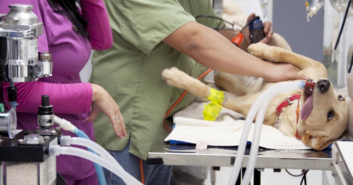
    For this reason, pregnant animals should always be preoxygenated prior to induction – with as much of the patient preparation completed – to reduce the risk of hypoxaemia. This is one of the main reasons the time from induction to delivery of the puppies should be as short as possible.

    Sensitivity to anaesthetic agents

    Secondly, pregnant animals have an increased sensitivity to anaesthetic agents. Blood volume and cardiac output also increase dramatically during pregnancy; therefore, if blood loss occurs and blood pressure is not maintained, significant hypotension can occur.

    Any medication that crosses the blood-brain barrier will equally cross the placental barrier; therefore, the effect of medications can be reduced by a few things. Firstly, the use of local anaesthetics (such as epidurals) can be employed to minimise inhalation anaesthetics, thus their hypotensive effects. Always use minimal drug dosages that achieve the desired effect. Short-acting, rapidly metabolised drugs and reversible drugs should be used whenever possible.

    Don’t premedicate

    Premedication of caesarean patients is strictly avoided at our hospital. Acepromazine can result in hypotension and has a long duration of action, while opioids can cause potent respiratory depressants in unborn fetuses as it crosses the placenta.

    Puppies and kittens born heavily narcotised or sedated will have bradycardia and may not take spontaneous breaths, further increasing the risk of mortality.

    Speedy delivery

    IMAGE: Pilipipa / Adobe Stock
    Once the patient has been induced, the speed of delivering the fetuses is of paramount importance. Image © Pilipipa / Adobe Stock

    Once the patient has been induced, the speed of delivering the fetuses is of paramount importance. Inhalant anaeshetics causes maternal vasodilation and decreases uterine blood flow, as well as neonatal depression.

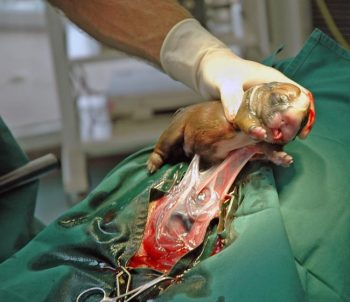
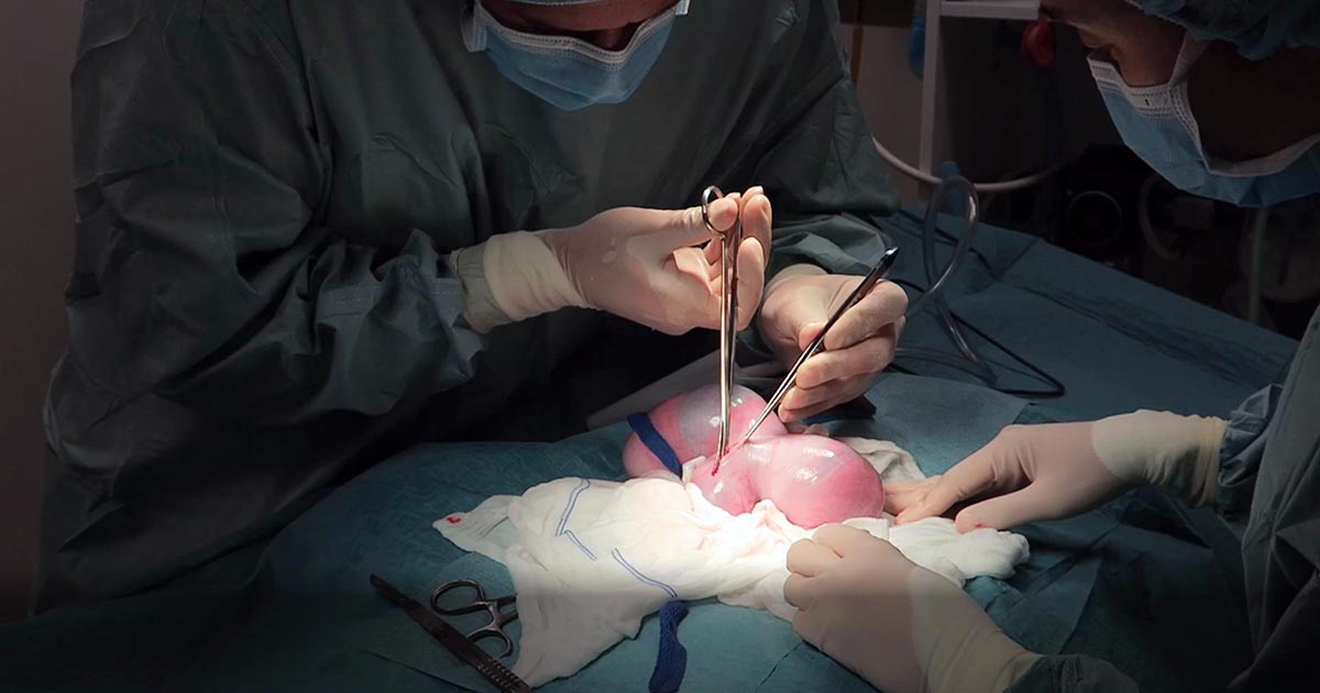
    Making a large abdominal incision is highly advised, despite the fact it may take longer to close, as it enables faster and more gentle manipulation of a large fetus-filled uterus.

    The traditional caesarean technique involves a single incision in the uterine body. Fetuses should be gently squeezed towards the incision. In patients with many fetuses, especially large-breed dogs, making a single uterine body incision may significantly delay delivery of the fetuses. Concern also exists with excessive traction and manipulation of uterine blood vessels when trying to manipulate the fetuses to the uterine body incision. In these cases, additional incisions in the uterine horns can be made.

    With this method, surgical time for closure will be longer and considered carefully in patients where future breeding is likely, as the risks of adhesions and uterine rupture in subsequent pregnancies increases compared to the single uterine body incision method.

    Closure of the uterine wall should always be in two layers – firstly, an appositional simple continuous pattern; followed by a second inverting (Cushing or Lembert) pattern.

    Post-fetal removal

    Once the fetuses have been removed, a few medications can be given safely intraoperatively.

    Firstly, opioids are safe at this time. Fast analgesia can be achieved when the opioid is given IV. Oxytocin can also be administered during this time, but beware uterine involution and contraction will be immediate; therefore, close attention needs to be paid to the uterine sutures to ensure they have not become loose.

    NSAIDs should be avoided in lactating queens and bitches, as most are excreted in the milk. Safety data has not been established in lactating animals, while previous animal studies have shown an adverse effect on the fetus.

    Tramadol, a synthetic opiate-like (μ receptor) agonist, has high analgesic effects. Tramadol and its active metabolite are known to enter maternal milk, albeit at very low levels. No animal reproduction studies exist to establish its safety in use in neonates, but it is an analgesic considered safe to use in young animals.

    Conclusion

    Caesarean section is the one emergency surgical procedure where speed is of essence.

    With prompt stablisation, pre-induction surgical preparation, fast delivery of fetuses and avoidance of certain medications, the chances of survival of the already distressed fetuses can dramatically increase.

  • Dystocia, pt 2: diagnostics

    Dystocia, pt 2: diagnostics

    Part one of this series covered the stages of labour and indications dystocia is present.

    Once the bitch presents to the clinic, a few basic diagnostic checks need completing to determine the status of the bitch/queen and the fetuses.

    Physical examination

    The first is a thorough physical examination, starting with the bitch or queen:

    • Demeanour, hydration status, vital signs, mucous membrane colour, capillary refill time and temperature are important.
    • Pregnancy anaemia is not uncommon; however, for patients with a haemorrhagic discharge, it is important to know their cardiovascular status.
    • A thorough abdominal palpation should be carried out to assess comfort level and palpation for the presence of fetuses. Palpating fetuses can be difficult and cannot confirm if no fetuses are present.
    • A digital vaginal examination should be performed. Feathering response – also known as the Ferguson reflex in human medicine – is the neuroendocrine reflex where the self-sustained cycle of uterine contractions is initiated by firm pressure on the dorsal aspect of the vestibulovaginal wall. If this is absent, the patient is unlikely to progress with the parturition unaided.
    • Palpation of fetuses in the canal can help decide whether surgical management is required. Obvious fetal malposition, malposture or malpresentation, or fetopelvic disparity, will be indications of caesarean. Abnormal pelvic diameter is also another reason to not proceed with medical management. To confirm these suspicions, abdominal radiography is required.
    • Radiographs will also help determine the number of fetuses to be expected, the signs of fetal death (presence of gas surrounding the fetus) and aforementioned fetomaternal abnormalities. I always repeat radiographs after the expected number of neonates is passed, to make sure I have not miscounted at the start.

    Ultrasound

    Panel 1. Heart rate ranges to help indicate stress of fetuses

    Dogs:

    • normal – 180 to 220 beats per minute (bpm)
    • Stressed – 160bpm
    • Real concern – less than 160bpm

    Cats:

    • normal – more than 220pbm
    • fetal stress – less than 180bpm

    The second important diagnostic tool is ultrasound.

    Fetal heart rates are good indicators of fetal stress. Some heart rate ranges that can help provide information about the status of the fetuses are detailed in Panel 1. These ranges vary between sources, but are good guidelines.

    Ultrasounds can also help visualise the maturation status of the fetuses. At-term fetuses should have normal hepatic, renal and intestinal development. Intestinal peristalsis should be evident in at-term fetuses.

    Other diagnostics

    Other diagnostics may be indicated for patients, depending on the status of the bitch/queen:

    • If the patient is stable, but dystocia is present, a minimum database would include PCV/total protein, electrolytes, glucose, ionised calcium, lactate and acid-base balance.
    • Serum ionised calcium levels are important, as they influence the strength of contractions and how much supplementation is required.
    • Hypoglycaemia needs to be ruled out as a cause of dystocia, especially when large litters are involved.
    • If the patient is unstable or systemically unwell, include complete blood count, blood smears and biochemistry.
    • Physiological pregnancy anaemia can be present. The presence of regenerative response can help differentiate this from acute haemorrhage.
    • Abnormal leukocyte panel, especially with the presence of degenerative left shift, can indicate the presence of an infection – especially if toxic changes are present in the neutrophil.

    Part three will briefly look at the medical management of dystocia and when surgical intervention is required.

  • Dystocia, pt 1: labour stages

    Dystocia, pt 1: labour stages

    Now most female canine patients are spayed, it comes as no surprise reproductive emergencies are not as common.

    One confusion seems to be not knowing how to determine a true dystocia emergency – especially when given advice over the telephone – from the process of normal parturition.

    Another concern is how to confidently form a diagnostic pathway to determine the cause of dystocia – especially for reasons other than obvious physical abnormalities (for example, fetopelvic disparity and fetal malposition).

    Often, once we decide to go down the medical treatment pathway – whether the result of findings or owner/financial constraint – no one is confident as to what medication should be used and how often drugs can be given safely.

    This series of blogs will address these issues in a step-by-step manner. Hopefully, by the end, you will be confident in the diagnosis and management of dystocia.

    Labour stages

    Before moving on to the signs of dystocia, let’s go through the signs of labour.

    First stage labour

    First stage labour is characterised by panting, tremoring, nesting behaviour, a drop in core temperature – usually a drop by almost 1°C 24 hours prior to second stage labour – and a drop of progesterone to below 2mg/ml.

    Dog and puppy.
    Third stage labour occurs generally within 15 minutes after passing a puppy or kitten. Image © foto ARts / Adobe Stock
    • dogs: approximately 6 to 12 hours
    • cats: approximately 6 to 24 hours

    Second stage labour

    Second stage labour is landmarked by the water breaking, visible abdominal contractions, and the allantoic/amniotic sac or fetal parts visible from the vulva.

    If vulval discharge is present, they should be clear. Excessive amount of bright red haemorrhage, green or black discharge prior to delivery, or purulent material can indicate a pathological process requiring immediate veterinary attention.

    • dogs: approximately 3 to 6 hours
    • cats: approximately 6 to 24 hours

    Third stage labour

    Third stage labour this is when passage of all the placenta has occurred, generally within 15 minutes after passing a puppy or kitten.

    Clues

    Now we understand the normal progression of parturition, a few clues exist in the history that could suggest dystocia may be present.

    Some breeders will often know the ovulation timing of the patient – especially if AI was performed. Tests such as progesterone levels, luteal hormone (LH) levels, cytology and vaginoscopy are some ways where it can help time the ovulation.

    The normal gestation length should not be any longer than 66 days from the LH surge or, if the ovulation history is unknown, 72 days from the last known breeding.

    History of prior dystocia is a warning, as most animals with prior parturition difficulties are more likely to develop dystocia again.

    The same goes for animals that have previously required a caesarean. Their risk of requiring future caesareans is high, with further risk of uterine rupture if dystocia happens again.

    Image © Pilipipa / Adobe Stock
    Animals that have previously had a caesarean are at high risk of requiring future caesareans, with further risk of uterine rupture if dystocia happens again. Image © Pilipipa / Adobe Stock

    Intervention signs

    Owners often telephone after the failure of normal progression of delivery. The signs that always require immediate intervention are:

    • more than 4 hours have passed from the rupture of the first chorioallantois
    • more than 2 hours between delivery
    • more than 30 minutes of strong abdominal contraction and no delivery
    • presence of green or black discharge before delivery
    • large amount of bright red haemorrhage
    • abnormal amount of pain during contractions
    • collapse of the bitch or distracted mothering

    Any of these signs require immediate presentation to the veterinarian. Delivery of stillborn puppies is also an indication where veterinary attention is indicated.

    Finally, if owners are concerned, it is best to advise veterinary assessment rather than try to convince them everything is okay based on what they describe over the telephone.

  • Pancreatitis, pt 2: treatment and prognosis

    Pancreatitis, pt 2: treatment and prognosis

    Last week we covered diagnosing pancreatitis and the challenges associated with doing so. This week we look at management.

    The treatment of pancreatitis largely involves supportive care and monitoring for potential complications. Here, we recap the fundamentals.

    IV fluids

    IV fluids are critical in the acute phase to restore perfusion to core organs and correct hydration deficits. Once achieved, the goal is then to cover maintenance requirements and ongoing losses.

    Hypoproteinemia can develop due to a combination of haemodilution, increased losses in to inflammatory exudates or into gastrointestinal tract, and decreased production due to reduced intake.

    Colloidal therapy or plasma can be considered, but enteral nutrition is more effective and has numerous other benefits.

    Pain relief

    Pain relief largely involves the use of opioids.

    Pure opioid agonists, such as methadone, fentanyl constant rate infusions (CRIs) or fentanyl patches, are most commonly seen in dogs. In cats, partial agonists, such as buprenorphine, are favoured as the condition is generally less painful compared to dogs.

    NSAIDs should be avoided due to concerns of poor perfusion, concurrent kidney and gastrointestinal involvement.

    Antiemetic therapy

    Metoclopromide CRI and maropitant are the mainstays. Antacids, such as esomeprozole, are included to help reduce the risk of gastric ulceration from stress.

    Early enteral nutrition

    Numerous studies have demonstrated the benefit of early enteral nutrition and patients should be encouraged to eat a low-fat diet as soon as possible. Enteral nutrition has been shown to be more beneficial than withholding food as it reduces recovery times and helps maintain enterocyte health.

    If there has been a protracted period of anorexia then a nasogastric or nasoesophageal tube should be placed. A nasoesophageal tube has the added benefit of allowing excessive gastric secretions to be suctioned, as well as for feeding. Due the chronic nature of the disease in cats, oesophagostomy tubes are often placed so that nutritional support can be delivery for prolonged periods of time.

    Antibiotics

    Antibiotics are generally not indicated in canine pancreatitis unless markers of sepsis or if a septic exudate are found (this is an indication for exploratory surgery). In which case, triple antibiotic therapy consisting most commonly of amoxicillin, enrofloxacin and metronidazole are indicated.

    It has been reported 35% of feline pancreatitis patients have bacterial infections; therefore, antibiotics are often included in the treatment of feline pancreatitis.

    Exploratory laparotomy

    This is indicated when the following has occurred:

    • Extra-hepatic biliary obstruction that does not resolve with medical management.
    • Septic abdomen (as mentioned above).
    • Pancreatic abscessation.

    Prognosis

    The prognosis for pancreatitis is variable and dependent on the severity of the disease.

    In mild to moderate canine pancreatitis (which is the most common), the prognosis is generally good as they will normally respond well to supportive therapy and resolution of clinical signs occurs within several days of initiating treatment. However, with severe acute, necrotising pancreatitis, the prognosis is guarded as it can progress into systemic inflammatory response syndrome, disseminated intravascular coagulation and multiple organ failure – which has a high mortality rate.

    The prognosis of acute feline pancreatitis is generally guarded due to the chronic nature of the disease and the propensity to involve multiple organ systems.

  • Pancreatitis, pt 1: diagnosis

    Pancreatitis, pt 1: diagnosis

    Pancreatitis is one of the most common exocrine conditions seen in small animal practice. It is caused by premature activation of enzymes (zymogens) within the pancreas leading to autodigestion, and can result in severe morbidity with the potential to lead to mortality.

    To this day, the diagnosis of pancreatitis remains a challenge – especially in feline patients – and relies on the use of a combination of history, signalment, clinical signs and diagnostic findings.

    Presenting signs

    In dogs, overweight middle aged to older (more than five years old) are at higher risk. Miniature schnauzers, Yorkshire terriers and silky terriers are some breeds identified as higher risk. Approximately 90% present with anorexia and vomiting, 50% with abdominal pain and 30% with diarrhoea.

    In cats, no common age range exists, although domestic shorthair and longhair cats are the most commonly affected. They present more commonly with anorexia and lethargy; 30% presenting with vomiting and 25% with abdominal pain.

    Diagnostic changes

    Pancreatitis-Image---Pancreatitis
    Cases can be confirmed with ultrasound.

    A summary of the commonly seen diagnostic changes are included below, many of which due to the nature of the disease are non-specific:

    • Hyperlipaemia and an inflammatory leukogram can be present, but are both non-specific.
    • Hyperlipasaemia and hyperamylasaemia are commonly seen, but the sensitivity and specificity of both are only about 50%.
    • Elevation in alkaline phosphatase and bilirubin can indicate pancreatic bile obstruction. Other changes may be present and can indicate wider organ system involvement – azotemia, for example.
    • Canine pancreatic specific lipase immunoreactivity (cPLI) has a high sensitivity, but poor specificity – approximately 50%. This means, if real pancreatitis is present, it will show positive; but a positive result will only actually be real pancreatitis 50% of the time. A negative result, however, can be interpreted as “highly unlikely for pancreatitis to be present”. A positive cPLI should be confirmed with ultrasound, which is the gold standard for canine pancreatitis.
    • Feline pancreatic lipase immunoreactivity (fPLI) has been reported to have a sensitivity of 67% and specificity at 91%. This means, 90% of the time, a positive indicates real pancreatitis – but these figures vary between studies. To make things even more difficult, cats with pancreatitis can have normal fPLI/spec fPL and normal ultrasonographic findings.
    • Common changes on ultrasound with acute pancreatitis include free abdominal fluid (generally a non-septic exudate) and hypoechoic pancreas surrounded by hyperechoic peripancreatic fat. As mentioned above, feline pancreatitis often has no visible changes.
    • Radiography may show reduced serosal detail around the pancreatic regions – again, this is not specific or sensitive, but is helpful at ruling out other differentials, such as foreign bodies.

    Summary

    In summary, canine pancreatitis is less of a diagnostic challenge compared to its feline counterpart. There is no single test that can accurately confirm the presence of feline pancreatitis, apart from in biopsies via exploratory laparotomy – which are understandably invasive and costly.

    Next week, we will cover the fundaments of managing pancreatitis patients.

  • Systemic antibiotics – a brief guide for new grads

    Systemic antibiotics – a brief guide for new grads

    A lot of information is available regarding different antibiotics and, for the newest generation of vets, the pressure to use them correctly and responsibly is greater than ever.

    One of main challenges when you start clinical practice is knowing the most appropriate antibiotic for common presenting conditions.

    Below is a rough guide for antibiotic selection according to body system. However, make sure you stick to the following rules:

    1. Limit antibiotic use to animals that actually require them – resist the urge to dispense them due to pressure from owners or when you feel there is nothing else to turn to.
    2. What is the likely type of bacteria you are aiming to target (such as anaerobes, Gram-positives and Gram-negatives)? Collect samples from lesions/discharge or effusions/blood and urine, and see if there is evidence of bacteria under the microscope.
    3. Use the most narrow spectrum antibiotic as possible.
    4. Perform a culture and sensitivity whenever possible – especially if a case does not respond to your first line antibiotic.
    5. Avoid using fluoroquinolones, third and fourth generation cephalosporins and amikacin without evidence of resistance from culture and sensitivity results.
    6. Use an appropriate dosage regime and make sure the owners have the capacity to administer them accordingly.

    Skin

    • Try topical chlorhexidine alone if surface pyoderma
    • Clindamycin
    • Cephalexin
    • Amoxicillin-clavulanic acid

    Upper respiratory tract

    • Doxycycline
    • Amoxicillin-clavulanic acid

    Lower respiratory tract

    • Amoxicillin-clavulanic acid
    • Ampicillin

    GI tract

    • Metronidazole (research questions the use of antibiotics for diarrhoea cases)
    • Tylosin (chronic diarrhoea)

    Urogenital tract

    • Remember that cystitis in cats is often stress-related rather than due to infection
    • Amoxicillin-clavulanic acid
    • Trimethoprim-sulpha (penetrates the prostate)

    Mastitis

    • Amoxicillin-clavulanic acid


  • Thoracentesis, part 2: sample work

    Thoracentesis, part 2: sample work

    Last week we gave some hints and tips about how to perform a thoracocentesis. This week we look at what to do with the sample you collected and where to go to next.

    Looking at the sample is not enough, there are several things you need to do to make sure you are getting the most information from the collected sample. This includes:

    • Fluid cell counts
    • Total protein assessment
    • Packed cell volume
    • Glucose
    • Lactate (if it is an exudate)
    • In-house cytology
    • Collect a sample for culture and sensitivity, and also external cytology assessment

    With this information you can narrow down your list of differentials; often enough it can give you a diagnosis.

    Here is the list I use. Note, it is not exhaustive and assumes you have taken three-view thoracic radiographs as part of the initial diagnosis.

    Transudate

    • Haemorrhagic effusion.
      Haemorrhagic effusion.

      Clear appearance – characterised by low protein and low cellularity

    • Transudates are caused by reduced oncotic pressure
    • Total nucleated cell counts = <0.5x10e9/L
    • Total protein = <25g/L

    Differentials to consider

    • Liver disease
    • Protein-losing nephropathy
    • Protein-losing enteropathy

    Additional diagnostics

    • Cytology and culture of fluid
    • Haematology and biochemistry
    • +/- dynamic liver testing
    • Urinalysis, urine protein/creatinine ratio, culture and sensitivity

    Modified transudate

    • Yellow/serosanguinous/cloudy appearance
    • Caused by increased hydrostatic pressure leading to passive leakage of proteins and fluid into the pleural space
    • Total nucleated cell counts = 3.5-5x10e9/L
    • Total protein = variable, ~25-50g/L

    Differentials to consider

    • Increased capillary hydrostatic pressure and pericardial disease
    • Diaphragmatic hernia
    • Neoplasia
    • Lymphatic obstruction, such as neoplasia, diaphragmatic hernia and abscess
    • Increased permeability of vessels (blood and lymphatics), such as FIP

    Additional diagnostics

    • Cytology and culture of fluid
    • Haematology and biochemistry
    • Cardiac auscultation and ultrasound
    • +/- CT

    Exudate

    • Turbid appearance – Very proteinaceous liquid, froths when shaken
    • Fluid is a mix of plasma and inflammatory mediators, and is caused by either septic or aseptic inflammation
    • Total nucleated cell counts = >3.0x10e9/L
    • Total protein = >30g/L

    Aseptic exudate

    • Non-degenerate neutrophils and activated mesothelial cells predominate
    • Non-infectious cause

    Differentials

    • Inflammation: FIP (can have high globulins), liver disease, lung torsion and hernia
    • Neoplasia

    Additional diagnostics

    • Haematology and biochemistry
    • Cytology and culture of fluid
    • +/- ultrasound/CT
    • Further testing for FIP

    Septic exudate

    • Degenerate neutrophils predominate: nuclear swelling and pale staining
    • Intracellular or extracelluar microorganisms
    • Culture and sensitivity: aerobic and anaerobic
    • Pleural fluid [glucose] < serum [glucose]
    • Pleural fluid [lactate] > serum [lactate]

    Differentials to consider

    • Ruptured abscess
    • Foreign body inhalation or penetrating injury
    • Fungal infection

    Additional diagnostics

    • Haematology and biochemistry
    • Cytology and culture of fluid
    • +/- ultrasound/CT

    Chyle

    Thoracocentesis-Chyle
    Chyle.

    Opaque (milky) to pink.

    Differentials to consider

    • Rupture or obstruction of lymphatic flow
    • Neoplasia, traumatic and idiopathic
    • Secondary to heart failure (especially in cats)
    • Pseudochyle (usually formed by lymphoma)

    Additional diagnostics

    • CBC and biochemistry
    • Cytology and culture of fluid
    • Fluid [TAG] > serum
    • Large number of lymphocytes and other inflammatory cells
    • +/- ultrasound/CT

    Haemorrhage

    • Red blood cells
    • True haemorrhagic; for example, not iatrogenic: should not see platelets or erythophagocytosis on smears and sample should not clot
    • Time frame
    • Assess history
    • Compare fluid PCV/total protein (TP) to peripheral PCV/TP:
    1. <1% – non-significant
    2. 1% to 20% – neoplasia, trauma, pneumonia
    3. >50% – haemothorax
    • Other tips:
    1. If PCV/TP is similar = recent bleed, if PCV is low and TP normal = chronic
    2. If PCV is increasing or is higher than peripheral then active bleeding
    3. Presence of erythrophagocytosis = chronic

    Differentials to Consider

    • Trauma
    • Neoplasia
    • Coagulopathies
    • Ruptured granuloma

    Diagnostics

    • Activated clotting time, activated partial thromboplastin time, prothrombin time, blood smear and other coagulation tests, see “coagulopathy”
    • Blood smear
    • CBC and biochemistry
    • +/- ultrasound/CT

    Good luck with your next thoracocentesis. I hope this information was useful.

  • Seizures, part 3: management

    Seizures, part 3: management

    In the third and final part of this series, we look at managing seizures in pets, both in an emergency setting and in the longer term.

    When presented with a patient in status epilepticus, appropriate emergency treatment begins with:

    • Providing oxygen therapy.
    • Placing an IV catheter, if possible.
    • Administering diazepam as an 0.5mg/kg to 1mg/kg IV bolus, rectally at 2mg/kg or intranasally at 0.5mg/kg.
    • Intubating, if required to maintain a patent airway.
    • Cooling, if hyperthermic.
    • Giving mannitol at 0.5mg/kg to 1mg/kg slowly IV if seizure activity lasts more than 15 minutes or there is any reason to suspect cerebral oedema.
    • Collecting full bloods – test glucose, electrolyte and calcium levels first.
    • If on phenobarbital, collecting a sample for baseline testing.

    It is important to remember patients may continue to paddle slowly after seizure activity has finished, but if the eyelids are twitching, they are still seizing.

    If the seizures are controlled by these first steps, give a 4mg/kg dose of phenobarbital and commence supportive therapy with IV fluids, including correction of any electrolyte and metabolic derangements.

    If at first…

    If these first emergency steps fail to get the seizures under control, the following steps can be attempted:

    1. Diazepam 0.5mg/kg to 1mg/kg IV bolus – can be repeated every five minutes for up to three doses.
    2. Propofol 2mg/kg to 4mg/kg IV titrated to effect to stop motor activity.
    3. Phenobarbital slow IV 4mg/kg if already on maintenance therapy, mg/kg to 8mg/kg if not – can be repeated at 20 to 30 minute intervals.
    4. +/- Diazepam (or midazolam) continuous rate infusion (CRI) at 0.5mg/kg/hr.
    5. Propofol CRI following titrated dose, at 0.2mg/kg to 0.5mg/kg/min – continue for six hours then wean down slowly over next six hours.
    6. Levetiracetam at 20mg/kg to 60mg/kg IV titrated can be used instead of propofol (this is safer if hepatic disease is present).

    Ongoing treatment

    The recommendations for when to start long-term treatment are summarised as follows:

    1. Structural lesion present or prior history of brain disease or injury.
    2. Acute repetitive seizures or status epilepticus (ictal event ≥5 minutes or ≥3 or more generalised seizures within a 24-hour period).
    3. ≥2 or more seizure events within a six-month period.
    4. Prolonged severe, or unusual postictal periods.

    Chronic therapy in patients with ongoing seizures aims to reduce the frequency to an acceptable and manageable level. The drug used to achieve this is often down to clinician preference; one or a combination of the following can be used:

    • Phenobarbital – solo and combination therapy, drug monitoring is required along with regular monitoring of liver enzymes and function is particularly important.
    • Potassium bromide (not cats) – combination therapy, drug monitoring is required and can cause pancreatitis.
    • Imepitoin – solo or combination therapy, does not require drug monitoring.
    • Levetiracetam – solo or combination therapy, does not require drug monitoring.

    Regular testing of blood levels of anti-epileptics is important, although it does not indicate whether the drug should be working or not, it does help provide additional information when investigating when control is inadequate and to prevent toxic side effects.

  • Seizures, part 2: the differentials

    Seizures, part 2: the differentials

    In part one of this series we discussed the important questions to ask when taking a history from owners of dogs and cats that are having seizures. In this part, we look at the differential diagnoses for these cases.

    There are many ways to classify the different causes of seizures, but the simplest is as follows:

    • Structural – where intracranial pathology is causing the seizures.
    • Reactive – where an extracranial issue is causing a seizure response in a normal brain.
    • Idiopathic – a diagnosis of exclusion where we are unable to identify a reason for the disturbances in brain activity.

    Structural

    Intracranial differential diagnoses include:

    • inflammatory processes (meningoencephalitis), such as steroid responsive meningitis-arteritis
    • viral diseases (for example, distemper)
    • metabolic storage diseases
    • neoplasia
    • vascular accidents involving clots or bleeds
    • hydrocephalus
    • trauma

    Reactive

    Extracranial differentials include:

    • hepatic encephalopathy due to hepatic failure or a portosystemic shunt
    • various toxicities, such as lead, chocolate, caffeine, ethylene glycol, parasiticides and slug/snail bait
    • metabolic issues, such as hypoglycaemia, hypocalcaemia and thiamine deficiency

    Idiopathic

    If diagnostic investigations (including advanced imagery, such as MRI) are unable to identify an underlying cause of recurrent seizures, this is referred to as idiopathic epilepsy.

    To break down this list of differentials into a more relevant and concise list is to consider the most common differentials according to signalment.

    In dogs less than a year old:

    • portosystemic shunts
    • inflammatory conditions of the brain
    • distemper
    • hydrocephalus or storage disease
    • toxicity

    In dogs one to five years old:

    • idiopathic epilepsy
    • inflammatory
    • toxicity
    • cerebral neoplasia

    In dogs of five years or older:

    • cerebral neoplasia
    • inflammatory
    • toxicity
    • idiopathic epilepsy
    • metabolic disease
    • vascular issues

    In cats:

    • toxoplasmosis
    • FIP, FeLV and FIV
    • audiogenic reflex seizures (older cats)
    • neoplasia
    • trauma
    • toxins
  • Seizures, part 1: the questions to ask

    Seizures, part 1: the questions to ask

    Clients often panic when they think their pet is having a seizure and can skip over vital information.

    Often, what an owner describes as a “fit” may actually be syncope, collapse from anaphylaxis or internal haemorrhage (for example, neoplasia), a vestibular event or a behavioural condition.

    True seizures

    True seizures can be divided into two groups:

    • Generalised (grand mal) seizures, which involve both cerebral hemispheres and result in loss of consciousness, incontinence and muscle activity.
    • Focal/partial (petit mal) seizures, which originate from a focal region in the brain. These can also result in alterations in consciousness, but more typically only manifest in the form of repetitive twitching or limb movement.

    Once you have established the owner is likely describing a true seizure, there are many important questions to ask to narrow down your differential diagnoses and treatment options.

    The important questions

    So, as part of a thorough history, always ask:

    Was the pet conscious during the episode?

    This will help to determine whether the seizure was generalised or focal.

    How long did the episode last?

    Status epilepticus is when a continuous seizure lasts more than five minutes or when the patient has not recovered fully before another seizure occurs. This can result in severe secondary brain injury.

    How many episodes has the pet had in the past?

    Epilepsy is the condition of recurrent seizures. This can be further classified as primary and symptomatic epilepsy, with symptomatic being secondary to an underlying cause (such as head trauma or a brain tumour).

    How close together were the episodes?

    • Cluster seizures are when an animal has more than two or three episodes within a 24-hour period.
    • If a patient presents first time with a cluster, this carries a poorer prognosis in dogs, but has no influence in cats.
    • Clusters are generally an indication for commencing long-term management.

    How was the pet before and after the episode?

    • Seizures often come with predicting (pre-ictal) and recovery (post-ictal) events.
    • In the pre-ictal phase, the patient may act strangely (for example, agitated or clingy) and may vomit.
    • Alterations in consciousness prior to a seizure usually indicate an intracranial cause.
    • The post-ictal phase can last anywhere between minutes and days, and animals are usually disorientated and/or lethargic. They may also appear blind.

    Has the pet demonstrated any other strange activity recently?

    • For example, if an animal has also been circling to one side, you can start to predict the location of the lesion.
    • Cats more commonly present with partial seizures compared to generalised – this is seen as stereotypic behaviours and bursts of activity.

    Has the pet been exposed to any toxins or chemicals?

    Seizures caused by toxins (such as snail bait) generally do not stop and start, but are continuous.

    In the next part of this series, we will look at differential diagnoses for seizures and highlight the differences between dogs and cats.