Tag: Cats

  • It’s time to hydrate: nutritious hydration solution hits veterinary wholesalers!

    It’s time to hydrate: nutritious hydration solution hits veterinary wholesalers!

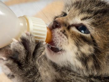
    They’re not eating. They’re not drinking. And they’re turning their nose up at everything you offer. Add post-operative food aversions to a need to initiate voluntary fluid and food intake, and it can feel like an uphill battle of wills.

    What have you got that supports hydration, is easily digestible and is palatable enough to encourage eating and drinking in your hospitalised and at-home patients?

    Furr Boost has the answer…

    It started with the bladder

    Encouraging her poorly beagle Phoebe to drink is a battle Louise Toal faced after her vet diagnosed urinary issues. She just wasn’t interested in drinking. Luckily for Phoebe, Louise is a Food Technologist and began experimenting with protein shakes for dogs, to bolster hydration and combat inappetence. This is when Furr Boost was born.

    It’s more than a hydration drink; it supports healthy digestion, immunity, metabolism and anxiety… plus, it’s delicious.

    Furr Boost has taken the pet market by storm and even braved the Dragon’s Den dragons. Now, this highly palatable, nutritious hydration solution has hit your wholesalers for use in clinic!

    What is Furr Boost?

    In short, it’s a nutritional superfood smoothie for your canine and feline patients. Packed with 100% natural, functional ingredients and no fillers. It’s a low fat, but irresistible blend of meat, fruit and vegetables to use as a drink, food topper or boredom breaker.

    Low in purine, protein, sodium and phosphorous, it’s ideal for encouraging inappetent patients to eat and hydrate post-operatively, during hospitalisation, and at home.

    Hydration

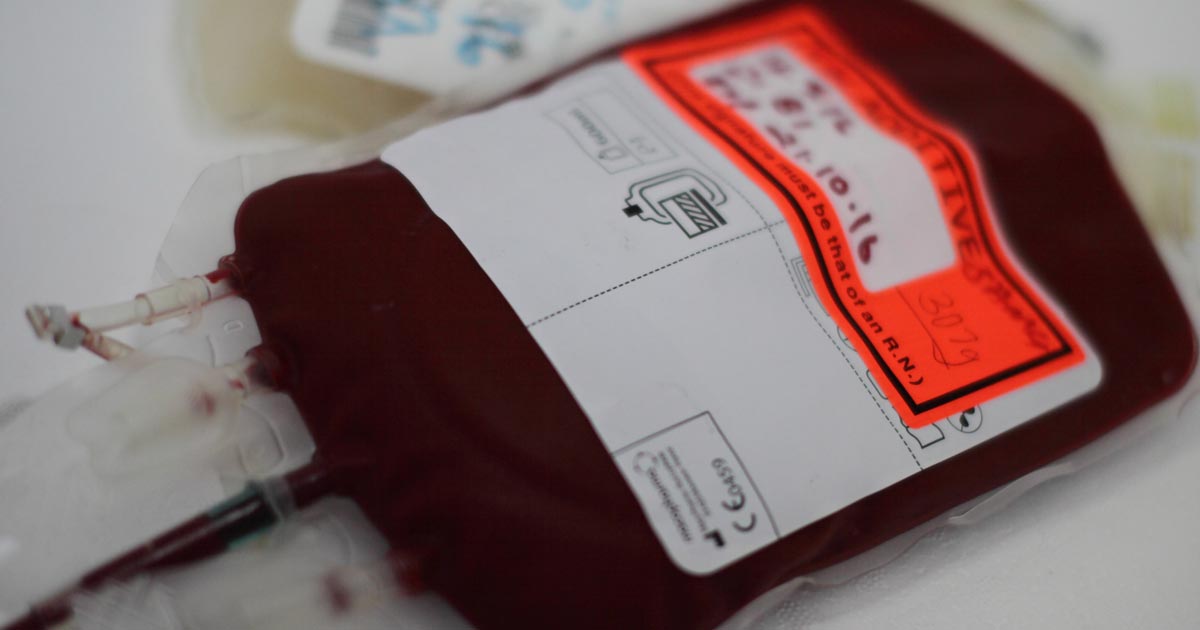
    Water… but tasty! More than 75% moisture content to replenish lost fluid and nutrients.

    Healthy metabolism

    Contains low-fat protein sources plus a blend of oils to provide energy for metabolism and to support a normal recovery.

    Immunity

    Packed with ingredients to support a robust immune system.

    Digestion

    Pre-biotics and dietary soluble fibre aid normal digestion and maintain healthy gut motility.

    Anxiety

    B vitamins and water combat anxiety. Use frozen or on lick mats to distract anxious patients.

    Skin and coat

    Full of naturally occurring fatty acids and omega 3 and 6 for hydrated, healthy skin and a glossy coat.

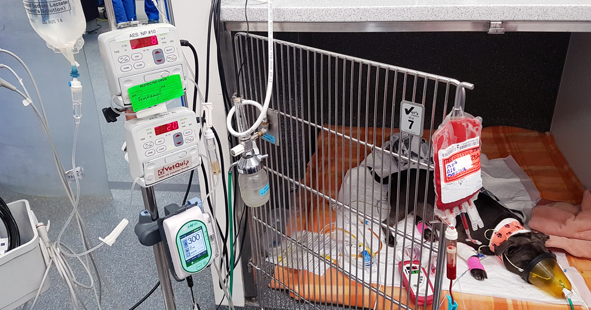
    Furr Boost case study – meet Ozzy

    Meet Ozzy the Saluki who was diagnosed with canine meningitis on 24 June, weighing in at 16Kg and was stabilised by the wonderful medical team who treated him.

    Owner Jayne was very worried for her beloved pet and could see the visible signs of not only his weight loss after his ordeal, but also his mood.

    Jayne said: “When he came home he was so skinny, dehydrated and reluctant to engage with food and drink despite what I tried. Then I discovered Furr Boost and imagine my delight when he absolutely lapped it up with real enthusiasm. He absolutely loves it!

    “I was struggling to get him interested in eating anything to try and build him up while he was taking seven steroids a day. A Furr Boost a day has aided his progress I am sure. He is still on four steroids a day but his weight is now a healthier 22kg. Hoping for dose to reduce to two per day next week.

    “It’s been a long job and he is very flat with the medication, which hopefully will start to improve. I was just so grateful to find your drink when things were looking very grim for him.”

    While Furr Boost is mainly for hydration, the all-natural and human-grade ingredients are used to entice the dog to drink or eat if used as a topper. Our highly palatable formula is irresistible to even the fussiest of dogs and is packed with oils and nutraceuticals to get dogs back on their feet. Our drinks can be used in their pourable form or, for dogs who are not drinking, watered down by up to 50% to push fluids*.

    * We recommend dogs drink 50ml of fluid per body weight and in extreme cases fluid intake should always be monitored.

    Where does Furr Boost fit into your practice?

    Whether it’s in the kennels, in recovery, or to recommend to your clients, Furr Boost is a highly palatable, convenient source of hydration and nutrition. Give as a drink to replenish lost fluids and nutrients, or feed it to encourage voluntary enteral eating and drinking, and to support normal gut motility.

    If patients are food-averse, or the palatability of recommended diets is causing food refusal, add Furr Boost as a topper to soften kibble and entice their tongues back into action, with a range of lip-smackingly delicious flavours.

    Furr Boost is suitable for:

    • all dogs and cats over the age of eight weeks
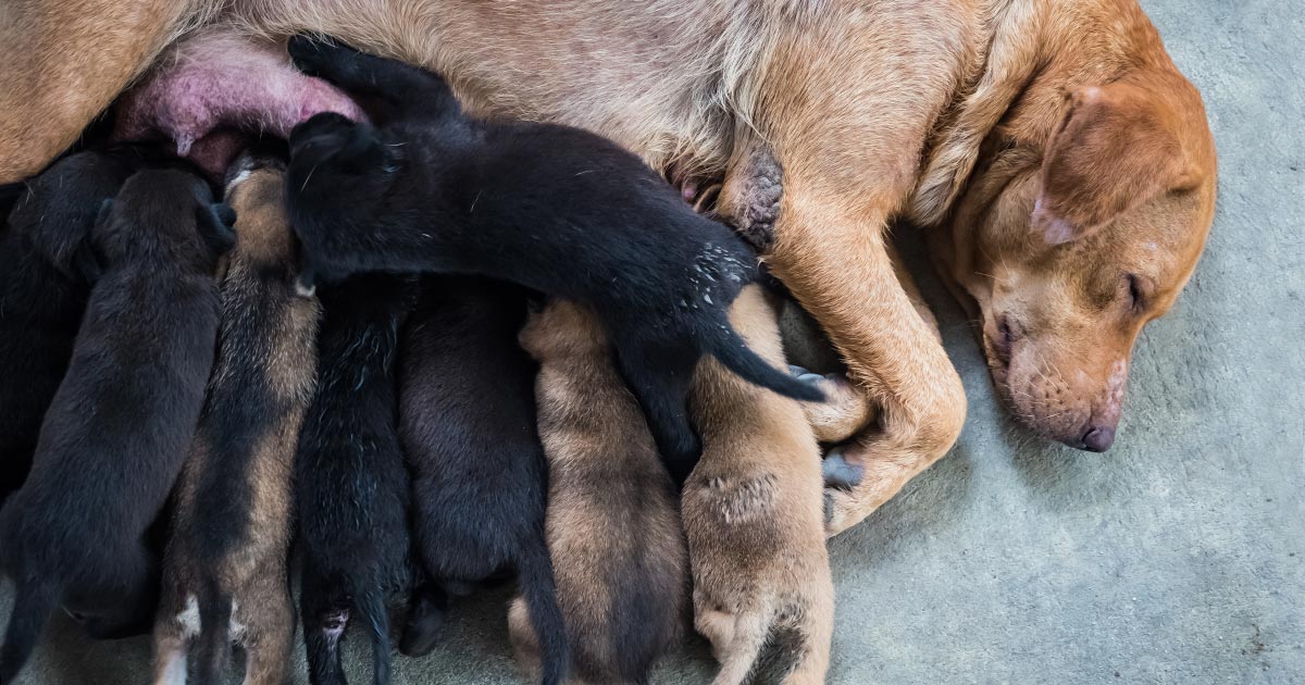
    • pregnant and whelping bitches and queens
    • recovery
    • dogs requiring a low sodium, protein, phosphorous or purine diet
    • maintaining hydration
    • supporting weight management or low-fat diets
    • encouraging inappetent dogs
    • end-of-life support
    • distraction and enrichment, particularly for anxious or immobile patients

    Get your free sample at The London Vet Show

    Have you got a patient in mind that Furr Boost could support? Would you like to test it in your clinic?

    Come along to stand F66 at the London Vet Show on 14-15 November 2024 to grab your free carton!

    Not going to be at the show?

    No worries, Furr Boost is now available via MWI Animal Health, Covetrus, and IVC Evidensia, or you can order directly at www.furrboost.com/veterinary where our team is also available to answer any questions.

  • Highlighting oesophageal foreign bodies on radiographs

    Highlighting oesophageal foreign bodies on radiographs

    before
    Lateral feline chest radiograph before contrast.

    QUICK TIP: Need to know if there is an oesophageal foreign body but can’t be certain on radiographs?

    We have all been in the scenario where we are unsure whether there is an oesophageal foreign body on the radiographs we have just taken.

    You might think of using a contrast medium to help, and the first that always comes to mind is barium. However, my personal first choice is a iodinated contrast medium – urografin, for example.

    Advantages

    Why? Their use is typically limited to myelograms or intravenous contrast studies but they can be given orally as well, for the assessment of oesophageal foreign bodies.

    after
    Lateral feline chest radiograph after contrast.

    The advantage of using this over barium is that if this dye is accidentally aspirated it does not cause pneumonia like barium can.

    How to

    Using iodinated contrast medium is simple:

    • Given orally non-diluted, Dogs: 5-10ml, Cats: 5ml – you can give more if necessary
    • Immediately repeat the radiographs
    • If there is anything in the oesophagus, it will be highlighted

    Tip of the Week author Gerardo Poli is the author of The MiniVet Guide to Companion Animal Medicine – now available in the UK.

  • Oh, CR*P! Using point-of-care C-reactive protein tests

    Oh, CR*P! Using point-of-care C-reactive protein tests

    Few companies now offer affordable point-of-care tests for canine C-reactive protein (CRP). As we did when we recently received our new box of CRP slides, you might soon be asking the question: what do we even do with this stuff?

    Here’s what we’ve learnt…

    CRP is one of the acute phase proteins produced by the liver in response to inflammation. Healthy patients have very low levels of CRP, but a systemic inflammatory condition will cause an increase in CRP within four to six hours. Conversely, increased levels will decrease rapidly on resolution of inflammation. This provides an almost real time measure of inflammation that is more responsive and reliable than the white blood cell response.

    In other words, CRP can indicate the presence of inflammation before the patient’s white blood cell count gives any clues, or before it becomes pyrexic – and, unlike the white blood cell count, stress and steroids do not affect CRP levels.

    Uses

    So, how do we use it?

    • I love it for early pickups of problems in those grey area cases: the dog seems okay on clinical examination, but something about it bothers me. A normal or mildly increased CRP test will make me sleep more easy, while a surprise high reading will prompt me to admit for full diagnostics, or at least get the patient in for a follow-up CRP the next day. Conversely, a localised problem – such as an abscess – combined with a normal CRP test might mean you can hold off on antibiotics and just recheck CRP in 24 hours.
    • It’s great for monitoring response to treatment. If my plan is working then I’d expect CRP to show a significant decrease by day two or three. If it’s not dipping by then, I need to reassess my treatment plan. Do I need to change antibiotics? Scan it again? Maybe we need to consider surgery? It can also be a good prognosticator. Research has shown failure of CRP to decrease significantly (around a 3× decrease) by around day three is generally bad news for patients with inflammatory conditions such as pancreatitis and immune-mediated haemolytic anaemia.
    • We are starting to play with it for post-surgical monitoring. Any surgery will cause inflammation with an increase in CRP levels, but in an uncomplicated postoperative period, you should expect levels to start decreasing by day three to five. A base line CRP 24 hours after surgery with a recheck on day three should pick up early signs of postoperative problems such as infection, and prompt investigation or intervention.
    • A potentially nifty use for it that we haven’t yet had the opportunity to use is in differentiating inflammatory lamenesses (arthritis, infection, injury) from a neurological causes – that is, is it arthritis or a nerve problem?

    Limitations

    • Remember, it’s very sensitive, so will increase with almost any inflammation. A mild upper respiratory infection or a bad gingivitis will likely induce some changes, so it’s important not to over-interpret (keep in mind that the magnitude of the increase in CRP does generally correspond with the severity of the inflammatory response). A pancreatitis case where the CRP fails to drop does not always mean death is looming – you may have just missed the concurrent skin disease. Always interpret CRP values in concert with your clinical examination.
    • Be aware that pregnancy and intense exercise can increase CRP values.
    • Not all serious conditions have an inflammatory component. CRP will be unchanged in most veterinary cases of heart disease; in common hormonal disease, such as adrenal disease and uncomplicated diabetes; urinary obstructions; many localised cancers; epilepsy and many others. Don’t presume that just because CRP is normal, everything is fine.
    • No similar test exists for cats.

    Sit up and say…

    My favourite way to explain how to use this test is by using its highly appropriate acronym – any unexpected increase should make you sit up and say: “Oh CR*P! What am I missing?”

  • Managing thrombocytopenia in cats and dogs: part 2

    Managing thrombocytopenia in cats and dogs: part 2

    Last week we discussed the causes and diagnostic pathway for investigating immune-mediated thrombocytopenia. This week we will go through the management of this condition.

    Despite the fact red blood cells are not actually being destroyed, a severe anaemia can develop from blood loss due to coagulopathy – a common reason for why they present to emergency practices. The management of these patients is broken down into three main areas:

    • improving oxygen delivery
    • commencing immunosuppression
    • management of the underlying cause (if identified)

    Optimising oxygen delivery in the acute phase is going to keep them alive long enough for immunosuppression to work. This is achieved through IV fluids to help improve perfusion and blood transfusions to replace red blood cells. If fresh whole blood is available, it can assist in increasing platelet numbers, but generally it is not very effective.

    Platelet transfusions using platelet-rich plasma can be considered if it is available. Plasma transfusion is not effective at managing the coagulopathy as it is due to a loss of platelets, not a loss of coagulation factors.

    Immunosuppression

    Large areas of ecchymotic haemorrhage on the skin are a quite obvious sign of thrombocytopenia.
    Large areas of ecchymotic haemorrhage on the skin are a quite obvious sign of thrombocytopenia.

    Immunosuppression therapy is often commenced concurrently as the patient is being stabilised.

    The first choice is either dexamethasone 0.5mg/kg IV every 24 hours if the patient is not stable enough for oral medications; otherwise, once stable, start prednisolone at 2mg/kg by mouth per day divided every 12 hours.

    Other immunosuppressive agents include:

    • Azathioprine – 2mg/kg by mouth every 24 hours then 0.5mg/kg by mouth every other day. The main concerns are bone marrow suppression and hepatoxicity – also, it is very toxic in cats.
    • Ciclosporin – 5mg/kg to 10mg/kg by mouth divided twice a day; cats 5mg/kg by mouth every 24 hours.
    • Chlorambucil could also be used at a dose of 0.1mg/kg/day to 0.2mg/kg/day by mouth if the response to prednisolone is insufficient.

    Management

    Management of the underlying cause should be commenced if a cause is identified, but this is often not the case.

    Other management options include:

    • Vincristine can be trialled to increase platelet number as it stimulates the release of platelets from the bone marrow.
    • Gastroprotectants can be considered if gastrointestinal bleeding has occurred – these include proton pump inhibitors and sucralfate.
    • Strict confinement, potentially sedatives and minimal blood sampling are important to minimise injury that may result in further bleeding and blood loss.
    • Antithrombotic therapy is not part of standard management as, unlike immune-mediated haemolytic anaemias, thrombotic events rarely occur.

    When it comes to monitoring, platelet counts are performed daily until more than 40 × 109/L – this can take up to two weeks to occur.

    Once above this level, take weekly counts until the numbers have normalised. Once they have, taper immunosuppressive medications over four to six months, with 20% dose reduction every couple weeks, generally with the adjunctive immunosuppressants first and prednisolone last.

  • Ionised hypocalcaemia, pt 4: controversies and prognostic indications

    Ionised hypocalcaemia, pt 4: controversies and prognostic indications

    Ionised hypocalcaemia (iHCa) is a well-known electrolyte abnormality in critical human patients, which is also beginning to be recognised in our critical feline and canine patients.

    The exact mechanism for the development of iHCa is still unknown – making prevention difficult, if at all possible. Controversy also exists as to whether treating iHCa is of any benefit, especially in non-clinical cases.

    Despite these issues, serum concentration is proving to be an accurate prognostic indicator for the morbidity and mortality rates of some of the more critical patients.

    Research

    Over the past 30 years, significant resources have been put into trying to demystify the pathophysiological causes of iHCa in critically ill people; however, the exact mechanisms are still to be determined.

    Some proposed mechanisms include:

    • abnormal parathyroid hormone secretion or function
    • abnormal vitamin D synthesis or function
    • hypomagnesaemia
    • calcium chelation
    • alkalaemia
    • calcium sequestration in tissue or cells
    • an increase in calcitonin precursors (procalcitonin)

    In a canine study where endotoxaemia was induced, it was found hypovitaminosis D was associated with iHCa (Holowaychuk et al, 2012).

    Veterinary studies

    The true incidence of iHCa in critically ill canine and feline patients is yet to come to a consensus, due to the limited veterinary studies.

    In one retrospective study, 90% of 55 cats with septic peritonitis was reported to have iHCa (Kellett-Gregory et al, 2010), while only 24% of septic dogs (n=58) was reported to have iHCa (Luschini et al, 2010).

    Regardless of the true incidence, the commonness of this change questions whether a need exists to treat iHCa, especially cases in the mild or non-clinical categories.

    No consensus

    At this stage, no consensus exists to either support or prohibit the treatment of hypocalcaemia in critically ill patients.

    Well-designed prospective studies are scarce in human literature and non-existent in the veterinary field; no evidence-based guidelines are available for treatment.

    Based on logic, arguments for the administration of calcium to critically ill patients include:

    • iHCa during hospitalisation is a negative predictor for morbidity and mortality of patients.
    • Hypocalcaemia can cause decreased myocardial contractility.
    • In hypotensive patients dependent on vasopressors or inotropic agents, the supplementation of calcium may be beneficial.

    Arguments against calcium supplementation include:

    • Calcium accumulation within cells predisposes to hypoxia and ischaemia-reperfusion injury.
    • Increased mortality in experimental models of sepsis when calcium is supplemented, on top of the lack of evidence to support this act.

    Prognostic use

    Serum calcium concentrations – or, rather, the trend of it in hospital – appears to be of valuable prognostic indicators.

    Kellett-Gregory et al (2010) found although no direct associations existed between the presence or severity of iHCa at the time of patient admission, a positive correlation existed between the lowest iCa post-hospitalisation, and the length of hospitalisation and duration of intensive care stay.

    Of the cats that had iHCa, those that failed to return to a normal ionised calcium (iCa) during hospitalisation had a significantly lower rate of survival to discharge. Interestingly, iHCa was not associated with the status of hypotension, coagulopathy or arrhythmias, so cannot be used to predict the occurrence of these.

    These findings were echoed by Luschini et al (2010), where low mean ionised calcium and lowest documented ionised calcium concentrations were found to be associated with a poor outcome. The severity and duration of iHCa appears to be important in determining prognosis in these patients.

    Conclusion

    Controversy exists regarding whether treatment of mild iHCa in critically ill patients is recommended; however, one thing we now know is serum iCa concentration is a reliable predictor of mortality and morbidity in canine and feline patients.

    References

  • Ionised hypocalcaemia, pt 3: acute treatment and management

    Ionised hypocalcaemia, pt 3: acute treatment and management

    Treatment of ionised hypocalcaemia (iHCa) is reserved for patients with supportive clinical signs, then divided into acute and chronic management.

    Since the most common cases of clinical hypocalcaemia in canine and feline patients are acute to peracute cases, this blog will focus on the acute treatment and management of hypocalcaemia.

    Clinical signs

    The severity of clinical signs of iHCa is proportional to the magnitude, as well as the rate of decline in ionised calcium (iCa) concentration.

    The normal reference range for iCa is 1.2mmol/L to 1.5mmol/L in dogs and 1.1mmol/L to 1.4mmol/L in cats. Serum iCa concentrations in younger dogs and cats are, on average, 0.025mmol/L to  0.1mmol/L higher than adults.

    Mild iHCa (0.9mmol/L to 1.1mmol/L) – as seen in critically ill dogs and cats with diabetic ketoacidosis, acute pancreatitis, protein-losing enteropathies, sepsis, trauma, tumour lysis syndrome or urethral obstructions – often has no observable clinical signs.

    Moderately (0.8mmol/L to 0.9mmol/L) to severely (lower than 0.8mmol/L) affected animals – in the case of eclampsia and those with parathyroid disease – often display severe signs.

    Early signs of iHCa are often non-specific, and include:

    • anorexia
    • rubbing of the face
    • agitation
    • restlessness
    • hypersensitivity
    • stiff and stilted gait

    As the serum iCa concentration further decreases, patients often progress to:

    • paresthesia
    • tachypnoea
    • generalised muscle fasciculations
    • cramping
    • tetany
    • seizures

    In cats, the gastrointestinal system can also be affected, presenting as anorexia and vomiting.

    Treatment

    The need for treatment of hypocalcaemia is dependent on the presence of clinical signs, rather than a specific cut-off of serum concentration of iCa itself.

    Moderate to severe iHCa should always be treated. Mild hypocalcaemia, on the other hand, may not be necessary, especially if it is well tolerated. It should be remembered the threshold for development of clinical signs is variable, and treatment may benefit critical cases with an iCa concentration of less than 1.0mmol/L.

    Treatment is divided into the acute treatment phase and chronic management.

    In the tetanic phase, IV calcium is required – 10% calcium gluconate (equivalent to 9.3mg/ml) administered at 0.5ml/kg to 1.5ml/kg dosing to effect. This should be administered slowly with concurrent ECG monitoring. Infusion of calcium needs to be stopped if bradycardia develops or if shortening of the QT interval occurs.

    Some suggest calcium gluconate (diluted 1:1 with 0.9% sodium chloride) of half or the full IV dose can be given SC and repeated every six to eight hours until the patient is stable enough to receive oral supplementation. However, be aware calcium salts SC can cause severe necrosis or skin mineralisation.

    Calcium chloride should never be given SC, as it is a severe perivascular irritant.

    Correcting iCa

    Irrespective of the chronicity of the treatment, the rule of thumb is correction of calcium should not exceed 1.1mmol/L.

    Correction of iCa to normal or hypercalcaemic concentration should always be avoided, as this will result in the desensitisation of the parathyroid response, predisposing renal mineralisation and formation of urinary calculi.

    Some of the more common calcium supplementation medications – both parenteral and oral formulas – are detailed in Table 1. Supplementation of magnesium may also benefit some patients, as it is a common concurrent finding in critically ill patients with iHCa.

    Table 1. Common calcium supplementation medications
    Drug Calcium Content Dose Comment
    Parenteral calcium
    Calcium gluconate
    (10% solution)
    9.3mg/ml
    i) slow IV dosing to effect (0.5ml/kg to 1.5ml/kg); acute crisis, 50mg/kg to 150mg/kg over 20 to 30 minutes
    ii) 5mg/kg/hr to 15mg/kg/hr IV or 1,000mg/kg/day to 1,500mg/kg/day (or 42mg/kg/hr to 63mg/kg/hr)
    Stop if bradycardia or shortened QT interval occurs.
    Infusion to maintain normal Ca level
    SC calcium salts can cause severe skin necrosis/mineralisation.
    Calcium chloride
    (10% solution)
    27.2mg/ml 5mg/kg/hr to 15mg/kg/hr IV Do not give SC as severe perivascular irritant
    Oral calcium
    Calcium carbonate
    (many sizes)
    40% tablet 5mg/kg/day to 15mg/kg/day
    Calcium lactate
    (325mg, 650mg)
    13% tablet 25mg/kg/day to 50mg/kg/day
    Calcium chloride
    (powder)
    27.2% 25mg/kg/day to 50mg/kg/day May cause gastric irritation
    Calcium gluconate (many sizes) 10% 25mg/kg/day to 50mg/kg/day

    Next time…

    The next blog will look at the pathophysiology behind iHCa among critically ill animals. It will also look at the controversy regarding treatment of non-clinical iHCa cases and the prognostic indications of iCa concentrations.

  • Ionised hypocalcaemia, pt 2: eclampsia

    Ionised hypocalcaemia, pt 2: eclampsia

    As discussed in part one of this blog series, a myriad of disease processes can lead to ionised hypocalcaemia (iHCa).

    Despite this, only hypocalcaemia caused by eclampsia and hypoparathyroidism (primary or iatrogenic – post-surgical parathyroidectomy) are severe enough to demand immediate parenteral calcium administration.

    Hypoparathyroidism is quite rare, so this blog will not explore the detailed pathophysiology behind this syndrome. However, it is worthwhile mentioning – aside from primary hypoparathyroidism – no other disease state requires long-term calcium supplementation.

    Eclampsia, on the other hand, is the most common cause of clinical hypocalcaemia in dogs and cats. Multiple factors can predispose animals to the development of this phenomenon, so understanding the pathophysiology behind this potentially fatal disease will not only help with future diagnosis and treatment, but also help prevent this issue.

    Periparturient occurrence

    Eclampsia – also known as puerperal tetany or periparturient hypocalcaemia – occurs in the periparturient period anywhere from the final few weeks of gestation to four weeks postpartum, with the latter being the more common time frame of manifestation.

    The serum concentration of ionised calcium (iCa) is often less than 0.9mmol/L in bitches or less than 0.8mmol/L in queens. It presents as muscle fasciculation and tetany, but not usually in seizure since most patients maintain consciousness. Exceptions occur when these patients are left untreated – these patients may develop refractory seizures, cerebral oedema and death.

    The increased muscle activity generates a lot of heat and uses a significant amount of glucose; therefore, hyperthermia and hypoglycaemia are common sequelae in patients with delayed presentations.

    Reduced iCa

    Eclampsia occurs as a result of reduced iCa in the extracellular compartment. In lactation-associated hypocalcaemia, it is the result of the body’s inability to maintain serum iCa through increased osteolytic activity and gastrointestinal calcium absorption, and reduced renal calcium excretion to compensate for the loss of calcium through milk production.

    Other factors often predispose animals to developing eclampsia. These can include poor periparturient nutrition, excessive calcium supplementation and large litter size.

    Excessive calcium supplementation in the prenatal period causes parathyroid gland atrophy, preventing parathyroid hormone release – resulting in reduced gastrointestinal calcium absorption and osteoclastic activity, and increased kidney calcium loss.

    Clinical signs

    Clinical signs can progress rapidly and become fatal if left untreated.

    In the early phases, non-specific signs can present as:

    • facial pruritus
    • hyperaesthesia
    • panting
    • tremors
    • muscle fasciculations
    • paresis
    • ataxia

    Within a few hours, these clinical signs rapidly progress to rigidity, and tonic and clonic spasms with opisthotonos. By this stage, animals will develop severe tachycardia, tachypnoea and hyperthermia. Without treatment, a high mortality rate exists.

    kitten
    “Early supplementation of puppies and kittens with commercial milk formula will significantly reduce the lactation demand on the dam.” Image © Dobroslav / Adobe Stock

    Patients presenting with eclampsia require immediate medical intervention, as well as concurrent supportive therapy. The acute management of clinical iHCa is the same, regardless of the cause, and will be discussed in detail in part three.

    Supportive therapies required to manage and prevent a patient relapsing in eclampsia often include active cooling and glucose supplementation. In cases that seizure, anti-seizure medications – such as diazepam and barbiturates – and mannitol for cerebral oedema may be required.

    Prevention

    Even before getting to the stage where an animal requires treatment, all effort must be taken to prevent a dam from developing hypocalcaemia. This can be easily achieved by improving the calcium content of the food during the perinatal period, as well as reducing the milk demand by early weaning kittens or puppies. This is likely particularly helpful for those with a history of eclampsia or with large litters.

    From the second half of gestation, it is recommended a commercial formulation of puppy/kitten food (1% to 1.8% calcium and 0.8% to 1.6% phosphorus) is to be fed to the dam without any additional minerals or vitamin supplementation.

    Postpartum calcium is similar to the second half of gestation, requiring a diet containing at least 1.4% calcium with a 1:1 ratio with phosphorus (most balanced growth formula for puppies and kittens).

    Less demand

    Early supplementation of puppies and kittens with commercial milk formula will significantly reduce the lactation demand on the dam. Together with this, starting at aged three to four weeks, solids can be introduced at this time. These techniques will be particularly helpful to those with a history of previous eclampsia or those with large litter sizes.

    Aside from the parenteral calcium supplementation required, other supportive therapy – such as active cooling, IV fluid therapy and glucose supplementation – may be required.

    Long term, the dam’s nutritional content of calcium must be optimal from the second half of gestation. All additional calcium or other vitamins and mineral supplementations should not occur prior to parturition.

    In the postpartum dam with a history of eclampsia or that is at risk, changing to a nutritionally balanced commercial food aim for growing puppies and kittens is ideal. Early weaning – or abrupt weaning if hypocalcaemia is severe – may be required in severe cases or those with a high risk of relapse/development.

  • Ionised hypocalcaemia, pt 1: introduction

    Ionised hypocalcaemia, pt 1: introduction

    Low ionised calcium (iCa) is a widely recognised electrolyte disturbance in critically ill human patients who have undergone surgery, are septic, have pancreatitis, or have sustained severe trauma or burns.

    Similar changes occur in our critical canine and feline patients, though less well documented.

    Calcium plays a vital role in a myriad of physiological processes in the body, so any deviation from the very narrowly controlled range is associated with severe repercussions.

    Low iCa has many causes; however, this three-part blog will only focus on the more common and peracute to acute causes. It will also discuss the treatment of low iCa and the controversy behind treatment of iCa in critically ill patients.

    Forms

    Calcium in the serum or plasma exists in three forms:

    • ionised or free calcium
    • protein-bound calcium
    • complexed or chelated calcium (bound to phosphate, bicarbonate, sulfate, citrate and lactate)

    iCa is the biologically active fraction of calcium and is not to be confused with total calcium (tCa). A lack of concordance exists between the two. Adjustment formulas are inaccurate, even with the correction of the tCa to serum total protein or albumin concentration, and should not be used to predict iCa.

    The normal reference range for iCa in dogs is 1.2mmol/L to 1.5mmol/L; in cats, it is 1.1mmol/L to 1.4mmol/L.

    Function

    An example of low ionised calcium.
    An example of low ionised calcium.

    Calcium is essential in maintaining normal physiological processes in the body. iCa regulates:

    • vascular tone
    • myocardial contraction
    • homeostasis

    In addition, it is needed for:

    • enzymatic reactions
    • nerve conductions
    • neuromuscular transmission
    • muscle contraction
    • hormone release
    • bone formation
    • resorption

    In critical patients, particularly those with severe trauma or sepsis, vascular tone and coagulation is particularly important. For this reason, iCa is tightly kept in a narrow range and regulated by the interactive feedback loop that involves iCa, phosphorous, parathyroid hormone, calcitriol and calcitonin.

    Diseases and causes

    Diseases commonly associated with low iCa in dogs and cats include:

    • acute kidney failure
    • acute pancreatitis
    • diabetic ketoacidosis
    • eclampsia
    • ethylene glycol intoxication
    • protein-losing enteropathies
    • sepsis
    • trauma
    • urethral obstruction
    • parathyroid diseases
    • tumour lysis syndrome

    Situations altering the fraction of extracellular calcium seen on a regular basis include:

    • acid-base disturbances
    • lactic acidosis
    • protein loss or gain
    • increased free fatty acids

    Iatrogenic causes include:

    • citrate (anticoagulant) administration during blood transfusions
    • phosphate
    • bicarbonate
    • sulfate administration

    Low iCa can also develop during cardiopulmonary resuscitation, quickly declining with increased duration.

    • Part two will go into more depth regarding the most common causes of low iCa that require acute treatment, the treatment involved, controversies surrounding treatment of non-clinical low iCa, and prognostic indications.
  • Blood transfusions, pt 2: what do I give?

    Blood transfusions, pt 2: what do I give?

    To make the most of a precious resource, donated blood is often separated into two components: red blood cells (packed red cells) and plasma (fresh frozen plasma, most commonly).

    Haematocrit tube
    Haematocrit tube from a patient with immune mediated haemolytic anaemia and a slide demonstrating autoagglutination.

    This not only extends the life span of plasma component, but it also means you can pick and administer which component you need the most as not all anaemic patients need the same product.

    Which product?

    Here are three different examples of patients with different blood product requirements:

    Immune mediated haemolytic anaemia

    This a prime example of a patient that can have an anaemia with transfusion triggers, but are rarely coagulopathic. The key component required here is packed red cells.

    Internal bleed

    This is a grey zone – an anaemia with transfusion triggers can occur, but not always a coagulopathy. A coagulopathy may develop later if the bleed continues. So, packed red cells can be used initially, and the plasma administered after, if required.

    Rodenticide intoxication

    typing kit
    Commercial feline blood typing kit.

    These patients will often need both red blood cells and plasma. So, whole blood – either fresh or stored – can be used, or a unit of packed red cells combined with a unit of fresh frozen plasma.

    Blood typing and crossmatching

    Before administration of the product there is one critical step. Blood typing is important – not only for cats, but also dogs – as it helps rule out blood type incompatibilities and can significantly reduce the risk of an acute transfusion reaction. Easy-to-use commercial blood typing kits (pictured) are available.

    Just because you blood typed doesn’t mean there is no value in crossmatching. Other factors or uncommon red blood types, such as DAL (dogs) and Mik (cats) can cause acute reactions.

    Both these tests do not 100% rule out the possibility of an acute reaction, so, whenever a blood product is being administered, the patient needs to be monitored closely.

    Next week, we cover how much to give.

  • Blood transfusions, pt 1: clinical signs

    Blood transfusions, pt 1: clinical signs

    I get asked frequently when is the right time to transfuse an anaemic patient?

    The difficulty lies in the fact not all anaemic patients require blood transfusions. Just because a patient has pale mucous membranes does not mean the patient needs a transfusion.

    The term commonly brought up during the discussion is “transfusion triggers present”.

    What constitutes a “transfusion trigger”?

    A couple of different definitions exist: classically, it is the PCV or haemoglobin level at which a transfusion is indicated in an individual animal – essentially, if it gets below a certain number, transfusion is required – but it is not always that simple. Just because the PCV is 15%, it doesn’t always mean a transfusion is required.

    When the PCV drops low enough, clinical signs of reduced oxygen delivery to the tissues start to develop, these include:

    • decreased exercise tolerance
    • weakness
    • dull mentation
    • tachycardia
    • tachypnoea
    • elevated lactate levels when shock has been addressed

    Rapid or slow?

    These clinical signs are influenced by the speed at which the anaemia has developed.

    If the anaemia has occurred rapidly due to internal bleeding from trauma or a ruptured organ, these clinical signs can present in a matter of minutes, depending on how big the bleed is. This means a transfusion might be indicated when the PCV is still 25%, especially if further rapid blood loss is likely.

    If the anaemia developed over days to weeks (slow red cell destruction or anaemia of chronic disease, for example) transfusion triggers might not be present until the PCV drops below 15%, as the body has had time to compensate.

    Summary

    So, in summary, the decision-making process involves asking the questions:

    • What is the PCV?
    • How fast has the anaemia developed?
    • Are there clinical signs of reduced oxygen delivery?
    • Is further loss likely?

    When you combine the core aspects of each of the questions above, “transfusions triggers” change from absolute numbers to this:

    • PCV under 15% with clinical signs of reduced oxygen delivery.
    • Rapid PCV drop to under 20% in dogs and 15% in cats.
    • PCV under 25% and surgery or anaesthesia is required, and/or rapid ongoing blood loss is occurring.

    Blood products you should use and why will be covered in a future post.

    Note: Haemoglobin levels should also be assessed in conjunction with the PCV.