Tag: Bilirubin

  • Pancreatitis, pt 1: diagnosis

    Pancreatitis, pt 1: diagnosis

    Pancreatitis is one of the most common exocrine conditions seen in small animal practice. It is caused by premature activation of enzymes (zymogens) within the pancreas leading to autodigestion, and can result in severe morbidity with the potential to lead to mortality.

    To this day, the diagnosis of pancreatitis remains a challenge – especially in feline patients – and relies on the use of a combination of history, signalment, clinical signs and diagnostic findings.

    Presenting signs

    In dogs, overweight middle aged to older (more than five years old) are at higher risk. Miniature schnauzers, Yorkshire terriers and silky terriers are some breeds identified as higher risk. Approximately 90% present with anorexia and vomiting, 50% with abdominal pain and 30% with diarrhoea.

    In cats, no common age range exists, although domestic shorthair and longhair cats are the most commonly affected. They present more commonly with anorexia and lethargy; 30% presenting with vomiting and 25% with abdominal pain.

    Diagnostic changes
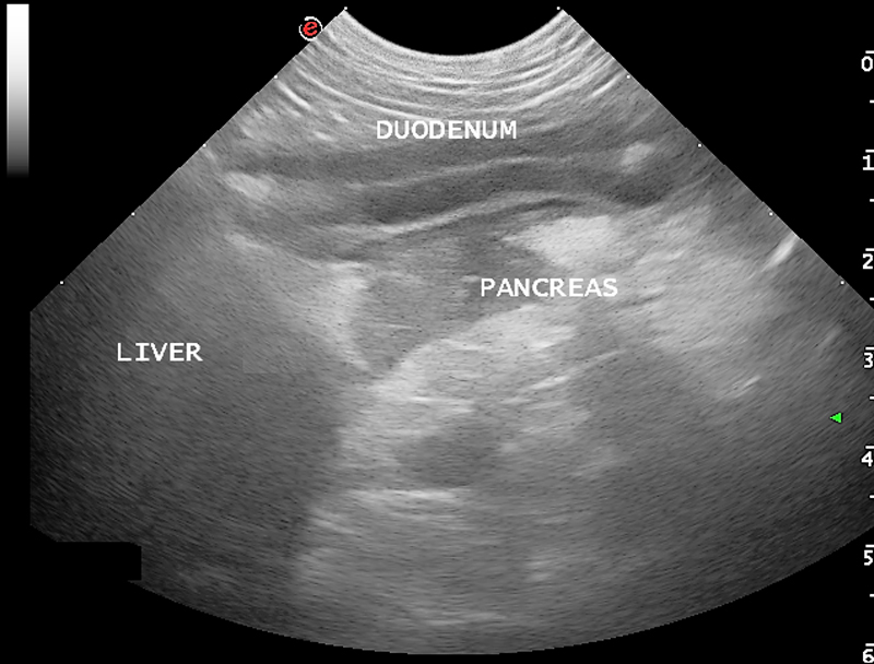
    Pancreatitis-Image---Pancreatitis
    Cases can be confirmed with ultrasound.

    A summary of the commonly seen diagnostic changes are included below, many of which due to the nature of the disease are non-specific:

    • Hyperlipaemia and an inflammatory leukogram can be present, but are both non-specific.
    • Hyperlipasaemia and hyperamylasaemia are commonly seen, but the sensitivity and specificity of both are only about 50%.
    • Elevation in alkaline phosphatase and bilirubin can indicate pancreatic bile obstruction. Other changes may be present and can indicate wider organ system involvement – azotemia, for example.
    • Canine pancreatic specific lipase immunoreactivity (cPLI) has a high sensitivity, but poor specificity – approximately 50%. This means, if real pancreatitis is present, it will show positive; but a positive result will only actually be real pancreatitis 50% of the time. A negative result, however, can be interpreted as “highly unlikely for pancreatitis to be present”. A positive cPLI should be confirmed with ultrasound, which is the gold standard for canine pancreatitis.
    • Feline pancreatic lipase immunoreactivity (fPLI) has been reported to have a sensitivity of 67% and specificity at 91%. This means, 90% of the time, a positive indicates real pancreatitis – but these figures vary between studies. To make things even more difficult, cats with pancreatitis can have normal fPLI/spec fPL and normal ultrasonographic findings.
    • Common changes on ultrasound with acute pancreatitis include free abdominal fluid (generally a non-septic exudate) and hypoechoic pancreas surrounded by hyperechoic peripancreatic fat. As mentioned above, feline pancreatitis often has no visible changes.
    • Radiography may show reduced serosal detail around the pancreatic regions – again, this is not specific or sensitive, but is helpful at ruling out other differentials, such as foreign bodies.

    Summary

    In summary, canine pancreatitis is less of a diagnostic challenge compared to its feline counterpart. There is no single test that can accurately confirm the presence of feline pancreatitis, apart from in biopsies via exploratory laparotomy – which are understandably invasive and costly.

    Next week, we will cover the fundaments of managing pancreatitis patients.

  • Icteric serum

    Icteric serum

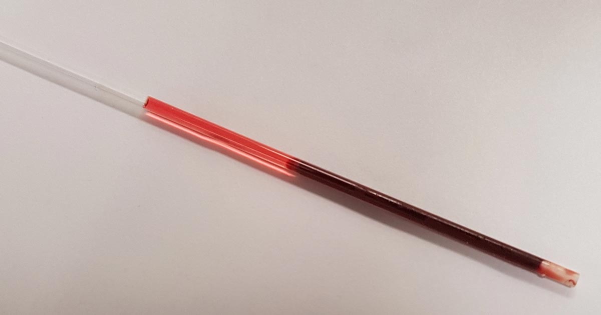
    The final discolouration of the serum we are going to cover is icteric serum.

    Icteric serum
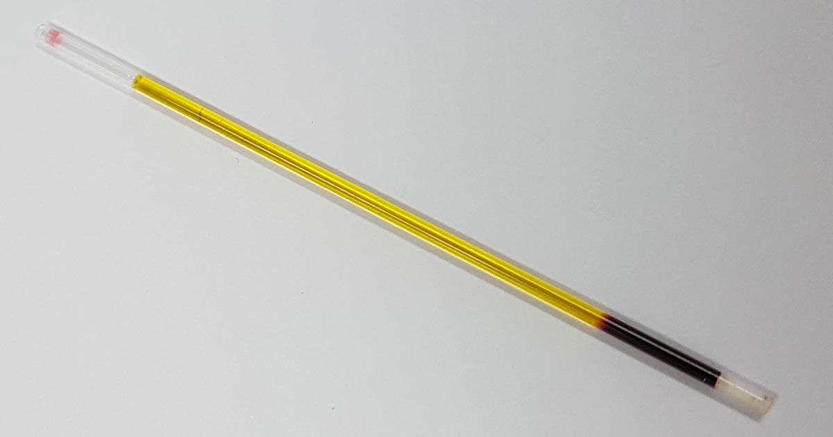
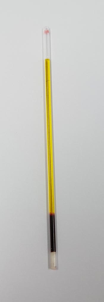
    Icteric serum is caused by the presence of excess bilirubin in the blood stream.

    Icteric serum is caused by the presence of excess bilirubin in the blood stream as a result of increased production (pre-hepatic) or inappropriate excretion (hepatic and post-hepatic).

    The most common cause of pre-hepatic icterus is haemolytic anaemia, while hepatic disease and biliary tract obstruction are the most common causes for hepatic and post-hepatic icterus, respectively.

    Tips on where to start

    If icterus and concurrent anaemia exist, my first suspicion would be some kind of pre-hepatic cause. The most common causes are immune-mediated haemolytic anaemia and infectious haemolytic anaemia, such as haemotropic mycoplasma and babesiosis.

    Other causes can include snake envenomation and oxidative injury from heavy metal toxicity or onion ingestion.

    Regarding hepatic and post-hepatic causes, unfortunately it is not always clear-cut. Both are commonly associated with elevation in both alanine transaminase (ALT) and alkaline phosphatase (ALKP), and, although no specific changes are pathognomonic for hepatic or post-hepatic disease, the pattern of change may help identify the origin of the cause. ALT is released from the inside of hepatocytes, and in higher amounts when cell damage occurs.

    Hepatic hints

    Some pointers on what you can do to help differentiate:

    • Compare the ALT and ALKP elevation; if one is in order of magnitudes higher than the other then it can help point to an origin.
    • If the cause is of hepatic origin, one would expect the ALT to be significantly more elevated than the ALPK. Likewise, this is usually true in reverse for post-hepatic causes. However, it should be noted in chronic hepatic diseases, where active damage to hepatocytes is comparatively lower, a mild increase in ALT and marked increase in ALPK does not preclude disease of hepatic origin. Therefore, biopsies should always be used for definitive diagnosis.
    • If other biochemistry parameters such as albumin, glucose and cholesterol are low, or prolonged clotting times are present, the case for a hepatic origin is strengthened.
    • The gallbladder and bile duct can be assessed using abdominal ultrasonography. The presence of a dilated bile duct, or evidence supportive of pancreatitis, is highly suggestive of a post-hepatic cause.

    Finally, it is important to be aware of the impact on hyperbilirubinaemia on laboratory testing. Hyperbilirubinaemia generally causes decreased cholesterol, triglyceride, creatinine, lipase, total protein and gamma-glutamyltransferase levels.

  • Lipaemia – the bane of biochemistry

    Lipaemia – the bane of biochemistry

    Last week we covered haemolysed samples – this week we’re looking at lipaemic samples.

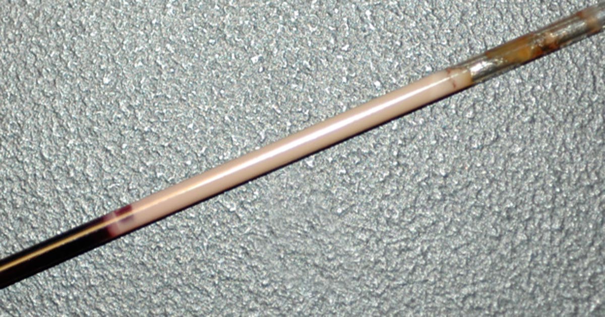
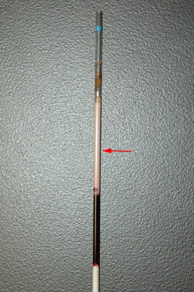
    Lipaemic samples are caused by an excess of lipoproteins in the blood, creating a milky/turbid appearance that interferes with multiple biochemical tests and can even cause haemolysis of red blood cells.

    lipaemic sample
    A severely lipaemic sample (red arrow). IMAGE: eClinPath.com (CC BY-NC-SA 4.0).

    Lipaemia can follow recent ingestion of a meal – especially one high in fat. Although not pathognomonic for any diseases, its presence can help increase the suspicion of certain diseases, including:

    • pancreatitis
    • diabetes mellitus
    • hypothyroidism
    • hyperadrenocorticism
    • primary hyperlipidaemia (in some specific breeds, such as the miniature schnauzer)

    It warrants further investigation in patients that have been ill and inappetent.

    Irksome interpretations

    Lipaemia can dramatically impact laboratory testing and is often troublesome in critically ill patients, making interpretation of biochemistry particularly difficult, if not impossible.

    Lipaemia can affect different analysers in different ways, most commonly causing:

    • Falsely increased calcium, phosphorus, bilirubin, glucose and total protein (via refractometer) and some liver parameters such as alkaline phosphatase, alanine aminotransferase, aspartate aminotransferase, haemoglobin concentration, and mean corpuscular haemoglobin concentration.
    • Falsely decreased sodium, potassium, chloride, albumin and bicarbonate.

    Tube tips

    Assessment of a centrifuged haematocrit tube before running a biochemistry panel can help reduce wasted biochemistry consumables.

    If the sample is lipaemic in the haematocrit tube then maybe try some of the following tips.

    • If blood tests are planned in advance, try fasting the patient beforehand for 12 to 24 hours.
    • Repeat sampling a couple of hours later may yield a less lipaemic sample.
    • Collecting and centrifuging a larger amount of blood (3ml to 5ml, for example) can sometimes yield enough clear sample between the lipid layer and red blood cells.
    • Refrigeration of the sample can help the separation.
    • Extract lipids using polar solvents, such as polyethylene glycol.
    • Centrifugation at higher than normal speeds (if possible) can also assist in clearing the layer.
  • PCV/total solids interpretation: serum colour

    PCV/total solids interpretation: serum colour

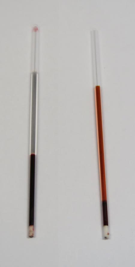
    When interpreting the often misinterpreted and underused PCV and total solids test, it is important to take note of the serum colour as this may give clues into the diagnosis.

    PCV tubes
    Normal serum colour (left) compared to a patient with immune-mediated haemolytic anaemia. The serum is haemolysed and anaemia is present.

    The most common abnormalities seen in clinic are icteric, haemolysed and lipaemic serum.

    Clear serum can also be of importance – especially when you interpret it with blood counts and urine colour.

    Haemolysis

    The most common abnormality of serum colour changes is haemolysis. In my experience, the most common cause is suboptimal collection technique. To confirm this, simply collect another sample and repeat.

    If it is repeatable, and concurrent anaemia or pigmenturia is present, it warrants further investigation.

    Intravascular haemolysis can be caused by:

    • immune-mediated haemolytic anaemia
    • blood transfusion reactions
    • infectious diseases such as Mycoplasma haemofelis, Babesia canis, Ehrlichia canis, FeLV and others
    • Heinz bodies from the ingestion of heavy metal, onions or paracetamol
    • hypophosphataemia
    • macroangiopathic disease (neoplasia, for example)
    • envenomation – typically, snake bites

    Testing issues

    Haemolysis can also affect other laboratory testing. It can lead to an artefactual increase in glucose, phosphorus, bilirubin, total protein, fructosamine and triglycerides, and a decrease in sodium (pseudohyponatraemia), cholesterol, calcium, potassium and albumin.

    Extravascular haemolysis often does not cause haemolysed serum as it is generally slower and the body is able to clear the haemoglobin before it can lead to discolouration of the serum.

  • Making sense of effusions (part 1): is your patient septic?

    Making sense of effusions (part 1): is your patient septic?

    Interpreting effusion samples can be confusing, so try to think of effusions as if you were collecting a blood sample.

    Septic effusion
    Septic effusion.

    Many of the in-clinic diagnostic tests that apply to blood samples also apply to effusions, such as:

    • PCV/total protein
    • smears
    • glucose
    • lactate
    • potassium
    • creatinine
    • bilirubin

    It’s not enough to only check the protein concentration of the effusion then classify it as either a transudate, modified transudate or exudate and leave it at that – there is more information left to extract from that sample.

    Challenging diagnosis

    Determining if an effusion is septic can be a challenge. Here are the steps I take.

    analysis
    Abdominal and peripheral blood gas analysis.
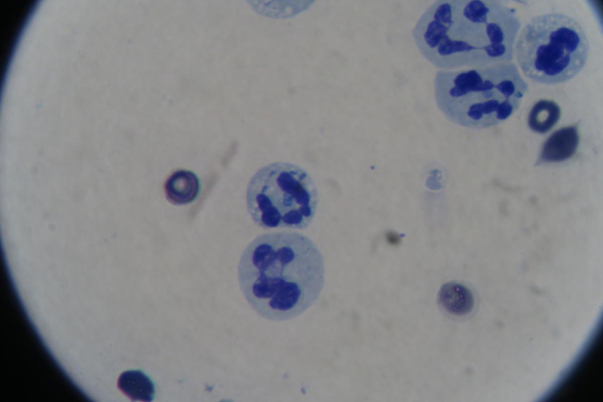
    1. Perform a cytological examination of your effusion in the smear and look for inflammatory cells and the presence of bacteria. Look inside the cells as well as outside. If you don’t see bacteria it does not mean it isn’t a septic effusion, and only a couple bacteria are needed for me to call it septic.
    2. Glucose and lactate: You need to compare the glucose levels in the effusion with blood glucose levels. If the effusion glucose level is less than serum glucose, it is more likely you have a septic exudate. This makes sense in that bacteria would metabolise glucose in the effusion, leading to lower glucose levels. A by-product of metabolism is, of course, lactate. Therefore, you next need to check the lactate levels in the effusion and compare it to the serum lactate level. If lactate level in the effusion is more than the serum lactate level, then again you have more evidence you are dealing with a septic exudate.

    Try to measure glucose and lactate from both blood and effusion samples at the same time on the same machine. Keep in mind glucose and lactate values are less accurate for monitoring for the presence of bacteria in post-surgical patients.