Tag: Anorexia

  • Ionised hypocalcaemia, pt 3: acute treatment and management

    Ionised hypocalcaemia, pt 3: acute treatment and management

    Treatment of ionised hypocalcaemia (iHCa) is reserved for patients with supportive clinical signs, then divided into acute and chronic management.

    Since the most common cases of clinical hypocalcaemia in canine and feline patients are acute to peracute cases, this blog will focus on the acute treatment and management of hypocalcaemia.

    Clinical signs

    The severity of clinical signs of iHCa is proportional to the magnitude, as well as the rate of decline in ionised calcium (iCa) concentration.

    The normal reference range for iCa is 1.2mmol/L to 1.5mmol/L in dogs and 1.1mmol/L to 1.4mmol/L in cats. Serum iCa concentrations in younger dogs and cats are, on average, 0.025mmol/L to  0.1mmol/L higher than adults.

    Mild iHCa (0.9mmol/L to 1.1mmol/L) – as seen in critically ill dogs and cats with diabetic ketoacidosis, acute pancreatitis, protein-losing enteropathies, sepsis, trauma, tumour lysis syndrome or urethral obstructions – often has no observable clinical signs.

    Moderately (0.8mmol/L to 0.9mmol/L) to severely (lower than 0.8mmol/L) affected animals – in the case of eclampsia and those with parathyroid disease – often display severe signs.

    Early signs of iHCa are often non-specific, and include:

    • anorexia
    • rubbing of the face
    • agitation
    • restlessness
    • hypersensitivity
    • stiff and stilted gait

    As the serum iCa concentration further decreases, patients often progress to:

    • paresthesia
    • tachypnoea
    • generalised muscle fasciculations
    • cramping
    • tetany
    • seizures

    In cats, the gastrointestinal system can also be affected, presenting as anorexia and vomiting.

    Treatment

    The need for treatment of hypocalcaemia is dependent on the presence of clinical signs, rather than a specific cut-off of serum concentration of iCa itself.

    Moderate to severe iHCa should always be treated. Mild hypocalcaemia, on the other hand, may not be necessary, especially if it is well tolerated. It should be remembered the threshold for development of clinical signs is variable, and treatment may benefit critical cases with an iCa concentration of less than 1.0mmol/L.

    Treatment is divided into the acute treatment phase and chronic management.

    In the tetanic phase, IV calcium is required – 10% calcium gluconate (equivalent to 9.3mg/ml) administered at 0.5ml/kg to 1.5ml/kg dosing to effect. This should be administered slowly with concurrent ECG monitoring. Infusion of calcium needs to be stopped if bradycardia develops or if shortening of the QT interval occurs.

    Some suggest calcium gluconate (diluted 1:1 with 0.9% sodium chloride) of half or the full IV dose can be given SC and repeated every six to eight hours until the patient is stable enough to receive oral supplementation. However, be aware calcium salts SC can cause severe necrosis or skin mineralisation.

    Calcium chloride should never be given SC, as it is a severe perivascular irritant.

    Correcting iCa

    Irrespective of the chronicity of the treatment, the rule of thumb is correction of calcium should not exceed 1.1mmol/L.

    Correction of iCa to normal or hypercalcaemic concentration should always be avoided, as this will result in the desensitisation of the parathyroid response, predisposing renal mineralisation and formation of urinary calculi.

    Some of the more common calcium supplementation medications – both parenteral and oral formulas – are detailed in Table 1. Supplementation of magnesium may also benefit some patients, as it is a common concurrent finding in critically ill patients with iHCa.

    Table 1. Common calcium supplementation medications
    Drug Calcium Content Dose Comment
    Parenteral calcium
    Calcium gluconate
    (10% solution)
    9.3mg/ml
    i) slow IV dosing to effect (0.5ml/kg to 1.5ml/kg); acute crisis, 50mg/kg to 150mg/kg over 20 to 30 minutes
    ii) 5mg/kg/hr to 15mg/kg/hr IV or 1,000mg/kg/day to 1,500mg/kg/day (or 42mg/kg/hr to 63mg/kg/hr)
    Stop if bradycardia or shortened QT interval occurs.
    Infusion to maintain normal Ca level
    SC calcium salts can cause severe skin necrosis/mineralisation.
    Calcium chloride
    (10% solution)
    27.2mg/ml 5mg/kg/hr to 15mg/kg/hr IV Do not give SC as severe perivascular irritant
    Oral calcium
    Calcium carbonate
    (many sizes)
    40% tablet 5mg/kg/day to 15mg/kg/day
    Calcium lactate
    (325mg, 650mg)
    13% tablet 25mg/kg/day to 50mg/kg/day
    Calcium chloride
    (powder)
    27.2% 25mg/kg/day to 50mg/kg/day May cause gastric irritation
    Calcium gluconate (many sizes) 10% 25mg/kg/day to 50mg/kg/day

    Next time…

    The next blog will look at the pathophysiology behind iHCa among critically ill animals. It will also look at the controversy regarding treatment of non-clinical iHCa cases and the prognostic indications of iCa concentrations.

  • Hyponatraemia, pt 1: clinical signs

    Hyponatraemia, pt 1: clinical signs

    Hyponatraemia is a relatively common electrolyte disturbance encountered in critically ill patients, and the most common sodium disturbance of small animals.

    In most cases, this is caused by an increased retention of free water, as opposed to the loss of sodium in excess of water.

    Low serum sodium concentration

    Hyponatraemia is defined as serum concentration lower than 140mEq/L in dogs and lower than 149mEq/L in cats.

    The serum sodium concentration measured is not the total body sodium content, but the amount of sodium relative to the volume of water in the body. For this reason, patients with hyponatraemia can actually have decreased, increased or normal total body sodium content.

    This series will look briefly at the modulators of the sodium and water balance, clinical signs associated with hyponatraemia, the most common causes in small animals, the pathophysiology behind these changes, and treatment and management.

    ECF volume

    hyponatraemia
    An example of hyponatraemia.

    Sodium is the main osmotically active particle in the extracellular fluid (ECF), so is the main determining factor of the ECF volume. Any disease process that alters the patient’s ECF volume will lead to hyponatraemia, such as:

    • dehydration
    • polyuria
    • polydipsia
    • vomiting
    • diarrhoea
    • cardiac diseases
    • pleural or peritoneal effusion

    The modulators of water and sodium balance are also different, so should be thought of as different processes.

    Water balance is modulated by thirst and antidiuretic hormone, and the effect of this is to maintain normal serum osmolality and serum sodium concentration.

    Modulators of sodium balance aim to maintain normal ECF volume. It adjusts this by altering the amount of renal sodium excretion; an expansion of ECF volume will lead to an increased sodium excretion, while a reduction in ECF volume will lead to increased sodium retention.

    Rate and magnitude

    The clinical signs of hyponatraemia are both dependent on the magnitude of the decrease and the rate at which it developed.

    In mild or chronic patients, no visible clinical signs can exist. In severe (lower than 125mEq/L) and acute cases, clinical signs exhibited are typically neurological, reflecting cerebral oedema. Possibilities include:

    • lethargy
    • anorexia
    • weakness
    • incoordination
    • disorientation
    • seizures
    • coma

    Patients with acute hyponatraemia – for example, water intoxication – are more likely to show clinical signs, compared to those with chronic hyponatraemia, because the brain takes time (at least 24 to 48 hours) to produce idiogenic osmoles, osmotically active molecules that help shift free water out of brain cells.

    Therefore, any acute hyponatraemia that develops within a 24 to 48-hour period tend to show clinical signs, whereas chronic cases are less likely.

    • Next week’s blog will look into the different causes of hyponatraemia and how they result in sodium loss.
  • Breaking away from a vet’s diet of fast food

    Breaking away from a vet’s diet of fast food

    A while ago, I wrote about anorexia in vets… the stripped down, bare meaning being the clinical sign of “not eating”. As a student, I witnessed vets on placements routinely forgoing lunch or existing on a diet consisting entirely of Pot Noodles or fast food – one vet I shadowed had either a McDonalds or KFC on four of the five days.

    Then, I could appreciate the lack of time and energy for cooking, but still couldn’t imagine being able to stomach so much junk food. I could not understand how you could work effectively and remain healthy while pouring so much crap into your body – and, of course, you can’t!

    Isn’t it ironic that part of our job is to advise on diet and nutrition for clients’ four-legged friends, yet we don’t take our own advice?

    Realisation dawns

    Takeaway boxes
    Image © miketea88 / Adobe Stock.

    As a student, there would be rare occasions I wouldn’t get lunch until 4pm or would just order a pizza through tiredness (or, more likely, from being hungover). But, on the whole, I had a pretty good diet. I exercised a lot and was organised enough to make lunch 90% of the time, so I was never stuck without food.

    Yet now, as a new grad, I totally get the unhealthiness; it’s not really a matter of choice, but more a matter of pure exhaustion.

    I found myself going without lunch on numerous occasions, mostly due to being stuck on farm all day TB testing, but sometimes due to being swamped with surgeries. Having not been able to stop for food all day, my first exploratory laparotomy was done after inhaling a cupcake – not the most nutritious of lunches.

    24/7 shopping

    There have been weeks I have consumed more takeaways or McMuffins than I am proud of, purely down to a lack of time and effort. I’m too tired to shop for food, or cook it, and I don’t want to spend all weekend meal-prepping for the week ahead, which is what my more organised, student self would have done.

    It also doesn’t help that shops close early on a Sunday in England. I was definitely spoilt in Scotland, where 24-hour opening actually means 24 hours, 7 days a week.

    If I cooked like I did when I was a student, my day would literally be work, cook, eat, sleep. But, to be honest, it’s not much better anyway – more like work, pick up takeaway, eat, sleep.

    Maintaining a work-food balance

    I do manage to get out on the bike at weekends, but not during the week, and as a former gym frequenter at uni, it gets to me sometimes that I’m becoming seriously unfit. Perhaps the answer is to get up early and go to the gym before work, but that’s not in my nature… I tried early running once, and all it did was make the day feel really long by 9am.

    Mental health and well-being are constantly in the veterinary media at the moment, and, while I can empathise with my colleagues who lead the lifestyle of fast food, I’m not condoning it. This is no way to carry on. We need to try to achieve an acceptable work-life balance and, at the very least, a good work-food balance, which is something I am apparently not very good at yet.

    I’m told it gets easier. Whether that means you get over being so tired all the time or just get used to it and somehow manage to power through, I’m not sure, but I hope it does – and I hope I find the energy to improve my diet.

  • Eating disorders and the veterinary profession

    Eating disorders and the veterinary profession

    The general public associates the word “anorexia” with the eating disorder characterised by refusing to eat to lose weight, which, in human medicine, has the more specific name of “anorexia nervosa”. As vets, we use the word the term “anorexic” in the slightly different sense of being a clinical sign our patients exhibit – defined as “a lack or loss of appetite”.

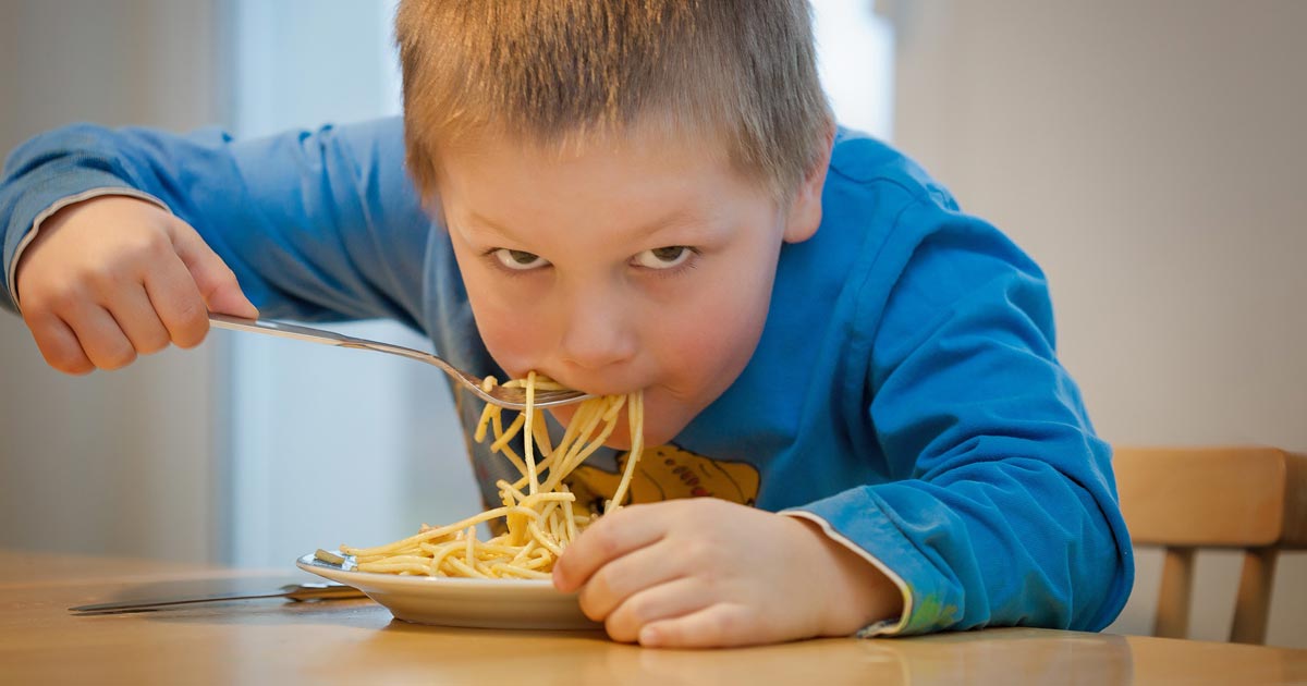
    Kid eating noodles.
    Is the veterinary profession practising what it preaches when it comes to nutrition?

    When referring to vets themselves, however, these definitions blur together a little, but I believe many vets frequently exhibit clinical signs that may or may not be part of an eating disorder.

    While there is a lack of hard evidence or figures for eating disorders within the veterinary profession, it is estimated 10% of UK veterinary students suffer from eating disorders (not limited to anorexia nervosa), which is higher than the figure for the general UK population, which sits at 6.4%. (vetlife.org.uk).

    Another branch

    We are all made very aware of the mental health statistics and suicide risk of vets, and eating disorders are another branch of that tree.

    Despite the lack of evidence to back up the theory, based on anecdotes alone, I’m willing to bet eating disorders, or even intermittent “anorexia” (the clinical sign), are more prevalent in qualified vets than the general population.

    I’ve lost count of the number of times I’ve been on EMS and spent the entire day in the car with the same vet, going from call to call and not seeing them eat once.

    I’m probably on the other end of the scale; I tend to get indigestion and heartburn from excess stomach acid production if I don’t eat regularly enough, so I tend to stress about the next time I’m going to eat (which turns into a vicious cycle because those symptoms also develop as a result of that stress). Therefore, I always try to ensure I have a packed lunch so I never get caught out.

    On the road

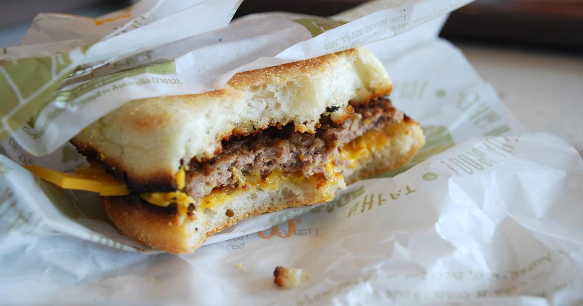
    Burger in a car.
    Veterinary professionals on the road can often find themselves picking up fast food, which will be of long-term detriment to their health.

    The number of ambulatory equine or farm vets who don’t appear to carry food in their cars is astounding.

    They often rely on getting time to stop for food – which, inevitably, results in them not eating for the whole working day or picking up unhealthy fast food or snacks, which isn’t really much better.

    When discussing my observations with fellow students, many of them have similar tales, and the problem is not limited to ambulatory practice.

    While many vets and nurses in small animal practice will have a slightly more routine structure to their working lives, there’s always the odd mad day, week or month when they just can’t seem to grab a minute to supply their digestive system between consults, surgeries and emergencies.

    Despite my own claim of always being prepared to avert such situations, sometimes they’re unavoidable.

    I’ve found myself a few select times during rotations when I’ve been so rushed off my feet, trying to get everything done or see clients, that I haven’t eaten lunch until well after 4pm. Then, it’s likely I’ve got to that stage of being “past hungry”, but, as it is at that point I normally get headachey and feel a bit weak, I force something down despite not really wanting it any more.

    Can’t or won’t?

    Now, there’s a subtle difference between anorexia (a “lack of appetite”) and physically not having a chance to eat despite the grumbling in your stomach and the agonising knowledge there’s a pretty decent lunch waiting in the fridge in the next room – but it’s a fine line.

    If a client has been waiting 20 minutes for you already, surely another two won’t hurt while you inhale a sandwich? Are vet staff just too busy to eat sometimes or are they not finding the time themselves? No matter how stressed and busy you are, you should still be able to satisfy the basic human right of being able to eat.

    I believe the problem of the profession not eating properly is a combination of possible eating disorders, stress related anorexia and the working environment.

    Take responsibility

    Peanut butter.
    Finding time to eat is crucial, says Jordan, for the health of both the profession and its patients.

    I have previously expressed my opinion of the poor work-life balance within the UK veterinary profession, and ignoring rest breaks that are a legal requirement in almost any other field – while not entirely to blame – certainly doesn’t make it easy for vets to look after themselves.

    On the other hand, vets need to take personal responsibility for their own health and find time to eat during the working day – if you don’t look after number one, you won’t be providing optimal care to your patients.

    As much as skipping one meal might seem like a short-term solution to helping a patient that little bit sooner, it will be at the detriment of your clinical ability in the long term.

    Evidently, this is not a clear cut problem and, as such, there’s no fix-all solution.

    However, I think vocalising these issues is a good starting point if we wish to become a more healthy, sustainable profession in the future.