Tag: Anaphylaxis

  • Blood transfusions, pt 3: how much?

    Blood transfusions, pt 3: how much?

    Now that you know how to spot the signs of when a blood transfusion is needed and what blood product to administer, this article will focus on the volume of blood to give.

    What PCV should I aim for?
    bloodpt3To start us off, no real benefit exists in increasing a PCV above 30%, unless you are anticipating further losses. The reason being is oxygen delivery to tissues is optimised at that level and administering more does not add significant benefit.

    How much volume to give?

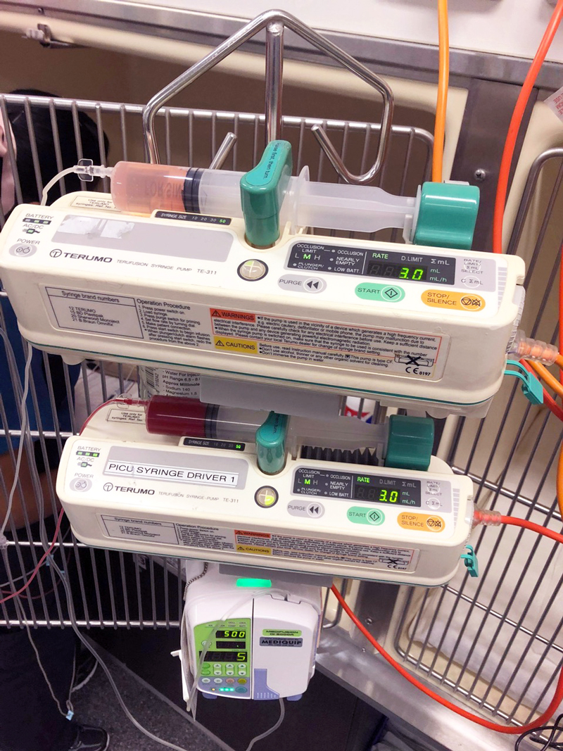
    There are several formulas for determining how much red blood cells are required to be given, some more simpler and less accurate than others.  As a rough guide, if increasing PCV by 1, you need 2ml/kg of whole blood or 1ml/kg of packed red blood cells.

    This gives me a starting volume, I administer that volume and recheck. Often, I will have to give more, but it depends greatly on the underlying disease, concurrent fluid therapy and ongoing blood losses.

    How to administer?

    Use a 170µm blood filter to collect any micro clots. Often, the blood is run through with a isotonic crystalloid, if so avoid administration with calcium containing crystalloid, such as Hartmann’s or lactated Ringers, as the calcium can result in activation of platelets and clots to form in the blood; so run with 0.9% saline or Plasmalyte 148.

    Packed red blood cells are usually diluted with at least 100mls of 0.9% saline. The generally rule is to administer the transfusion within 4 hours and change the fluid lines after to reduce the risk of bacterial contamination. Recheck PCV and coagulation times 30 minutes post-transfusion.

    How fast?

    Rate of administration really depends on degree of urgency. If the patient is suffering from acute haemorrhagic shock then you can administer blood as a bolus. The risk here is acute anaphylaxis if that patient is off a different blood type or if this is not their first transfusion.

    If it is not a crisis then administration should be started off slowly so monitoring for acute reactions can be performed. Start slow at 2ml/kg/hr for 15 minutes and monitor for reactions then progressively double the rate until the desired rate is reached.
  • Seizures, part 1: the questions to ask

    Seizures, part 1: the questions to ask

    Clients often panic when they think their pet is having a seizure and can skip over vital information.

    Often, what an owner describes as a “fit” may actually be syncope, collapse from anaphylaxis or internal haemorrhage (for example, neoplasia), a vestibular event or a behavioural condition.

    True seizures

    True seizures can be divided into two groups:

    • Generalised (grand mal) seizures, which involve both cerebral hemispheres and result in loss of consciousness, incontinence and muscle activity.
    • Focal/partial (petit mal) seizures, which originate from a focal region in the brain. These can also result in alterations in consciousness, but more typically only manifest in the form of repetitive twitching or limb movement.

    Once you have established the owner is likely describing a true seizure, there are many important questions to ask to narrow down your differential diagnoses and treatment options.

    The important questions

    So, as part of a thorough history, always ask:

    Was the pet conscious during the episode?

    This will help to determine whether the seizure was generalised or focal.

    How long did the episode last?

    Status epilepticus is when a continuous seizure lasts more than five minutes or when the patient has not recovered fully before another seizure occurs. This can result in severe secondary brain injury.

    How many episodes has the pet had in the past?

    Epilepsy is the condition of recurrent seizures. This can be further classified as primary and symptomatic epilepsy, with symptomatic being secondary to an underlying cause (such as head trauma or a brain tumour).

    How close together were the episodes?

    • Cluster seizures are when an animal has more than two or three episodes within a 24-hour period.
    • If a patient presents first time with a cluster, this carries a poorer prognosis in dogs, but has no influence in cats.
    • Clusters are generally an indication for commencing long-term management.

    How was the pet before and after the episode?

    • Seizures often come with predicting (pre-ictal) and recovery (post-ictal) events.
    • In the pre-ictal phase, the patient may act strangely (for example, agitated or clingy) and may vomit.
    • Alterations in consciousness prior to a seizure usually indicate an intracranial cause.
    • The post-ictal phase can last anywhere between minutes and days, and animals are usually disorientated and/or lethargic. They may also appear blind.

    Has the pet demonstrated any other strange activity recently?

    • For example, if an animal has also been circling to one side, you can start to predict the location of the lesion.
    • Cats more commonly present with partial seizures compared to generalised – this is seen as stereotypic behaviours and bursts of activity.

    Has the pet been exposed to any toxins or chemicals?

    Seizures caused by toxins (such as snail bait) generally do not stop and start, but are continuous.

    In the next part of this series, we will look at differential diagnoses for seizures and highlight the differences between dogs and cats.

  • Perfusion deficits and fluid resuscitation: a more in-depth look

    Perfusion deficits and fluid resuscitation: a more in-depth look

    A few weeks ago in the Tip of The Week, we discussed the four basic components of a fluid therapy plan – perfusion deficit, hydration deficit, maintenance requirements and ongoing losses.

    Let’s consider perfusion deficits.

    As an emergency clinician, correcting perfusion deficits is a crucial part of stabilising a patient. So what is a perfusion deficit? It either refers to a real or relative loss of intravascular fluid volume, or low blood pressure, leading to a decrease in perfusion of tissues and, ultimately, decreased oxygen delivery – ie, shock.

    What does this look like clinically?

    Pressure infuser
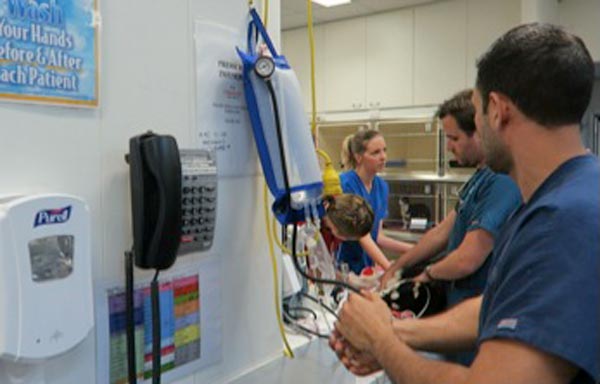
    Operating the pressure infusor at the triage bench.

    Clinical signs of perfusion deficits include:

    • pale gums
    • reduced capillary refill time
    • tachycardia
    • reduced pulse pressure
    • dull mentation
    • cold extremities and low core body temperatures

    A low normal body temperature in a critically ill patient should alert you to the possibility the patient may be experiencing early perfusion deficits, but is not yet severe enough to result in a low body temperature.

    What are these signs caused by?

    These signs are caused by activation of the sympathetic nervous system in response to reduced blood pressure (BP).

    BP is the product of cardiac output (CO) and systemic vascular resistance (SVR):

    BP = CO × SVR

    CO is the product of the heart rate (HR) and stroke volume (SV).

    CO = HR × SV

    Reduced perfusion results in reduced BP. The body increases BP by activating the sympathetic nervous system, resulting in a compensatory increase in HR and SV (beta adrenergic), which increases CO and vasoconstriction (alpha adrenergic) of the peripheral blood vessels to increase SVR and shunt the blood to the heart and brain.

    Peripheral vasoconstriction is seen clinically as pale gums, reduced capillary refill time, cool peripheries and low body temperature as blood is being shunted away from those peripheral capillary beds. It also results in reduced pulse pressures.

    In summary, clinical signs of perfusion deficits are signs of the body trying to compensate and push blood to where it is needed most.

    Blood pressure is normal, it can’t be in shock?

    Since the body’s compensatory mechanisms are geared towards preserving blood pressure, it often remains normal in patients with shock until the body cannot compensate any longer – decompensated shock.

    So, to me, normotension does not rule out perfusion deficits and hypotension is an indicator of severe perfusion deficits.

    Are IV fluid boluses safe for all patients in shock?

    Hartmanns fluid bolus
    Preparing a Hartmann’s fluid bolus.

    The vast majority of the time I would say yes, but you must ask one question – could this patient be in cardiogenic shock? IV bolus therapy would be contraindicated in most of these patients.

    Is it a small-breed dog presenting with a murmur and dyspnoea? Or a large-breed dog with an abnormal heart rhythm? If so then diuretics and anti-arrhythmic medications may be indicated rather then IV fluid boluses.

    How much volume can you give?

    I generally give buffered crystalloid fluid boluses of 10ml/kg over 5-10 minutes. I repeat this while monitoring for resolution of clinical signs of perfusion deficits. If I get to half a blood volume – 45ml/kg in a dog and 30ml/kg in a cat – I ask myself what could be causing this ongoing perfusion deficit. Could it be:

    • Distributive or vasodilatory shock from anaphylaxis or sepsis? In which case, vasopressors – such as adrenalin, dopamine and noradrenalin – are indicated.
    • Cardiogenic shock from dilated cardiomyopathy? Are diuretics, anti-arrthymic medications or positive inotropes required?
    • Restrictive shock from a gastric dilatation-volvulus or pericardial effusion? Is gastric decompression or pericardiocentesis required?
    • Does continued hypovolaemic shock exist? If so then continued fluid therapy is indicated, but this may cause significant haemodilution. Consider colloids or blood products, such as packed red blood cells, whole blood or plasma. Does it need emergency surgery or abdominal compression to stop an internal bleed?

    Perfusion deficits need to be corrected rapidly as continued oxygen delivery to tissues increases the risk of organ dysfunction and, ultimately, organ failure.