Tag: anaesthesia

  • The other side of the consult table, part 1

    The other side of the consult table, part 1

    There comes a time when even doctors and nurses have to make a visit to their local GP (perhaps somewhat begrudgingly), and I wonder if that evokes a similar feeling to when veterinary professionals take their own pets into an appointment?

    My own cat is going in to get her teeth cleaned in a matter of days, and although it is by no means her first trip to the vets, the act of taking her in feels slightly more surreal to me now than it did before I began my training and gained a more similar perspective to that of the vet or vet nurse behind the consult table.

    Familiar faces

    I’ve volunteered at my local practice for years and know the more senior members of staff rather well, but a newer face will obviously ask me the standard check-up questions and explain things the way they would with any other owner.

    To be honest, I never know whether to pretend it’s all new to me or admit I’m a third year vet student; I worry it sounds a bit off to just come out with it without being prompted – a bit like meeting Gordon Ramsay and blurting out that you, too, own several cook books and make a mean Shepherd’s pie.

    The hardest part

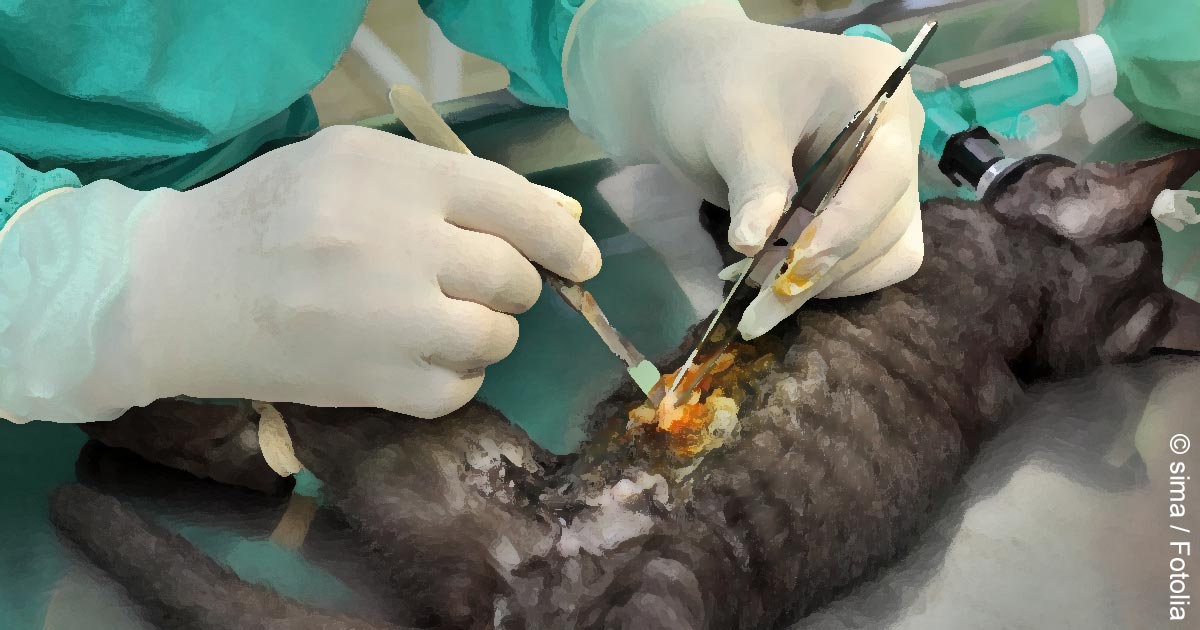
    The last time my cat, Bluebell had to undergo an operation I unfortunately had classes, and although the vet in question knew me well and offered to let me watch, I wasn’t able to – and with a small twist of irony, now that I am free as a bird, the logistics of COVID mean that I must once again sit this one out.

    Along with being the unfortunate messenger of the truly unknowable cost of a procedure to your parents (whose eyes widen at even your lowest estimates, though you try to explain it’s best to get it out of the way when she’s young and healthy), knowing the risks is probably one of the hard parts of being any medical professional – from hearing someone cough, and unconsciously jumping to the worst-case scenarios, to taking your pet in for routine surgery with the anaesthesia mortality statistics circa 2018 committed to memory.

    Not in control

    As ever, the advice you’d give to someone else never has quite the same effect when you try telling it to yourself, and when you’ve experienced the position of the person “in the driver’s seat”, so to speak, it can be hard to surrender control.

    COVID allowing, I would like to be in that operating room myself – and not just because it would be the first lot of EMS I’ve managed to wangle in the past nine months, but because, even if you are distanced from the world of veterinary medicine for any length of time, it never distances itself from you.

  • The art of veterinary medicine

    The art of veterinary medicine

    So, with less than three weeks until finals, my friends and I have finished rotations. Some of us have had job interviews and some have accepted job offers. This is all getting a bit real…

    pagerSupposedly, we’re ready to take on the outside world as real vets. We’ve got heads full of knowledge and hands that have meticulously repeated sutures, catheterisations, and injections to maintain muscle memory. But what we haven’t got is experience.

    Sure, we’ve consulted while on rotations and, before that, we had communication skills tutorials, and although these were realistic – with very good actors screaming at you for losing their hypothetical cat out the practice window or bursting into tears as you explain that their dog died under anaesthetic – they just aren’t quite the same.

    Gaining experience

    Any consultations we have done on rotations have been fairly straightforward, with the vet in the background to interject or, at least, within shouting/pager distance to check anything you’re unsure of.

    We have been involved in high stakes situations where things have gone wrong or an animal’s life has depended on the treatment and care we’ve contributed to – and as much as rotations are aimed at making you think and make your own decisions, you’re always steered in the right direction, or someone intervenes before you do something momentously stupid.

    We may feel like we’re carrying a good deal of responsibility at times but, at the end of the day, it hasn’t been our necks on the line.

    Cash concerns

    money
    “We are assigned a number of tasks throughout final year that make us consider cost […] but you never really have to have that conversation with the owner as such.” Image © Andy Dean / Fotolia.

    And what about money? A lot of our consulting in final year is done at charity clinics, where the treatment is often free or very cheap. This means treatment options are much more limited, so you learn how to approach things on a budget, but you never really have to have that conversation with the owner as such.

    Any consults done at the university’s small animal hospital are referrals – many of which are long-standing patients coming in for rechecks, so the owners have already been dealing with the insurance or have sorted payment with the reception staff.

    We are assigned a number of tasks throughout final year that make us consider cost, such as discussing the costs of anaesthesia and treatment options for certain conditions, or pricing farm visits in first opinion practice. But again, it isn’t really us, the students, making the final decisions based on the client’s budget.

    In at the deep end

    So we’ve got the veterinary science bit nailed (well, hopefully – finals pending). But going into that first day as a qualified vet is going to be nerve-wracking for everyone, and that’s when the education will really begin. That’s where we will learn the things no one can really teach you, nor can you pick up until you are the vet making the final decisions – your neck on the line.

    No one can teach you how to hold your tongue when the client opposite you is being completely irrational, or how to keep your composure when another breaks down in tears over the death of a pet, which you are really quite emotionally invested in too.

    Having the internal battle with yourself over what the ideal diagnostic or treatment protocol would be, versus what is realistically affordable, becomes something of an art – there’s no formula or calculation to work out the best approach. As for actually having the responsibility on your own shoulders, and not falling apart when things go wrong, that takes resilience.

    These are all things that will come with experience. It will be a steep learning curve, I’m sure, but essential if we’re to embark on the journey of life after vet school and master the art of veterinary medicine.

  • How to anaesthetise a reindeer

    How to anaesthetise a reindeer

    I’ve written before about omnicompetency, but the word is mostly used in the sense of vets being able to work in mixed practice and tackle the veterinary care of horses, dogs, cats and farm animals in the same day – certainly, the first thing to come to mind would not be a reindeer.

    However, on my recent equine placement, the staff were met with quite the challenge when a reindeer was referred in.

    Reindeer
    “Reindeer aren’t something you’d expect to see every day in practice,” says Jordan.

    With a history of acute coughing/regurgitation, the reindeer in question had a suspected food impaction in the cranial oesophagus. Conscious radiographs and an ultrasound scan (he was a very well-behaved reindeer) confirmed suspicions of foodstuff, but it didn’t seem to be in the oesophagus.

    Collaborative anaesthesia

    The equine team – with help from one of the farm vets and some phone calls to other colleagues and practices that had dealt with reindeer before – came up with an anaesthetic protocol and proceeded to surgery.

    The reindeer was induced with ketamine and xylazine before a gastroscope was used to try to visualise the larynx and trachea.

    There appeared to be a diverticulum or outpouching from the oesophagus at the level of the larynx, which is where the food impaction had settled.

    This discovery triggered a discussion as to whether our findings could be normal in some reindeer – similar to the Zenker’s diverticulum in people – since its appearance suggested a congenital, rather than acquired, defect.

    A gastroscope was used to aid placement of an endotracheal tube and the reindeer was, subsequently, maintained under anaesthesia with isoflurane. He was positioned carefully in consideration of the rumen and ventilated throughout the procedure, which was to incise into the pouch using a lateral approach and remove the impacted food material.

    Back to his reindeer games

    He recovered well from the anaesthesia and was happily bounding around a paddock before long, eating some specially imported moss provided by his owner.

    Reindeer aren’t something you’d expect to see every day in practice, but it was a great example of how veterinary knowledge can be adapted and applied to new situations, with the added benefit of working together with others with varying levels of experience to come up with a solution.

  • Standing surgery

    Standing surgery

    On my latest EMS placement at an equine hospital, I’ve seen a number of surgeries – some done under general anaesthesia (GA) and others under standing sedation.

    After getting over the fact a horse can stand half asleep while having its face drilled into and not really seem to care, I started wondering about the pros and cons of both approaches.

    Standing surgery
    “Sinus surgery to remove a bony mass – that is me in the pink scrubs holding the head,” says Jordan.

    Generally, standing sedation is accompanied by less haemorrhage and, therefore, increased visibility – in sinus surgery, for example. It also eliminates many risks associated with general anaesthesia. However, asepsis may be harder to maintain (for example, if the horse moves and the surgical site comes into contact with something that isn’t sterile, such as the stocks).

    Lower costs

    For the client, procedures conducted under standing sedation would be much cheaper than the costs incurred from general anaesthesia.

    During general anaesthesia, atelectasis contributes to the risks from an intraoperative point of view, as well as myositis and cardiac concerns (of which the risk can be considerably reduced by the use of acepromazine in the premedication protocol).

    A risk of injury also exists during recovery and knockdown, such as worsening incomplete fractures or other self-inflicted wounds, which can, to some degree, be prevented by carefully assisted knockdown and paying careful attention during recovery with the use of ropes.

    Achieving optimal sedation for standing surgery can, in some cases, be difficult. For example, the horse must be adequately sedated, but not so much it is swaying; this can be an issue for intricate surgeries, but may be more of a problem for diagnostic imaging (such as MRI or bone scintigraphy).

    In these cases, I have seen morphine used – opposed to the usual sedative culprits, such as detomidine, butorphanol and xylazine – and it seems to achieve sedation without so much swaying.

    Choosing correct method

    The choice of standing sedation versus GA depends on the type of surgery required, but a number of procedures can be done using either method.

    Last week, I saw tie-back surgeries (prosthetic laryngoplasties to correct laryngeal hemiplegia) done both ways, which made for an interesting comparison. The standing tie-back was considerably quicker, taking into account the time for knockdown and recovery, as well as surgical time.

    Both tie-backs were followed by a laser hobday procedure (ventriculoectomy), meaning both procedures were conducted under the same sedation in the standing horse, whereas the tie-back performed after GA had to be followed later the same day, after the horse had recovered sufficiently to undergo standing sedation for the laser.

    The second tie-back was a repeat of a previously failed procedure, hence GA was chosen to allow removal of the first prosthesis.

    The standing technique is still being tweaked, but, despite reports of postoperative infection in more cases than ideal, the easier access to the laryngeal cartilages while standing – and the avoidance of further risks associated with GA – contribute to continued work to perfect this method.

    Some surgeries, however, can still only be done properly via GA. Colic surgery, for example, requires significant abdominal access and, often, examination of the gastrointestinal tract. It is also highly recommended septic joint surgery and lavage is conducted under GA to ensure optimal sterility on closure of the joint.

    Conclusion

    Having now seen both types of surgery in the horse, it’s astonishing how quick standing surgery can be, and how much goes into the preparation and recovery for GA – even for the shortest of procedures. In one surgery, division of the aryepiglottic fold, causing epiglottic entrapment, took a matter of minutes  – if you didn’t count the couple of hours total taken for premedication, knockdown and recovery from GA.

    The choice very much depends on the procedure, and is assessed for each case. I do, however, think the advantages to standing surgery are significant and look forward to seeing more standing techniques developed in the future.

  • Flank approach to the bitch spay

    Jordan surgery
    An experienced vet could complete the entire procedure easily within 10 minutes. We “tentatively ambled” through our surgeries in 20.

    Having finally settled in one place in Jaipur, India, my friend and I were able to relax a little, safe in the knowledge we had two weeks of neutering for population control ahead of us.

    Being in an unfamiliar environment, and with our patients mainly being strays, we were prepared for very different methods of anaesthesia, variations on drugs we’re used to at home, and potentially questionable sterility. Even so, when the vet, stood with his scalpel at the ready, said “oh yes, we use the right flank method” as if it were the norm, we were a little surprised.

    At home, we’re so used to seeing flank cat spays and midline bitch spays, my gut reaction was “is that even anatomically possible?”. As it turns out, it is.

    The method

    A small incision (<2cm) is made on the right flank, first through the skin and then each of the 3 underlying muscles (transverse abdominis, external abdominal oblique and internal abdominal oblique). A spay hook is then used to exteriorise the right uterine horn.

    Once identified, the surgeon follows the horn to the ovary and applies tension caudally to break the suspensory ligament. A ligature (note single) is placed around the blood vessel and the ovary cut from it using the three clamp method in the same way as spays in the UK. The surgeon then follows the uterus to the cervix and along the left horn to the left ovary, where the procedure is repeated. A ligature is placed just above the cervix (again using the triple clamp method) and the uterus removed.

    Closing the incision comprises placing a horizontal mattress suture in each of the muscle layers, a cruciate suture in the subcutaneous fascia, and intradermal sutures in the skin.

    The positives

    While the very idea of flank spays in the bitch just seemed alien, this method seems to be successful and works well in a charity environment in a country where certain resources are unavailable.

    The reasons for choosing this method include easier wound checking, a shorter wound healing time (meaning the dogs can be re-released sooner) and less tension at the incision site, decreasing the risk of wound breakdown – essential for animals that, once released, are unlikely to be seen again.

    Jordan surgery
    Despite her initial surprise at the method used, Jordan admits the flank approach is the best compromise, considering the resources available.

    The surgeons at the charity have found, over the years, the single horizontal mattress suture seems to be the least aggravating to the body wall muscles, and intradermals are the closure of choice in any stray or vicious animal that would be difficult to get near to remove sutures.

    Another key advantage to the flank approach is speed; important for two reasons:

    • The sheer number of stray dogs to neuter to reach an adequate level of population control means faster surgery is required to reach the target numbers.
    • The surgical time under IV anaesthesia should be kept to a minimum to avoid prolonged or rocky recoveries and minimise side effects.

    The experienced vet could complete the entire procedure easily within 10 minutes (in a normal young bitch, opposed to a pregnant or in season girl), and we, tentatively ambling through our surgeries, could complete within 20.

    The negatives

    Disadvantages to this method include more potential bleeding due to incising through the three muscle layers, a possibility of more postoperative pain and increased difficulty in extending the incision if there are complications. The most important, however, is that recovery of a dropped or bleeding ovarian stump is extremely difficult (or near impossible).

    The anaesthesia protocol used is premed: xylazine, induction/maintenence; IV ketamine and IM meloxicam as pain relief. Hence, the speed of the flank approach will also minimise the number of top ups needed and reduce the anaesthetic hangover comparing to a technique (such as midline) that is more time consuming.

    Compromise

    The method seems to be the best compromise, considering the resources available. I think the overruling disadvantage is that, if you were concerned about a slipped ligature, the ovarian and uterine stumps would be virtually impossible to find again via the original incision.

    However, that said, the only postoperative death we saw during our time on postmortem had all ligatures intact.

    It was eye-opening to see an entirely different approach to a bitch spay, and while it may not be the same as the routine at home, I still felt that we gained a lot of surgical experience and developed transferable skills.

  • Out of Africa: two weeks in the wild

    Shamwari Game Reserve
    Shamwari Game Reserve, South Africa.

    Wildlife veterinary medicine is more than being a good shot with a dart gun and knowing what anaesthetics to fill the darts with.

    I know this because I was part of a group from Glasgow vet school that visited the Shamwari Game Reserve in South Africa to find out exactly what wildlife medicine entails.

    Minimal intervention

    Generally, as the animals on the reserve are considered wild, the main aim is to keep veterinary intervention to a minimum.

    The majority of the veterinary work is translocation (involving mainly sedation and/or anaesthesia) but there are a few exceptions, which are often dealt with in the reserve’s rehabilitation centre – particularly if the animal in question is rare or highly valuable.

    Although we learned a lot about anaesthetics (which was surprisingly easy to relate to practice at home), there’s far more to it than meets the eye.

    In-depth knowledge

    buffalo_Jordan-SinclairDuring our two-week stay we learned that an in-depth knowledge of animal behaviour, among other aspects of conservation, is the vital foundation of the veterinary work.

    For example, the length of time an animal will spend in transit and whether the animal will be woken up in the field will have an impact on drug choices – a prey animal needs a complete and rapid recovery in the field to avoid showing weakness to the herd or any predators in the vicinity, whereas an animal travelling halfway across South Africa would require longer lasting sedation.

    Species differences are also important to consider; drugs that are highly dangerous in some animals may be the sedative of choice in others.

    Additionally, the reaction to being darted needs to be carefully evaluated in terms of both the animals’ and the staff’s safety. We were particularly aware of this when working with buffalo and having to judge the right amount of sedation reversal to allow them to wake up completely, while giving ourselves enough time to dash into the truck and put a safe distance between us before they did so.

    A different perspective

    The team at Shamwari were eager to give us a practical, hands-on experience – which was great.

    Image courtesy Shamwari Conservation Experience.
    Image courtesy Shamwari Conservation Experience.

    Not only was it an awe-inspiring couple of weeks, but it was also incredibly useful in terms of consolidating knowledge and practical application of pharmacology – particular anaesthesia.

    Experiencing all aspects of conservation in addition to the veterinary work also gave a different perspective to veterinary medicine.

    It was exhilarating to be working with such beautiful and potentially dangerous animals – I could certainly feel my heart going through the roof when taking blood from a lion whose snores sounded suspiciously like growls.

  • Using PDS to “wire” mandibular symphyseal fractures in cats

    We have now started to use Polydioxanone (PDS) to stabilise symphyseal fractures in cats. We use 4 metric PDS passed under the mandible and around the canines in a figure of 8.

    In uncomplicated fractures, we have found this to be really effective and the fracture will heal within four to six weeks. If, like me, you have ever had trouble removing the wire at this time (see video below for example of a traditional wiring method), this technique may well be for you – not to mention the cat will not need further anaesthesia.

    Video: Avulsion fracture of the lower lip and the mandibular symphysis in a cat. Source, YouTube.

  • Anaesthetising rabbits

    Vet holding rabbit
    Image ©iStock.com/4774344sean

    It is generally accepted rabbit anaesthesia can be challenging, but we do a lot of rabbit neutering as part of our work for an animal charity and use a protocol that has proved very safe and effective.

    We also use yellow gauge catheters placed into an ear vein for IV administration.

    Vetergesic (buprenorphine) 0.07ml/kg
    Hypnovel (midazolam) 0.2ml/kg

    • Combine these drugs in one syringe and inject subcutaneously 20 minutes before procedure.
    • Rabbit will become quiet and relaxed.
    • Ketamine 0.2ml/kg diluted with 1:10 saline (that is, 0.2ml ketamine in 2ml saline).
    • Give this slow IV to effect.
    • Most rabbits use half a syringe, but occasionally I have had to use the whole syringe. If it is not having an effect, flush the catheter, and mask with isoflurane if required.
    • Rabbit will react if it goes extravascular;
      • once induced, spray Intubeaze and intubate – listen to breath sounds; there is usually no cough.
    • Rabbit can usually be maintained on 2% isoflurane.
    • Slower to wake up, but eating.
    • Hypnovel must be discarded within 24 hours, so better to try to book in all rabbits on the same day.
    • Premed can be used for x-rays; incisor trimming.
  • I blame the NHS

    "Those detached from the veterinary world still uphold the perception that vets are rich and set high prices to rip off the unsuspecting public."
    Original image ©iStockphoto.com/hidesy

    A couple of articles have been brought to my attention this week regarding the public perception of vets versus the reality.

    When somebody finds out that you want to be a vet or are studying veterinary medicine, there are a few arduous questions that usually follow:

    • “Is it seven years at university for that?”
    • “It’s hard to get into isn’t it?”

    And, of course:

    • “Well, vets earn loads of money don’t they?”

    Unfortunately, only one of those three assumptions is true. However, those detached from the veterinary world still uphold the perception that vets are rich and set high prices to rip off the unsuspecting public. What these people don’t understand is that a new veterinary graduate can expect to start on a salary of around £20k while working all living hours of the day, plus being on call.

    While each individual job varies, the reality is that we can expect to earn very little considering the length of intense training required and the high levels of stress and responsibility that come with the job.

    With experience and promotion to more superior roles – such as becoming a partner in a practice – the salary will increase, but often to less than half the average wage of a GP doctor. In general, vets accept this and are highly dedicated to the health of the animal they work with. If they were after a high salary, they’d be better off in a different career field entirely.

    But the public can’t be entirely blamed for their own ignorance – I think a lot of the problem lies with the National Health Service

    There is no NHS for pets...
    “There is no NHS for pets, and I think many people would do well to remember this.”

    We are blessed with a “free” health service in the UK, but this means those of us outwith the field of human medicine have very little understanding of how much treatments, operations or drugs cost.

    Perhaps if people knew how much these things would cost if they had to fund them privately, they’d have a greater appreciation of both the NHS itself and the veterinary care they pay for for their animals. After all, there is no NHS for pets, and I think many people would do well to remember this.

    My American friends tell me that clients in the US do seem to have a more grounded understanding of the cost of healthcare and are able to apply this to veterinary care without quite so much complaining.

    It’s also worth noting that the money people spend on their animals’ treatments does not go directly into the pocket of the surgeon, but contributes to the cost of anything required for the procedure, including medication, electricity, needles and syringes, catheters, x-ray plates, bedding, food, anaesthetic, licensing… this list goes on – and somewhere at the bottom of that list sit the wages of the hardworking and dedicated vet, who often only receives a grumbling about the expense in “thanks”.

    On a recent EMS placement at a small animal veterinary surgery, I witnessed a lot of this grumbling, and sometimes even full blown arguments about cost. Luckily it’s the few clients that are truly grateful and would do anything for their animals that make it all worth it.

    I find it highly offensive and disrespectful when I hear remarks that vets are “only in it for the money” because, if that were true, then we are not as intelligent as our education might suggest.

  • How to examine a patient’s rima glottidis for suspected laryngeal paralysis

    Intraoral view of a dog with bilateral laryngeal paralysis during inspiration. The arytenoid cartilages (a) are immobile and the vocal folds (b) are medially displaced. Credit: Daniela Murgia.
    Intraoral view of a dog with bilateral laryngeal paralysis during inspiration. The arytenoid cartilages (a) are immobile and the vocal folds (b) are medially displaced. Credit: Daniela Murgia.

    Anaesthesia normally depresses laryngeal movements, making diagnosis of laryngeal paralysis challenging. The animal should be anaesthetised to the point at which the mouth can be easily opened but a laryngeal reflex is still present.

    If jaw tone is such that you are afraid of being bitten during the examination, then the plane of anaesthesia is appropriate. If the GA is too deep, the patient will not have normal vocal fold movement and even a normal larynx may appear paralysed. In this case you should wait for drug redistribution allowing the patient to approach consciousness and repeat the examination.

    Exposure of the larynx is more readily accomplished with thiopental or propofol than with diazepam-ketamine.