Tag: Anaemia

  • Oh, CR*P! Using point-of-care C-reactive protein tests

    Oh, CR*P! Using point-of-care C-reactive protein tests

    Few companies now offer affordable point-of-care tests for canine C-reactive protein (CRP). As we did when we recently received our new box of CRP slides, you might soon be asking the question: what do we even do with this stuff?

    Here’s what we’ve learnt…

    CRP is one of the acute phase proteins produced by the liver in response to inflammation. Healthy patients have very low levels of CRP, but a systemic inflammatory condition will cause an increase in CRP within four to six hours. Conversely, increased levels will decrease rapidly on resolution of inflammation. This provides an almost real time measure of inflammation that is more responsive and reliable than the white blood cell response.

    In other words, CRP can indicate the presence of inflammation before the patient’s white blood cell count gives any clues, or before it becomes pyrexic – and, unlike the white blood cell count, stress and steroids do not affect CRP levels.

    Uses

    So, how do we use it?

    • I love it for early pickups of problems in those grey area cases: the dog seems okay on clinical examination, but something about it bothers me. A normal or mildly increased CRP test will make me sleep more easy, while a surprise high reading will prompt me to admit for full diagnostics, or at least get the patient in for a follow-up CRP the next day. Conversely, a localised problem – such as an abscess – combined with a normal CRP test might mean you can hold off on antibiotics and just recheck CRP in 24 hours.
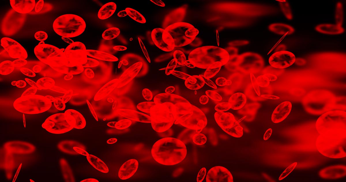
    • It’s great for monitoring response to treatment. If my plan is working then I’d expect CRP to show a significant decrease by day two or three. If it’s not dipping by then, I need to reassess my treatment plan. Do I need to change antibiotics? Scan it again? Maybe we need to consider surgery? It can also be a good prognosticator. Research has shown failure of CRP to decrease significantly (around a 3× decrease) by around day three is generally bad news for patients with inflammatory conditions such as pancreatitis and immune-mediated haemolytic anaemia.
    • We are starting to play with it for post-surgical monitoring. Any surgery will cause inflammation with an increase in CRP levels, but in an uncomplicated postoperative period, you should expect levels to start decreasing by day three to five. A base line CRP 24 hours after surgery with a recheck on day three should pick up early signs of postoperative problems such as infection, and prompt investigation or intervention.
    • A potentially nifty use for it that we haven’t yet had the opportunity to use is in differentiating inflammatory lamenesses (arthritis, infection, injury) from a neurological causes – that is, is it arthritis or a nerve problem?

    Limitations

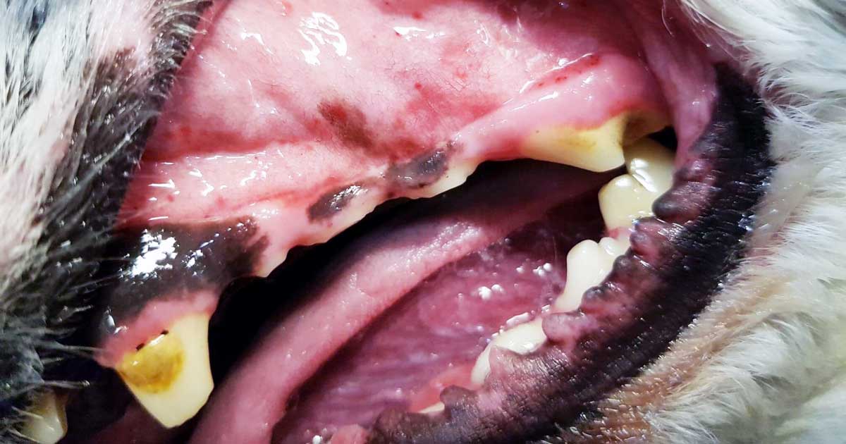
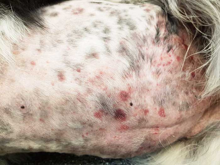
    • Remember, it’s very sensitive, so will increase with almost any inflammation. A mild upper respiratory infection or a bad gingivitis will likely induce some changes, so it’s important not to over-interpret (keep in mind that the magnitude of the increase in CRP does generally correspond with the severity of the inflammatory response). A pancreatitis case where the CRP fails to drop does not always mean death is looming – you may have just missed the concurrent skin disease. Always interpret CRP values in concert with your clinical examination.
    • Be aware that pregnancy and intense exercise can increase CRP values.
    • Not all serious conditions have an inflammatory component. CRP will be unchanged in most veterinary cases of heart disease; in common hormonal disease, such as adrenal disease and uncomplicated diabetes; urinary obstructions; many localised cancers; epilepsy and many others. Don’t presume that just because CRP is normal, everything is fine.
    • No similar test exists for cats.

    Sit up and say…

    My favourite way to explain how to use this test is by using its highly appropriate acronym – any unexpected increase should make you sit up and say: “Oh CR*P! What am I missing?”

  • Managing thrombocytopenia in cats and dogs: part 2

    Managing thrombocytopenia in cats and dogs: part 2

    Last week we discussed the causes and diagnostic pathway for investigating immune-mediated thrombocytopenia. This week we will go through the management of this condition.

    Despite the fact red blood cells are not actually being destroyed, a severe anaemia can develop from blood loss due to coagulopathy – a common reason for why they present to emergency practices. The management of these patients is broken down into three main areas:

    • improving oxygen delivery
    • commencing immunosuppression
    • management of the underlying cause (if identified)

    Optimising oxygen delivery in the acute phase is going to keep them alive long enough for immunosuppression to work. This is achieved through IV fluids to help improve perfusion and blood transfusions to replace red blood cells. If fresh whole blood is available, it can assist in increasing platelet numbers, but generally it is not very effective.

    Platelet transfusions using platelet-rich plasma can be considered if it is available. Plasma transfusion is not effective at managing the coagulopathy as it is due to a loss of platelets, not a loss of coagulation factors.

    Immunosuppression

    Large areas of ecchymotic haemorrhage on the skin are a quite obvious sign of thrombocytopenia.
    Large areas of ecchymotic haemorrhage on the skin are a quite obvious sign of thrombocytopenia.

    Immunosuppression therapy is often commenced concurrently as the patient is being stabilised.

    The first choice is either dexamethasone 0.5mg/kg IV every 24 hours if the patient is not stable enough for oral medications; otherwise, once stable, start prednisolone at 2mg/kg by mouth per day divided every 12 hours.

    Other immunosuppressive agents include:

    • Azathioprine – 2mg/kg by mouth every 24 hours then 0.5mg/kg by mouth every other day. The main concerns are bone marrow suppression and hepatoxicity – also, it is very toxic in cats.
    • Ciclosporin – 5mg/kg to 10mg/kg by mouth divided twice a day; cats 5mg/kg by mouth every 24 hours.
    • Chlorambucil could also be used at a dose of 0.1mg/kg/day to 0.2mg/kg/day by mouth if the response to prednisolone is insufficient.

    Management

    Management of the underlying cause should be commenced if a cause is identified, but this is often not the case.

    Other management options include:

    • Vincristine can be trialled to increase platelet number as it stimulates the release of platelets from the bone marrow.
    • Gastroprotectants can be considered if gastrointestinal bleeding has occurred – these include proton pump inhibitors and sucralfate.
    • Strict confinement, potentially sedatives and minimal blood sampling are important to minimise injury that may result in further bleeding and blood loss.
    • Antithrombotic therapy is not part of standard management as, unlike immune-mediated haemolytic anaemias, thrombotic events rarely occur.

    When it comes to monitoring, platelet counts are performed daily until more than 40 × 109/L – this can take up to two weeks to occur.

    Once above this level, take weekly counts until the numbers have normalised. Once they have, taper immunosuppressive medications over four to six months, with 20% dose reduction every couple weeks, generally with the adjunctive immunosuppressants first and prednisolone last.

  • Managing thrombocytopenia in dogs and cats: part 1

    Managing thrombocytopenia in dogs and cats: part 1

    Thrombocytopenia is a condition characterised by a decrease in platelet numbers, which is often caused by increased destruction of platelets or a decrease in production.

    Thrombocytopenia can manifest in many ways – the signs can be subtle and easily missed, such as small petechiae on gums, or quite obvious signs, such as large areas of ecchymotic haemorrhage on the skin.

    Ecchymotic haemorrhages are often attributed to disorders of secondary haemostasis, such as rodenticide intoxication, but it can also occur with thrombocytopenia depending on the severity and chronicity.

    Other common clinical signs include:

    • epistaxis
    • blood in stools, urine or vomit
    • pale mucus membranes
    • lethargy
    • weakness

    Therefore, the first step to managing a patient with a severe thrombocytopenic episode that has resulted in significant blood loss is to manage shock, if present, with IV fluids, then administer a red blood cell transfusion.

    Patient handling

    Careful patient handling is critical as these patients can bleed easily, leading to blown veins, large bruises that contribute to the development of anaemia and significant patient discomfort. Beyond initial patient stabilisation, the next step is to determine the underlying cause.

    Diagnosing thrombocytopenia is relatively straightforward with the demonstration of low platelet counts. Generally, bleeding does not occur until the platelet count drop below 40,000 thousands per cubic milliliter (k/uL). This can be determined by either a haematology machine or manually via blood smear analysis.

    When assessing blood smears, the general rule is one platelet per high-powered field on the monolayer is equal to 15,000k/uL. With either method, you must assess for platelet clumping on a blood smear as this can artefactually drop platelet numbers, leading to a false diagnosis.

    Causes

    PCR
    The thrombocytopenic diagnostic pathway includes assays such as PCR. Image © science photo / Adobe Stock

    The diagnostic pathway should not stop there. It needs to continue to determine the underlying cause.

    The most common cause of thrombocytopenia is immune-mediated destruction. This can be either a primary (diagnosis of exclusion) or secondary cause (such as Rickettsia infection, and drugs such as sulphonamides, toxins and neoplasia). Other less common causes include:

    • splenomegaly, which can lead to platelet sequestration
    • disseminated intravascular coagulation and acute blood loss, leading to platelet consumption
    • bone marrow disease, which results in reduced platelet production

    Signalment and history will refine such a diagnosis, as certain breeds are more prone to developing thrombocytopenia than others – for example, grey collies due to a defect in haematopoietic stem cells, and whippets and greyhounds, which traditionally have a lower platelet count than other breeds.

    The generally diagnostic pathway continues to include haematology and biochemistry, thoracic radiographs, abdominal ultrasound and depending regional prevalence testing for infectious organisms with PCR and ELISA assays.

    Next week I will cover the management principles of the thrombocytopenic patient.

  • Using lactate measurements in general practice

    Using lactate measurements in general practice

    Several easy and affordable ways exist to measure lactate in general practice, which means the clinical applications of monitoring lactate is no longer the reserve of specialist and emergency centres.

    But why and how should you be using it in general practice?

    What is lactate again?

    When oxygen is not effectively delivered to cells throughout the body – which, in our patients, will mostly be due to hypoperfusion (for example, hypovolaemia, vasodilatory shock and cardiac disease) – cells will switch from aerobic to anaerobic metabolism to stay alive.

    Think of anaerobic metabolism as the fuel-powered generator that kicks in during a power cut – it’s not as good, but it’ll keep the lights on for a while. However, unless the power comes back on, the generator will eventually also fail and plunge you into darkness.

    Lactate is the end product of this process of anaerobic metabolism. To be clear, lactate is not the bad guy – in fact, it plays an important role in keeping the cells going until they have access to sufficient oxygen again. It’s simply the bearer of bad news.

    Why should I care?

    Anaerobic metabolism – similar to a fuel generator being used during a power cut. Image © bildlove / Adobe Stock
    Anaerobic metabolism – similar to a fuel generator being used during a power cut. Image © bildlove / Adobe Stock

    Because lactate is the harbinger of doom; it’s the leading horseman of the apocalypse…

    When lactate is high, you should stop whatever else you are doing and pay attention to the patient in question – this patient is probably surviving on anaerobic metabolism. It is critically ill and possibly heading for a long walk in the great park in the sky…

    How should I use it?

    It is valuable in any patient with any serious illness or injury. Things that should make you consider checking lactate would be:

    • slow capillary refill time
    • any mucous membrane colour other than a nice, healthy pink
    • increased heart rate
    • weak or bounding pulses
    • significant dehydration
    • depressed mentation
    • history of major trauma
    • major infections
    • significant blood loss
    • any disease that has the potential to progress into a life-threatening condition

    …basically, any animal sick enough to make you worry about it possibly dying.

    Run it with your initial diagnostics to get a baseline level, and run it within 10 minutes of taking the sample.

    What do I do about my results?

    Normal range

    What do my results mean?

    < 2.5  ?

    3-4 ? ?

    4-6  ? ?

    > 6  ? ?

    (levels in mmol/L)

    If it’s within the normal range then check regularly – ideally until your patient is well on its way to a full recovery.

    Lactate levels may start increasing in response to hypoperfusion before the patient starts showing overt signs of deterioration – the fuel in the generator hasn’t run out yet – which makes it a very useful monitoring tool to detect problems early on.

    We find a six to eight-hourly check in very sick patients will pick up deterioration fast enough to give you time to react, while a twice-a-day check in more stable animals will suffice.

    Elevated

    If it is increased at any point, you need to focus immediately on trying to reduce it.

    This usually means starting with fluid boluses for shock. Recheck lactate one hour after initiating the appropriate therapy. Your goal is for the lactate value to be reduced by approximately 50% within one to three hours (ideally one hour) of initiating therapy. If it’s coming down nicely then keep checking every two to three hours.

    You want it to be back to normal within 24 hours (48 hours max).

    Nothing working?

    If it has not decreased as expected – or, especially, if it increases despite treatment – it means things are going seriously wrong. At this stage you need to:

    • Devote all your attention on trying to find and correct the underlying cause while you adjust your emergency therapy to address hypoperfusion.
    • Speak to the owners.
    • If you do not have the time, facilities or experience to deal with shock cases, you need to consider referring the patient to a specialist centre urgently for stabilisation, if this is an option available to you.

    Remember…

    Intense exercise, muscle tremors and seizures are associated with anaerobic muscle activity and can, therefore, cause significant increases in lactate levels (the cause of the “deep burn” when you’re dying in that CrossFit class). This can also occur when a patient resists when you are taking blood, or starts trembling in fear the moment it walks into the clinic, which can cause misleading lactate results.

    Puppies can have a higher “normal” level of plasma lactate up to seven months of age.

    Anaemia does not generally cause hyperlactataemia unless it is very severe, but by this stage you shouldn’t need lactate to tell you the patient is in serious trouble.

    Having said that, normal lactate levels that suddenly start climbing in a hospitalised “stable” anaemic patient could be the push you need towards giving that blood transfusion.

  • Blood transfusions, pt 2: what do I give?

    Blood transfusions, pt 2: what do I give?

    To make the most of a precious resource, donated blood is often separated into two components: red blood cells (packed red cells) and plasma (fresh frozen plasma, most commonly).

    Haematocrit tube
    Haematocrit tube from a patient with immune mediated haemolytic anaemia and a slide demonstrating autoagglutination.

    This not only extends the life span of plasma component, but it also means you can pick and administer which component you need the most as not all anaemic patients need the same product.

    Which product?

    Here are three different examples of patients with different blood product requirements:

    Immune mediated haemolytic anaemia

    This a prime example of a patient that can have an anaemia with transfusion triggers, but are rarely coagulopathic. The key component required here is packed red cells.

    Internal bleed

    This is a grey zone – an anaemia with transfusion triggers can occur, but not always a coagulopathy. A coagulopathy may develop later if the bleed continues. So, packed red cells can be used initially, and the plasma administered after, if required.

    Rodenticide intoxication

    typing kit
    Commercial feline blood typing kit.

    These patients will often need both red blood cells and plasma. So, whole blood – either fresh or stored – can be used, or a unit of packed red cells combined with a unit of fresh frozen plasma.

    Blood typing and crossmatching

    Before administration of the product there is one critical step. Blood typing is important – not only for cats, but also dogs – as it helps rule out blood type incompatibilities and can significantly reduce the risk of an acute transfusion reaction. Easy-to-use commercial blood typing kits (pictured) are available.

    Just because you blood typed doesn’t mean there is no value in crossmatching. Other factors or uncommon red blood types, such as DAL (dogs) and Mik (cats) can cause acute reactions.

    Both these tests do not 100% rule out the possibility of an acute reaction, so, whenever a blood product is being administered, the patient needs to be monitored closely.

    Next week, we cover how much to give.

  • Blood transfusions, pt 1: clinical signs

    Blood transfusions, pt 1: clinical signs

    I get asked frequently when is the right time to transfuse an anaemic patient?

    The difficulty lies in the fact not all anaemic patients require blood transfusions. Just because a patient has pale mucous membranes does not mean the patient needs a transfusion.

    The term commonly brought up during the discussion is “transfusion triggers present”.

    What constitutes a “transfusion trigger”?

    A couple of different definitions exist: classically, it is the PCV or haemoglobin level at which a transfusion is indicated in an individual animal – essentially, if it gets below a certain number, transfusion is required – but it is not always that simple. Just because the PCV is 15%, it doesn’t always mean a transfusion is required.

    When the PCV drops low enough, clinical signs of reduced oxygen delivery to the tissues start to develop, these include:

    • decreased exercise tolerance
    • weakness
    • dull mentation
    • tachycardia
    • tachypnoea
    • elevated lactate levels when shock has been addressed

    Rapid or slow?

    These clinical signs are influenced by the speed at which the anaemia has developed.

    If the anaemia has occurred rapidly due to internal bleeding from trauma or a ruptured organ, these clinical signs can present in a matter of minutes, depending on how big the bleed is. This means a transfusion might be indicated when the PCV is still 25%, especially if further rapid blood loss is likely.

    If the anaemia developed over days to weeks (slow red cell destruction or anaemia of chronic disease, for example) transfusion triggers might not be present until the PCV drops below 15%, as the body has had time to compensate.

    Summary

    So, in summary, the decision-making process involves asking the questions:

    • What is the PCV?
    • How fast has the anaemia developed?
    • Are there clinical signs of reduced oxygen delivery?
    • Is further loss likely?

    When you combine the core aspects of each of the questions above, “transfusions triggers” change from absolute numbers to this:

    • PCV under 15% with clinical signs of reduced oxygen delivery.
    • Rapid PCV drop to under 20% in dogs and 15% in cats.
    • PCV under 25% and surgery or anaesthesia is required, and/or rapid ongoing blood loss is occurring.

    Blood products you should use and why will be covered in a future post.

    Note: Haemoglobin levels should also be assessed in conjunction with the PCV.

  • Dystocia, pt 2: diagnostics

    Dystocia, pt 2: diagnostics

    Part one of this series covered the stages of labour and indications dystocia is present.

    Once the bitch presents to the clinic, a few basic diagnostic checks need completing to determine the status of the bitch/queen and the fetuses.

    Physical examination

    The first is a thorough physical examination, starting with the bitch or queen:

    • Demeanour, hydration status, vital signs, mucous membrane colour, capillary refill time and temperature are important.
    • Pregnancy anaemia is not uncommon; however, for patients with a haemorrhagic discharge, it is important to know their cardiovascular status.
    • A thorough abdominal palpation should be carried out to assess comfort level and palpation for the presence of fetuses. Palpating fetuses can be difficult and cannot confirm if no fetuses are present.
    • A digital vaginal examination should be performed. Feathering response – also known as the Ferguson reflex in human medicine – is the neuroendocrine reflex where the self-sustained cycle of uterine contractions is initiated by firm pressure on the dorsal aspect of the vestibulovaginal wall. If this is absent, the patient is unlikely to progress with the parturition unaided.
    • Palpation of fetuses in the canal can help decide whether surgical management is required. Obvious fetal malposition, malposture or malpresentation, or fetopelvic disparity, will be indications of caesarean. Abnormal pelvic diameter is also another reason to not proceed with medical management. To confirm these suspicions, abdominal radiography is required.
    • Radiographs will also help determine the number of fetuses to be expected, the signs of fetal death (presence of gas surrounding the fetus) and aforementioned fetomaternal abnormalities. I always repeat radiographs after the expected number of neonates is passed, to make sure I have not miscounted at the start.

    Ultrasound

    Panel 1. Heart rate ranges to help indicate stress of fetuses

    Dogs:

    • normal – 180 to 220 beats per minute (bpm)
    • Stressed – 160bpm
    • Real concern – less than 160bpm

    Cats:

    • normal – more than 220pbm
    • fetal stress – less than 180bpm

    The second important diagnostic tool is ultrasound.

    Fetal heart rates are good indicators of fetal stress. Some heart rate ranges that can help provide information about the status of the fetuses are detailed in Panel 1. These ranges vary between sources, but are good guidelines.

    Ultrasounds can also help visualise the maturation status of the fetuses. At-term fetuses should have normal hepatic, renal and intestinal development. Intestinal peristalsis should be evident in at-term fetuses.

    Other diagnostics

    Other diagnostics may be indicated for patients, depending on the status of the bitch/queen:

    • If the patient is stable, but dystocia is present, a minimum database would include PCV/total protein, electrolytes, glucose, ionised calcium, lactate and acid-base balance.
    • Serum ionised calcium levels are important, as they influence the strength of contractions and how much supplementation is required.
    • Hypoglycaemia needs to be ruled out as a cause of dystocia, especially when large litters are involved.
    • If the patient is unstable or systemically unwell, include complete blood count, blood smears and biochemistry.
    • Physiological pregnancy anaemia can be present. The presence of regenerative response can help differentiate this from acute haemorrhage.
    • Abnormal leukocyte panel, especially with the presence of degenerative left shift, can indicate the presence of an infection – especially if toxic changes are present in the neutrophil.

    Part three will briefly look at the medical management of dystocia and when surgical intervention is required.

  • Oxyhaemoglobin dissociation curve, pt 4: left and right shift

    Oxyhaemoglobin dissociation curve, pt 4: left and right shift

    In various disease and physiological states, the oxyhaemoglobin dissociation curve (OHDC) can shift either left or right. This indicates the increase, or decrease, of the haemoglobin’s (Hgb’s) affinity to oxygen, respectively.

    It is important to recognise the situations in which this happens, to manage patients effectively.

    Right shift

    A shift of the OHDC to the right indicates the Hgb has a reduced affinity to oxygen. This is normally seen in environments where oxygen needs to be released by the Hgb molecules – for example, muscles and placenta.

    Four major factors influence this:

    • low blood pH (lactic acid)
    • high temperature – especially working muscles
    • high partial pressure of carbon dioxide (PCO2)
    • increased 2,3-bisphosphoglycerate (2,3-DPG) – an intermediate of glycolysis

    2,3-DPG

    2,3-DPG is produced in red blood cells during glycolysis. Its production is increased for several conditions in the presence of diminished peripheral tissue oxygen availability, such as hypoxaemia, chronic lung disease, anaemia and congestive heart failure. It promotes oxygen to be released into the tissues and, therefore, makes it harder for oxygen to bind with Hgb in the lungs.

    The presence of 2,3-DPG can increase oxygen release to the tissue equivalent to that if the surrounding arterial partial pressure of oxygen (PaO2) was 10mmHg higher. This is why 2,3-DPG is increased during pregnancy to increase oxygen delivery to the growing fetus.

    Another example is during exercise – in the presence of high CO2 and more hydrogen ions (H+) from lactic acid (lowering the pH), the curve is shifted to the right to help increase oxygen release in the muscles.

    Left shift

    Oxygen dissociation – left and right shift
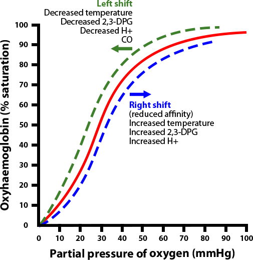
By the same token, a shift of the curve to the left means the Hgb has an increased affinity for oxygen, so is less likely to release it to the tissue. This normally occurs in the lungs.

    The factors this promotes are:

    • high blood pH (breathing off CO2 – an acid)
    • low temperature (ambient temperature usually lower than that of the lungs)
    • reduced PCO2 (ventilation)
    • decreased 2,3-DPG and the presence of fetal Hgb

    So, during exercise, in the presence of lower CO2 and less H+, the curve shifts to the left to help increase oxygen uptake into the Hgb.

    Carbon monoxide toxicity

    One pathological situation that causes a left shift of the curve is carbon monoxide (CO) toxicity.

    Not only does it cause the OHDC curve to shift to the left, but the Hgb is also 240 times more likely to bind to CO than oxygen. The situation is further complicated by pulse oximeters’ inability to differentiate CO-bound Hgb to O2-bound Hgb, therefore giving a false reading of normal partial pressure of oxygen in the face of severe hypoxaemia. Similarly, methemoglobinaemia has a similar effect to CO and causes a left shift of the OHDC.

    Conclusion

    Understanding the relationships between Hgb, oxygen saturation, PaO2 and the causes of the shifts of the curves will allow accurate assessments of patients.

    For example, a patient with unusual hypoxaemia derived from arterial blood gas – despite normal PaO2 and peripheral oxygen saturation – will immediately increase the suspicion of CO toxicity.

    Similarly, a patient presenting with shortness of breath, despite adequate ventilation and oxygen saturation, will suggest Hgb deficiency.

  • Icteric serum

    Icteric serum

    The final discolouration of the serum we are going to cover is icteric serum.

    Icteric serum
    Icteric serum is caused by the presence of excess bilirubin in the blood stream.

    Icteric serum is caused by the presence of excess bilirubin in the blood stream as a result of increased production (pre-hepatic) or inappropriate excretion (hepatic and post-hepatic).

    The most common cause of pre-hepatic icterus is haemolytic anaemia, while hepatic disease and biliary tract obstruction are the most common causes for hepatic and post-hepatic icterus, respectively.

    Tips on where to start

    If icterus and concurrent anaemia exist, my first suspicion would be some kind of pre-hepatic cause. The most common causes are immune-mediated haemolytic anaemia and infectious haemolytic anaemia, such as haemotropic mycoplasma and babesiosis.

    Other causes can include snake envenomation and oxidative injury from heavy metal toxicity or onion ingestion.

    Regarding hepatic and post-hepatic causes, unfortunately it is not always clear-cut. Both are commonly associated with elevation in both alanine transaminase (ALT) and alkaline phosphatase (ALKP), and, although no specific changes are pathognomonic for hepatic or post-hepatic disease, the pattern of change may help identify the origin of the cause. ALT is released from the inside of hepatocytes, and in higher amounts when cell damage occurs.

    Hepatic hints

    Some pointers on what you can do to help differentiate:

    • Compare the ALT and ALKP elevation; if one is in order of magnitudes higher than the other then it can help point to an origin.
    • If the cause is of hepatic origin, one would expect the ALT to be significantly more elevated than the ALPK. Likewise, this is usually true in reverse for post-hepatic causes. However, it should be noted in chronic hepatic diseases, where active damage to hepatocytes is comparatively lower, a mild increase in ALT and marked increase in ALPK does not preclude disease of hepatic origin. Therefore, biopsies should always be used for definitive diagnosis.
    • If other biochemistry parameters such as albumin, glucose and cholesterol are low, or prolonged clotting times are present, the case for a hepatic origin is strengthened.
    • The gallbladder and bile duct can be assessed using abdominal ultrasonography. The presence of a dilated bile duct, or evidence supportive of pancreatitis, is highly suggestive of a post-hepatic cause.

    Finally, it is important to be aware of the impact on hyperbilirubinaemia on laboratory testing. Hyperbilirubinaemia generally causes decreased cholesterol, triglyceride, creatinine, lipase, total protein and gamma-glutamyltransferase levels.

  • PCV/total solids interpretation: serum colour

    PCV/total solids interpretation: serum colour

    When interpreting the often misinterpreted and underused PCV and total solids test, it is important to take note of the serum colour as this may give clues into the diagnosis.

    PCV tubes
    Normal serum colour (left) compared to a patient with immune-mediated haemolytic anaemia. The serum is haemolysed and anaemia is present.

    The most common abnormalities seen in clinic are icteric, haemolysed and lipaemic serum.

    Clear serum can also be of importance – especially when you interpret it with blood counts and urine colour.

    Haemolysis

    The most common abnormality of serum colour changes is haemolysis. In my experience, the most common cause is suboptimal collection technique. To confirm this, simply collect another sample and repeat.

    If it is repeatable, and concurrent anaemia or pigmenturia is present, it warrants further investigation.

    Intravascular haemolysis can be caused by:

    • immune-mediated haemolytic anaemia
    • blood transfusion reactions
    • infectious diseases such as Mycoplasma haemofelis, Babesia canis, Ehrlichia canis, FeLV and others
    • Heinz bodies from the ingestion of heavy metal, onions or paracetamol
    • hypophosphataemia
    • macroangiopathic disease (neoplasia, for example)
    • envenomation – typically, snake bites

    Testing issues

    Haemolysis can also affect other laboratory testing. It can lead to an artefactual increase in glucose, phosphorus, bilirubin, total protein, fructosamine and triglycerides, and a decrease in sodium (pseudohyponatraemia), cholesterol, calcium, potassium and albumin.

    Extravascular haemolysis often does not cause haemolysed serum as it is generally slower and the body is able to clear the haemoglobin before it can lead to discolouration of the serum.