Tag: Albumin

  • Ionised hypocalcaemia, pt 1: introduction

    Ionised hypocalcaemia, pt 1: introduction

    Low ionised calcium (iCa) is a widely recognised electrolyte disturbance in critically ill human patients who have undergone surgery, are septic, have pancreatitis, or have sustained severe trauma or burns.

    Similar changes occur in our critical canine and feline patients, though less well documented.

    Calcium plays a vital role in a myriad of physiological processes in the body, so any deviation from the very narrowly controlled range is associated with severe repercussions.

    Low iCa has many causes; however, this three-part blog will only focus on the more common and peracute to acute causes. It will also discuss the treatment of low iCa and the controversy behind treatment of iCa in critically ill patients.

    Forms

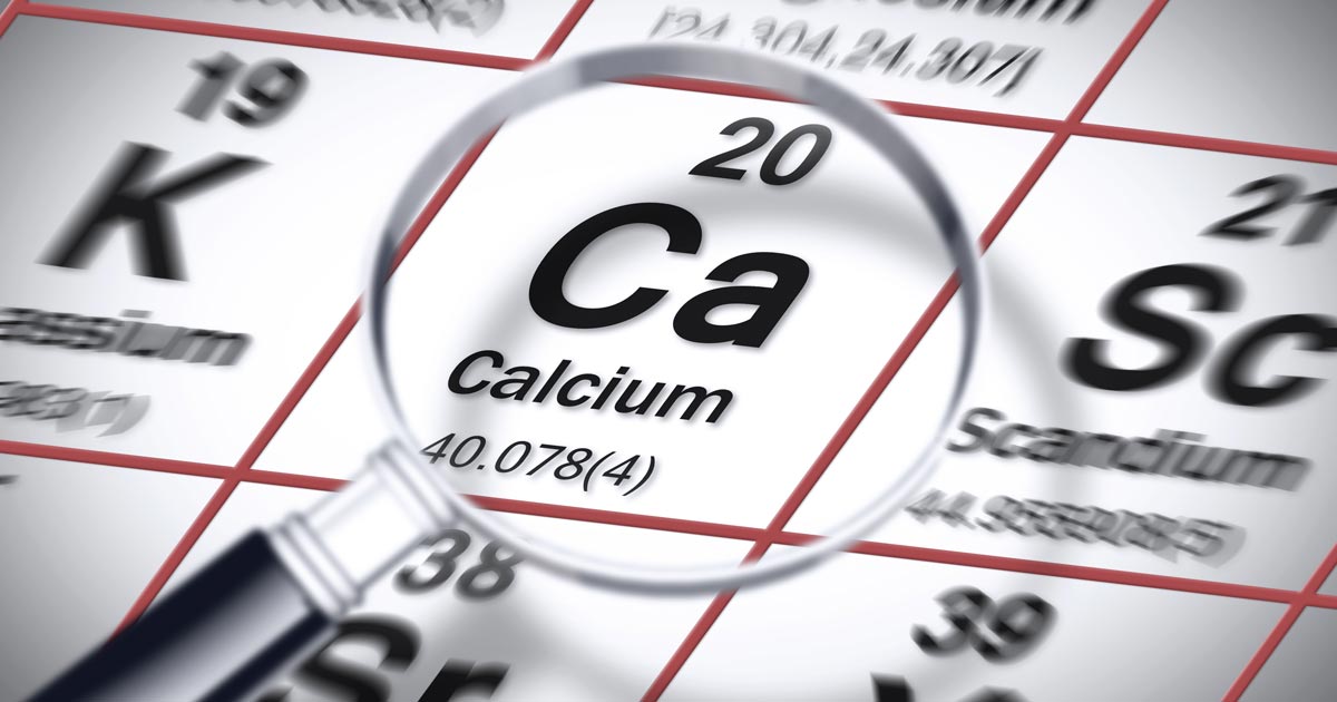
    Calcium in the serum or plasma exists in three forms:

    • ionised or free calcium
    • protein-bound calcium
    • complexed or chelated calcium (bound to phosphate, bicarbonate, sulfate, citrate and lactate)

    iCa is the biologically active fraction of calcium and is not to be confused with total calcium (tCa). A lack of concordance exists between the two. Adjustment formulas are inaccurate, even with the correction of the tCa to serum total protein or albumin concentration, and should not be used to predict iCa.

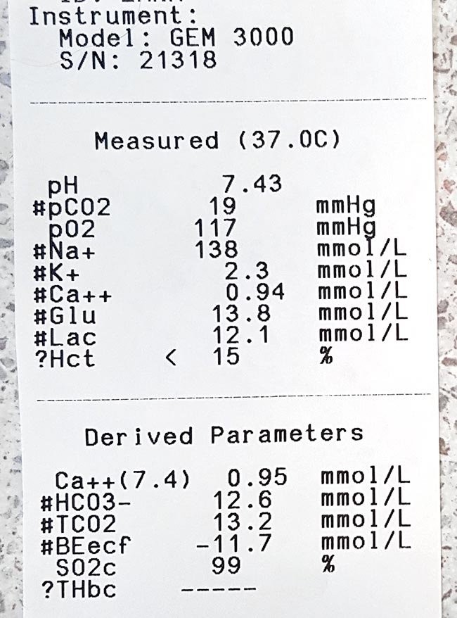
    The normal reference range for iCa in dogs is 1.2mmol/L to 1.5mmol/L; in cats, it is 1.1mmol/L to 1.4mmol/L.

    Function

    An example of low ionised calcium.
    An example of low ionised calcium.

    Calcium is essential in maintaining normal physiological processes in the body. iCa regulates:

    • vascular tone
    • myocardial contraction
    • homeostasis

    In addition, it is needed for:

    • enzymatic reactions
    • nerve conductions
    • neuromuscular transmission
    • muscle contraction
    • hormone release
    • bone formation
    • resorption

    In critical patients, particularly those with severe trauma or sepsis, vascular tone and coagulation is particularly important. For this reason, iCa is tightly kept in a narrow range and regulated by the interactive feedback loop that involves iCa, phosphorous, parathyroid hormone, calcitriol and calcitonin.

    Diseases and causes

    Diseases commonly associated with low iCa in dogs and cats include:

    • acute kidney failure
    • acute pancreatitis
    • diabetic ketoacidosis
    • eclampsia
    • ethylene glycol intoxication
    • protein-losing enteropathies
    • sepsis
    • trauma
    • urethral obstruction
    • parathyroid diseases
    • tumour lysis syndrome

    Situations altering the fraction of extracellular calcium seen on a regular basis include:

    • acid-base disturbances
    • lactic acidosis
    • protein loss or gain
    • increased free fatty acids

    Iatrogenic causes include:

    • citrate (anticoagulant) administration during blood transfusions
    • phosphate
    • bicarbonate
    • sulfate administration

    Low iCa can also develop during cardiopulmonary resuscitation, quickly declining with increased duration.

    • Part two will go into more depth regarding the most common causes of low iCa that require acute treatment, the treatment involved, controversies surrounding treatment of non-clinical low iCa, and prognostic indications.
  • Icteric serum

    Icteric serum

    The final discolouration of the serum we are going to cover is icteric serum.

    Icteric serum
    Icteric serum is caused by the presence of excess bilirubin in the blood stream.

    Icteric serum is caused by the presence of excess bilirubin in the blood stream as a result of increased production (pre-hepatic) or inappropriate excretion (hepatic and post-hepatic).

    The most common cause of pre-hepatic icterus is haemolytic anaemia, while hepatic disease and biliary tract obstruction are the most common causes for hepatic and post-hepatic icterus, respectively.

    Tips on where to start

    If icterus and concurrent anaemia exist, my first suspicion would be some kind of pre-hepatic cause. The most common causes are immune-mediated haemolytic anaemia and infectious haemolytic anaemia, such as haemotropic mycoplasma and babesiosis.

    Other causes can include snake envenomation and oxidative injury from heavy metal toxicity or onion ingestion.

    Regarding hepatic and post-hepatic causes, unfortunately it is not always clear-cut. Both are commonly associated with elevation in both alanine transaminase (ALT) and alkaline phosphatase (ALKP), and, although no specific changes are pathognomonic for hepatic or post-hepatic disease, the pattern of change may help identify the origin of the cause. ALT is released from the inside of hepatocytes, and in higher amounts when cell damage occurs.

    Hepatic hints

    Some pointers on what you can do to help differentiate:

    • Compare the ALT and ALKP elevation; if one is in order of magnitudes higher than the other then it can help point to an origin.
    • If the cause is of hepatic origin, one would expect the ALT to be significantly more elevated than the ALPK. Likewise, this is usually true in reverse for post-hepatic causes. However, it should be noted in chronic hepatic diseases, where active damage to hepatocytes is comparatively lower, a mild increase in ALT and marked increase in ALPK does not preclude disease of hepatic origin. Therefore, biopsies should always be used for definitive diagnosis.
    • If other biochemistry parameters such as albumin, glucose and cholesterol are low, or prolonged clotting times are present, the case for a hepatic origin is strengthened.
    • The gallbladder and bile duct can be assessed using abdominal ultrasonography. The presence of a dilated bile duct, or evidence supportive of pancreatitis, is highly suggestive of a post-hepatic cause.

    Finally, it is important to be aware of the impact on hyperbilirubinaemia on laboratory testing. Hyperbilirubinaemia generally causes decreased cholesterol, triglyceride, creatinine, lipase, total protein and gamma-glutamyltransferase levels.

  • Lipaemia – the bane of biochemistry

    Lipaemia – the bane of biochemistry

    Last week we covered haemolysed samples – this week we’re looking at lipaemic samples.

    Lipaemic samples are caused by an excess of lipoproteins in the blood, creating a milky/turbid appearance that interferes with multiple biochemical tests and can even cause haemolysis of red blood cells.

    lipaemic sample
    A severely lipaemic sample (red arrow). IMAGE: eClinPath.com (CC BY-NC-SA 4.0).

    Lipaemia can follow recent ingestion of a meal – especially one high in fat. Although not pathognomonic for any diseases, its presence can help increase the suspicion of certain diseases, including:

    • pancreatitis
    • diabetes mellitus
    • hypothyroidism
    • hyperadrenocorticism
    • primary hyperlipidaemia (in some specific breeds, such as the miniature schnauzer)

    It warrants further investigation in patients that have been ill and inappetent.

    Irksome interpretations

    Lipaemia can dramatically impact laboratory testing and is often troublesome in critically ill patients, making interpretation of biochemistry particularly difficult, if not impossible.

    Lipaemia can affect different analysers in different ways, most commonly causing:

    • Falsely increased calcium, phosphorus, bilirubin, glucose and total protein (via refractometer) and some liver parameters such as alkaline phosphatase, alanine aminotransferase, aspartate aminotransferase, haemoglobin concentration, and mean corpuscular haemoglobin concentration.
    • Falsely decreased sodium, potassium, chloride, albumin and bicarbonate.

    Tube tips

    Assessment of a centrifuged haematocrit tube before running a biochemistry panel can help reduce wasted biochemistry consumables.

    If the sample is lipaemic in the haematocrit tube then maybe try some of the following tips.

    • If blood tests are planned in advance, try fasting the patient beforehand for 12 to 24 hours.
    • Repeat sampling a couple of hours later may yield a less lipaemic sample.
    • Collecting and centrifuging a larger amount of blood (3ml to 5ml, for example) can sometimes yield enough clear sample between the lipid layer and red blood cells.
    • Refrigeration of the sample can help the separation.
    • Extract lipids using polar solvents, such as polyethylene glycol.
    • Centrifugation at higher than normal speeds (if possible) can also assist in clearing the layer.
  • PCV/total solids interpretation: serum colour

    PCV/total solids interpretation: serum colour

    When interpreting the often misinterpreted and underused PCV and total solids test, it is important to take note of the serum colour as this may give clues into the diagnosis.

    PCV tubes
    Normal serum colour (left) compared to a patient with immune-mediated haemolytic anaemia. The serum is haemolysed and anaemia is present.

    The most common abnormalities seen in clinic are icteric, haemolysed and lipaemic serum.

    Clear serum can also be of importance – especially when you interpret it with blood counts and urine colour.

    Haemolysis

    The most common abnormality of serum colour changes is haemolysis. In my experience, the most common cause is suboptimal collection technique. To confirm this, simply collect another sample and repeat.

    If it is repeatable, and concurrent anaemia or pigmenturia is present, it warrants further investigation.

    Intravascular haemolysis can be caused by:

    • immune-mediated haemolytic anaemia
    • blood transfusion reactions
    • infectious diseases such as Mycoplasma haemofelis, Babesia canis, Ehrlichia canis, FeLV and others
    • Heinz bodies from the ingestion of heavy metal, onions or paracetamol
    • hypophosphataemia
    • macroangiopathic disease (neoplasia, for example)
    • envenomation – typically, snake bites

    Testing issues

    Haemolysis can also affect other laboratory testing. It can lead to an artefactual increase in glucose, phosphorus, bilirubin, total protein, fructosamine and triglycerides, and a decrease in sodium (pseudohyponatraemia), cholesterol, calcium, potassium and albumin.

    Extravascular haemolysis often does not cause haemolysed serum as it is generally slower and the body is able to clear the haemoglobin before it can lead to discolouration of the serum.