Blog

  • It takes balls to neuter

    Sterilising a dog
    A close up of a vet performing sterilisation surgery on a dog. Image ©iStockphoto.com/Dan-Edwards

    As graduates, one of the most routine surgeries that we will be expected to be competent at is neutering. As students, when on work experience or EMS, we will have seen at least one of these procedures a day at small or mixed practices… often more.

    But routine does not necessarily mean easy, simple or without risk. When learning about reproductive anatomy, we were faced with a harsh truth: the concept of spaying is actually fairly terrifying, particularly as there is a considerable risk of a bitch bleeding to death.

    Spaying is not to be underestimated. Among the usual complications and risks involved in the use of general anaesthetic, there are also a few scary blood vessels to worry about.

    During the surgery, it is necessary for both pairs of ovarian and uterine arteries to be cut. It is of vital importance that these are ligatured (tied off) securely to prevent the likelihood of internal bleeding post-surgery. Neither of these are to be underestimated – the ovarian arteries are particularly important to ligature properly, since they branch directly from the aorta. A slipped ligature could result in serious problems, and could potentially result in the patient bleeding to death.

    The concept of spaying is actually fairly terrifying, particularly as there is a considerable risk of a bitch bleeding to death.Clients should always be made aware of surgical risks and all eventualities, but I would imagine that the last thing an owner would expect after taking their dog or cat to be neutered would be the death of their beloved pet post-surgery.

    This is quite a daunting prospect for the “most routine” surgery in practice. You can’t afford to be complacent – you really do have to get it right.

    As an avid traveller, I had always intended on getting involved in a neutering clinic in India for EMS, even before learning just how risky neutering can be if not done properly. Now, I will make sure to realise that aim, in order to get as much surgical practise as possible before graduating. Hopefully, it will help boost my confidence, so that I won’t be as concerned as I am currently about this “routine” surgery by the time I am a qualified vet.
  • Causes of sticky bottom syndrome in rabbits

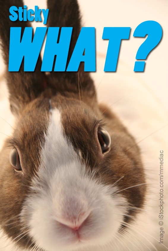
    Causes of sticky bottom syndrome in rabbits

    © iStockphoto.com/mmediac
    Original image
    © iStockphoto.com/mmediac

    Sticky bottom syndrome in rabbits is most often caused by feeding a badly balanced diet, or simply feeding too much food, resulting in over-production of caecal droppings. Rabbits will not eat the excess pellets.

    Fat and elderly rabbits (especially females with large dewlaps) are also at risk as they may not be able to reach their bottoms to practice coprophagia and to clean.

    Rabbits with dental disease may not eat their caecal pellets and won’t groom themselves properly either, also putting themselves at risk.

    Pet rabbits can get worms (usually if fed on green food collected from areas frequented by wild rabbits), but clinical problems are said to be rare. However, some rabbit owners report sticky bottom problems clear up after the rabbit is wormed.

  • Media in the veterinary world

    Marc Abraham
    Media savvy vet, Pup Aid founder and #wheresmum author Marc Abraham.

    In January, I attended my first AVS (the Association of Veterinary Students) congress. It was a fantastic event, featuring inspiring talks and lectures, fascinating practicals and the infamous AVS ball.

    I would strongly urge any vet student to attend congress at least once during their time at vet school – aside from all the events available during the weekend, it’s a great opportunity to meet students from other vet schools and make valuable contacts that you may well need in the future. It certainly opened my eyes and made me realise just how small the veterinary world can be.

    TV vet Mark Abraham opened the event with a talk to illustrate his achievements and work to date. He talked briefly about his Pup Aid campaign, which aims to prevent puppy farming in the UK and promote obtaining puppies from registered breeders or rescue centres only.

    In addition, he gave some valuable advice: fight for a cause you believe in and use the media to your advantage.

    He reiterated the importance of media within the veterinary world, which includes not only journals and papers within the profession, but other forms such as TV and social media that are more accessible to the general public.

    In particular, he emphasised the impact that Twitter has had on spreading the word about the Pup Aid campaign. With the support of well known celebrities “retweeting” the link to the online e-petition, Marc gained the required number of signatures, and puppy farming is currently being debated in Parliament as a result.

    Fight for a cause...

    A recent storyline on Eastenders involved breeding a dog and selling the puppies as a way to make money. This is a crucial example of the impact the media can have on public opinion. After seeing this, many viewers may be inspired to do the same in order to make a bit of spare cash, which is exactly the sort of attitude that the Pup Aid campaign is trying to abolish.

    In today’s age of technological advancement, there are so many media platforms available to bring the veterinary world into the public eye. We must use this to our advantage, whether that is to promote campaigns such as Pup Aid, raise awareness of veterinary issues or even to promote individual practices and the services they provide.

    The media is a powerful thing, and we must not forget that just because it sometimes feels like we live in a bubble that is the veterinary world.

    • Marc’s petition to ban the sale of young puppies and kittens without their mothers being present runs until 02/05/2014 – SIGN THE PETITION HERE!
  • When to extract retained deciduous teeth

    When to extract retained deciduous teeth

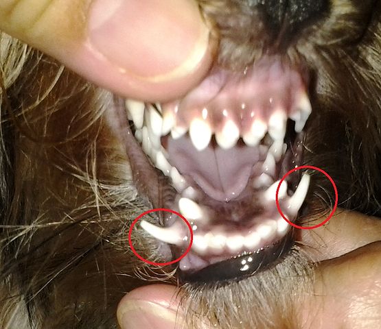
    Yorkie's retained deciduous or baby fangs.
    Yorkie’s retained deciduous or baby fangs, circled in red. Both lower permanent fangs did not grow right underneath the baby fangs. Photo by Socknye [CC-BY-3.0], via Wikimedia Commons.
    A common abnormality in canine dentition is retention of deciduous teeth.

    This occurs when the permanent tooth bud does not grow immediately beneath the deciduous tooth, and therefore does not cause the roots of the deciduous tooth to be resorbed.

    A retained tooth can cause the permanent tooth to erupt in an abnormal position or cause other types of problems, such as crowding, which may lead to debris accumulation between the teeth and greater plaque stagnation.

    In these cases, the retained tooth should be extracted.

  • Taurine and carnitine-responsive DCM in cocker spaniels

    Taurine and carnitine-responsive DCM in cocker spaniels

    Purebred American cocker spaniel. Image © iStockphoto.com/cynoclub
    Purebred American cocker spaniel. Image © iStockphoto.com/cynoclub

    American cocker spaniels with dilated cardiomyopathy (DCM) are often taurine deficient and responsive to taurine and carnitine supplementation.

    Whereas myocardial function does not return to normal in most cocker spaniels treated with taurine and carnitine, it can improve enough to allow discontinuation of cardiovascular drug therapy (furosemide, digoxin and angiotensin-converting enzyme inhibitors) and to maintain a normal quality of life for months to years.

  • Gravel sign

    Gravel sign

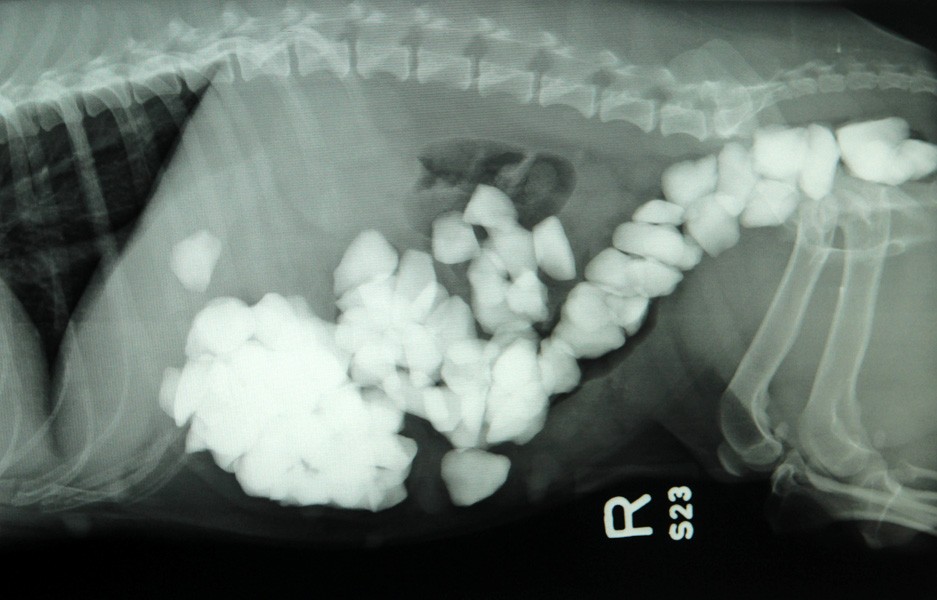
    gravel sign actually gravel
    The gravel sign that was actually gravel… Read Star the Jack Russell’s story at vetsonline.com.

    The gravel sign is seen radiographically as multiple, variously sized mineral fragments in the stomach or intestine, which build up as a result of poor gastric motility and/or emptying, and indicate a chronic stasis of food.

    It can be present in cases of both partial and complete obstruction.

    Heavier particles tend to settle in front of a partial obstruction, as fluid and less dense particles move beyond the obstruction.

  • PETS with rabies, in the UK?

    dog with rabies
    Close-up of a dog’s face during late-stage “dumb” paralytic rabies. Animals with “dumb” rabies appear depressed, lethargic, and uncoordinated. Gradually they become completely paralyzed. When their throat and jaw muscles are paralyzed, the animals will drool and have difficulty swallowing. Image credit: CDC/Barbara Andrews

    Lectures on legislation regarding veterinary-related issues are never the most interesting, often involving endless lists of dates and figures, but the Pet Travel Scheme (PETS) lecture this week had a slight edge to it.

    Among other important factors, such as microchipping and documentation, the focus was quite obviously on the recent changes to the rules regarding rabies.

    It was evident that abolishing the blood test was not a great move in the opinion of this particular lecturer. But he’s not alone – there seems to be a ripple of discomfort throughout the veterinary community in response to the new regulations.

    Pets are now free to travel within the EU just 21 days after having received one dose of the rabies vaccine, despite the incubation period of rabies being up to three months.

    Although Defra claims the risk of rabies entering the UK is still very low, there still seems to be an underlying concern within the veterinary world.

    It has been proposed that the potential increase in risk of exposure to rabies means that vets should be routinely receiving the rabies vaccination.

    I myself have had a course of rabies vaccinations. Not because I’m a vet student, but because I was travelling in South America, where there was a possibility of coming into contact with rabid animals.

    However, my understanding is that the vaccine does not prevent you developing rabies if bitten, but just puts it off for 24hrs, to give you time to get back to the UK to be appropriately treated. With this in mind, would providing the vaccine for those working in the veterinary profession in the UK be of any benefit if it is purely used to “buy time”?

    I’m not convinced that vaccinating vets would be an effective measure to take, but we must now include rabies as one of our differential diagnoses and be open to the possibility (however slim) of being presented with rabid animals in practice. Perhaps more advice about the risks and clinical signs of rabies would put the minds of vets and owners alike at rest.

  • Peripheral neuropathy in diabetic cats

    Peripheral neuropathy is the most common chronic complication of diabetes in cats.

    This degenerative disorder typically affects the hindlegs, causing weakness, loss of coordination, inability to jump and a plantigrade stance.

    No specific therapy for peripheral neuropathy in cats is available, but intensive blood glucose control decreases the risk and improves clinical signs in humans.

    Similarly, aggressive adjustment of insulin therapy may improve nerve function and reverse hindleg weakness and abnormal stance in diabetic cats. However, a cat’s response to the therapy varies and aggressive insulin management can, of course, increase the risk of hypoglycaemia.

    http://youtu.be/JAKpOvbHWSU&rel=0

    Video uploaded to YouTube by www.vetstoronto.com

  • Role play… at vet school?

    client communication
    Image © iStockphoto.com/DenGuy

    A role play class with professional actors brought in to help out would be commonplace on a course such as drama, and may sound a little odd for veterinary medicine. But is it really?

    Communication is a vital part of being a vet and can often be the “make or break” factor for client satisfaction. Often, owners won’t have any concept of your surgical skills or medical knowledge, but they will know instantly whether they trust their animals in your care within minutes of meeting you simply from the way you communicate.

    The class uses realistic scenarios with different types of “client” (the actors) that we are likely to come across in practice. These involved breaking the news that a pet has to be euthanised, dealing with tight horse owners that just think you’re trying to rip them off, and discussing alternative treatments for someone struggling to tablet their cat.

    Sometimes, it can be difficult if you don’t know enough about the condition the animals has in your scenario, but the important thing is how you convey the information, not necessarily the content of what you are saying.

    On the whole, the students got stuck in and enjoyed themselves. It was interesting to see how different students took different angles on the same scenario, proving there isn’t just one way to communicate effectively. The actors and supervising vet also gave us pointers on body language and tone of voice.

    Years ago, veterinary graduates were thrown into practice with very limited guidance on communication, which must have been terrifying. The interactive sessions give us the chance to make mistakes in an artificial environment, so that we (hopefully) won’t make them in the real world.

    I found the session incredibly helpful and think that communication is an immensely important skill to develop in order to give us the best chance at getting on with our clients in the future. Sessions like this should become a fundamental component of any veterinary course.

  • Dentition in chinchillas

    Dentition in chinchillas

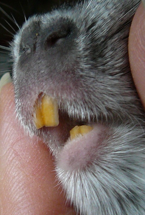
    Chinchilla teeth
    The teeth of a domestic chinchilla, by Salix [GFDL, CC-BY-SA-3.0 or CC-BY-2.5], via Wikimedia Commons.
    Chinchillas have 20 teeth from birth – 4 incisors and 16 molars.

    Teeth are open-rooted and, therefore, grow continuously, about 2-3 inches per year.

    The chinchilla should be provided with good quality hay, wood blocks or pumice stones to gnaw on and encourage tooth wear.

    The incisors are separated from the molars by a diastema.

    The teeth are covered by a hard layer of enamel made up of calcium, iron, phosphorous and other trace minerals.

    The normal tooth colour is dark yellow/orange – pale teeth are a sign of calcium deficiency.