Blog

  • Dystocia, pt 2: diagnostics

    Dystocia, pt 2: diagnostics

    Part one of this series covered the stages of labour and indications dystocia is present.

    Once the bitch presents to the clinic, a few basic diagnostic checks need completing to determine the status of the bitch/queen and the fetuses.

    Physical examination

    The first is a thorough physical examination, starting with the bitch or queen:

    • Demeanour, hydration status, vital signs, mucous membrane colour, capillary refill time and temperature are important.
    • Pregnancy anaemia is not uncommon; however, for patients with a haemorrhagic discharge, it is important to know their cardiovascular status.
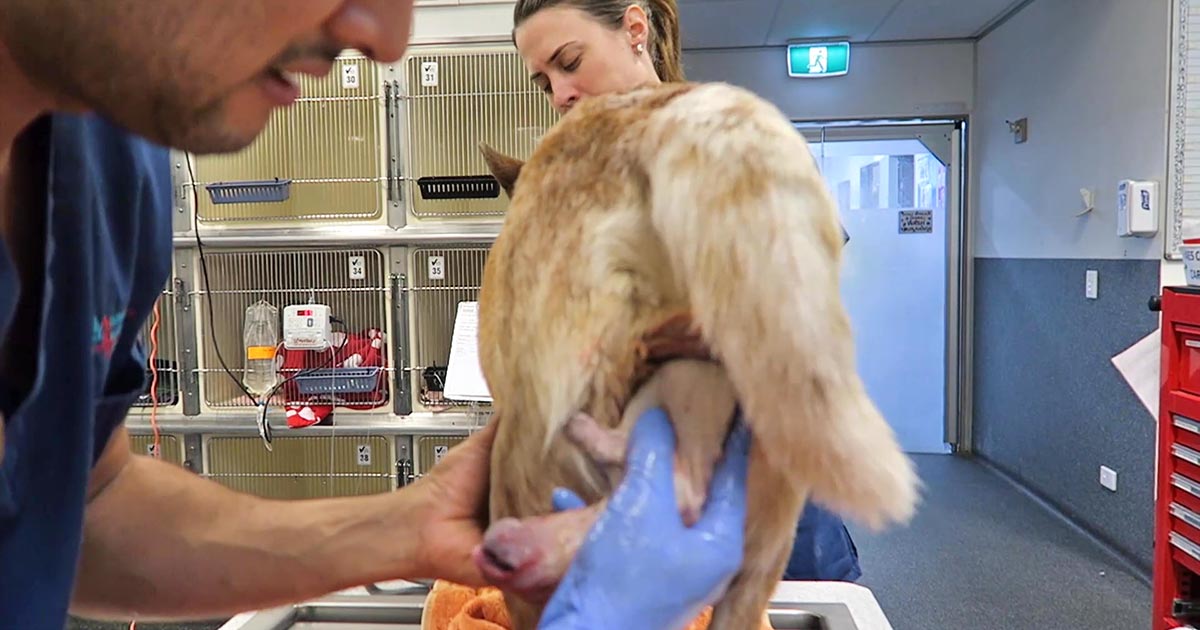
    • A thorough abdominal palpation should be carried out to assess comfort level and palpation for the presence of fetuses. Palpating fetuses can be difficult and cannot confirm if no fetuses are present.
    • A digital vaginal examination should be performed. Feathering response – also known as the Ferguson reflex in human medicine – is the neuroendocrine reflex where the self-sustained cycle of uterine contractions is initiated by firm pressure on the dorsal aspect of the vestibulovaginal wall. If this is absent, the patient is unlikely to progress with the parturition unaided.
    • Palpation of fetuses in the canal can help decide whether surgical management is required. Obvious fetal malposition, malposture or malpresentation, or fetopelvic disparity, will be indications of caesarean. Abnormal pelvic diameter is also another reason to not proceed with medical management. To confirm these suspicions, abdominal radiography is required.
    • Radiographs will also help determine the number of fetuses to be expected, the signs of fetal death (presence of gas surrounding the fetus) and aforementioned fetomaternal abnormalities. I always repeat radiographs after the expected number of neonates is passed, to make sure I have not miscounted at the start.
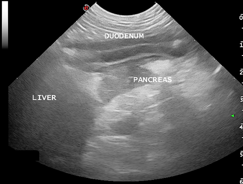
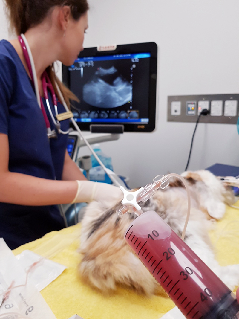
    Ultrasound

    Panel 1. Heart rate ranges to help indicate stress of fetuses

    Dogs:

    • normal – 180 to 220 beats per minute (bpm)
    • Stressed – 160bpm
    • Real concern – less than 160bpm

    Cats:

    • normal – more than 220pbm
    • fetal stress – less than 180bpm

    The second important diagnostic tool is ultrasound.

    Fetal heart rates are good indicators of fetal stress. Some heart rate ranges that can help provide information about the status of the fetuses are detailed in Panel 1. These ranges vary between sources, but are good guidelines.

    Ultrasounds can also help visualise the maturation status of the fetuses. At-term fetuses should have normal hepatic, renal and intestinal development. Intestinal peristalsis should be evident in at-term fetuses.

    Other diagnostics

    Other diagnostics may be indicated for patients, depending on the status of the bitch/queen:

    • If the patient is stable, but dystocia is present, a minimum database would include PCV/total protein, electrolytes, glucose, ionised calcium, lactate and acid-base balance.
    • Serum ionised calcium levels are important, as they influence the strength of contractions and how much supplementation is required.
    • Hypoglycaemia needs to be ruled out as a cause of dystocia, especially when large litters are involved.
    • If the patient is unstable or systemically unwell, include complete blood count, blood smears and biochemistry.
    • Physiological pregnancy anaemia can be present. The presence of regenerative response can help differentiate this from acute haemorrhage.
    • Abnormal leukocyte panel, especially with the presence of degenerative left shift, can indicate the presence of an infection – especially if toxic changes are present in the neutrophil.

    Part three will briefly look at the medical management of dystocia and when surgical intervention is required.

  • Dystocia, pt 1: labour stages

    Dystocia, pt 1: labour stages

    Now most female canine patients are spayed, it comes as no surprise reproductive emergencies are not as common.

    One confusion seems to be not knowing how to determine a true dystocia emergency – especially when given advice over the telephone – from the process of normal parturition.

    Another concern is how to confidently form a diagnostic pathway to determine the cause of dystocia – especially for reasons other than obvious physical abnormalities (for example, fetopelvic disparity and fetal malposition).

    Often, once we decide to go down the medical treatment pathway – whether the result of findings or owner/financial constraint – no one is confident as to what medication should be used and how often drugs can be given safely.

    This series of blogs will address these issues in a step-by-step manner. Hopefully, by the end, you will be confident in the diagnosis and management of dystocia.

    Labour stages

    Before moving on to the signs of dystocia, let’s go through the signs of labour.

    First stage labour

    First stage labour is characterised by panting, tremoring, nesting behaviour, a drop in core temperature – usually a drop by almost 1°C 24 hours prior to second stage labour – and a drop of progesterone to below 2mg/ml.

    Dog and puppy.
    Third stage labour occurs generally within 15 minutes after passing a puppy or kitten. Image © foto ARts / Adobe Stock
    • dogs: approximately 6 to 12 hours
    • cats: approximately 6 to 24 hours

    Second stage labour

    Second stage labour is landmarked by the water breaking, visible abdominal contractions, and the allantoic/amniotic sac or fetal parts visible from the vulva.

    If vulval discharge is present, they should be clear. Excessive amount of bright red haemorrhage, green or black discharge prior to delivery, or purulent material can indicate a pathological process requiring immediate veterinary attention.

    • dogs: approximately 3 to 6 hours
    • cats: approximately 6 to 24 hours

    Third stage labour

    Third stage labour this is when passage of all the placenta has occurred, generally within 15 minutes after passing a puppy or kitten.

    Clues

    Now we understand the normal progression of parturition, a few clues exist in the history that could suggest dystocia may be present.

    Some breeders will often know the ovulation timing of the patient – especially if AI was performed. Tests such as progesterone levels, luteal hormone (LH) levels, cytology and vaginoscopy are some ways where it can help time the ovulation.

    The normal gestation length should not be any longer than 66 days from the LH surge or, if the ovulation history is unknown, 72 days from the last known breeding.

    History of prior dystocia is a warning, as most animals with prior parturition difficulties are more likely to develop dystocia again.

    The same goes for animals that have previously required a caesarean. Their risk of requiring future caesareans is high, with further risk of uterine rupture if dystocia happens again.

    Image © Pilipipa / Adobe Stock
    Animals that have previously had a caesarean are at high risk of requiring future caesareans, with further risk of uterine rupture if dystocia happens again. Image © Pilipipa / Adobe Stock

    Intervention signs

    Owners often telephone after the failure of normal progression of delivery. The signs that always require immediate intervention are:

    • more than 4 hours have passed from the rupture of the first chorioallantois
    • more than 2 hours between delivery
    • more than 30 minutes of strong abdominal contraction and no delivery
    • presence of green or black discharge before delivery
    • large amount of bright red haemorrhage
    • abnormal amount of pain during contractions
    • collapse of the bitch or distracted mothering

    Any of these signs require immediate presentation to the veterinarian. Delivery of stillborn puppies is also an indication where veterinary attention is indicated.

    Finally, if owners are concerned, it is best to advise veterinary assessment rather than try to convince them everything is okay based on what they describe over the telephone.

  • Pancreatitis, pt 2: treatment and prognosis

    Pancreatitis, pt 2: treatment and prognosis

    Last week we covered diagnosing pancreatitis and the challenges associated with doing so. This week we look at management.

    The treatment of pancreatitis largely involves supportive care and monitoring for potential complications. Here, we recap the fundamentals.

    IV fluids

    IV fluids are critical in the acute phase to restore perfusion to core organs and correct hydration deficits. Once achieved, the goal is then to cover maintenance requirements and ongoing losses.

    Hypoproteinemia can develop due to a combination of haemodilution, increased losses in to inflammatory exudates or into gastrointestinal tract, and decreased production due to reduced intake.

    Colloidal therapy or plasma can be considered, but enteral nutrition is more effective and has numerous other benefits.

    Pain relief

    Pain relief largely involves the use of opioids.

    Pure opioid agonists, such as methadone, fentanyl constant rate infusions (CRIs) or fentanyl patches, are most commonly seen in dogs. In cats, partial agonists, such as buprenorphine, are favoured as the condition is generally less painful compared to dogs.

    NSAIDs should be avoided due to concerns of poor perfusion, concurrent kidney and gastrointestinal involvement.

    Antiemetic therapy

    Metoclopromide CRI and maropitant are the mainstays. Antacids, such as esomeprozole, are included to help reduce the risk of gastric ulceration from stress.

    Early enteral nutrition

    Numerous studies have demonstrated the benefit of early enteral nutrition and patients should be encouraged to eat a low-fat diet as soon as possible. Enteral nutrition has been shown to be more beneficial than withholding food as it reduces recovery times and helps maintain enterocyte health.

    If there has been a protracted period of anorexia then a nasogastric or nasoesophageal tube should be placed. A nasoesophageal tube has the added benefit of allowing excessive gastric secretions to be suctioned, as well as for feeding. Due the chronic nature of the disease in cats, oesophagostomy tubes are often placed so that nutritional support can be delivery for prolonged periods of time.

    Antibiotics

    Antibiotics are generally not indicated in canine pancreatitis unless markers of sepsis or if a septic exudate are found (this is an indication for exploratory surgery). In which case, triple antibiotic therapy consisting most commonly of amoxicillin, enrofloxacin and metronidazole are indicated.

    It has been reported 35% of feline pancreatitis patients have bacterial infections; therefore, antibiotics are often included in the treatment of feline pancreatitis.

    Exploratory laparotomy

    This is indicated when the following has occurred:

    • Extra-hepatic biliary obstruction that does not resolve with medical management.
    • Septic abdomen (as mentioned above).
    • Pancreatic abscessation.

    Prognosis

    The prognosis for pancreatitis is variable and dependent on the severity of the disease.

    In mild to moderate canine pancreatitis (which is the most common), the prognosis is generally good as they will normally respond well to supportive therapy and resolution of clinical signs occurs within several days of initiating treatment. However, with severe acute, necrotising pancreatitis, the prognosis is guarded as it can progress into systemic inflammatory response syndrome, disseminated intravascular coagulation and multiple organ failure – which has a high mortality rate.

    The prognosis of acute feline pancreatitis is generally guarded due to the chronic nature of the disease and the propensity to involve multiple organ systems.

  • Pancreatitis, pt 1: diagnosis

    Pancreatitis, pt 1: diagnosis

    Pancreatitis is one of the most common exocrine conditions seen in small animal practice. It is caused by premature activation of enzymes (zymogens) within the pancreas leading to autodigestion, and can result in severe morbidity with the potential to lead to mortality.

    To this day, the diagnosis of pancreatitis remains a challenge – especially in feline patients – and relies on the use of a combination of history, signalment, clinical signs and diagnostic findings.

    Presenting signs

    In dogs, overweight middle aged to older (more than five years old) are at higher risk. Miniature schnauzers, Yorkshire terriers and silky terriers are some breeds identified as higher risk. Approximately 90% present with anorexia and vomiting, 50% with abdominal pain and 30% with diarrhoea.

    In cats, no common age range exists, although domestic shorthair and longhair cats are the most commonly affected. They present more commonly with anorexia and lethargy; 30% presenting with vomiting and 25% with abdominal pain.

    Diagnostic changes

    Pancreatitis-Image---Pancreatitis
    Cases can be confirmed with ultrasound.

    A summary of the commonly seen diagnostic changes are included below, many of which due to the nature of the disease are non-specific:

    • Hyperlipaemia and an inflammatory leukogram can be present, but are both non-specific.
    • Hyperlipasaemia and hyperamylasaemia are commonly seen, but the sensitivity and specificity of both are only about 50%.
    • Elevation in alkaline phosphatase and bilirubin can indicate pancreatic bile obstruction. Other changes may be present and can indicate wider organ system involvement – azotemia, for example.
    • Canine pancreatic specific lipase immunoreactivity (cPLI) has a high sensitivity, but poor specificity – approximately 50%. This means, if real pancreatitis is present, it will show positive; but a positive result will only actually be real pancreatitis 50% of the time. A negative result, however, can be interpreted as “highly unlikely for pancreatitis to be present”. A positive cPLI should be confirmed with ultrasound, which is the gold standard for canine pancreatitis.
    • Feline pancreatic lipase immunoreactivity (fPLI) has been reported to have a sensitivity of 67% and specificity at 91%. This means, 90% of the time, a positive indicates real pancreatitis – but these figures vary between studies. To make things even more difficult, cats with pancreatitis can have normal fPLI/spec fPL and normal ultrasonographic findings.
    • Common changes on ultrasound with acute pancreatitis include free abdominal fluid (generally a non-septic exudate) and hypoechoic pancreas surrounded by hyperechoic peripancreatic fat. As mentioned above, feline pancreatitis often has no visible changes.
    • Radiography may show reduced serosal detail around the pancreatic regions – again, this is not specific or sensitive, but is helpful at ruling out other differentials, such as foreign bodies.

    Summary

    In summary, canine pancreatitis is less of a diagnostic challenge compared to its feline counterpart. There is no single test that can accurately confirm the presence of feline pancreatitis, apart from in biopsies via exploratory laparotomy – which are understandably invasive and costly.

    Next week, we will cover the fundaments of managing pancreatitis patients.

  • The dreaded client complaint

    The dreaded client complaint

    Regardless of how well you conduct yourself and how thorough you are in your work, it’s inevitable you’ll occasionally find yourself on the receiving end of a client complaint. This will always suck.

    When someone criticises you, it’s very natural to feel a strong negative emotional response. The emotional control centres of your brain interpret criticism as a direct threat to your safety, and will trigger the same response it would if you were under physical attack.

    But reactive and defensive behaviour will not help your cause – better to let your logical brain take over from your limbic system and institute a proactive plan in dealing with complaints.

    Five simple steps

    Here’s a proven five-step approach to dealing with client complaints:

    1. Listen

    Listen carefully to what the client has to say (preferably in person). Approach them with empathy and try to see it from their perspective.

    Avoid formulating a response in your head while they are still speaking, or focusing on the reasons why they are wrong and you are right. Try (and I mean really try) to understand why they are upset, then verbalise this to them: “I can see where you’re coming from – I understand why this looks bad and why you are upset.”

    2. Critically evaluate the merits of the complaint

    Sometimes people have an unreasonable point of view, but very often they don’t. They might have blown it completely out of proportion, or have a distorted understanding of a situation, but it’s rare someone complains where they don’t have some legitimate basis for the complaint – at least in their eyes.

    Remember: what seems trivial to you might be a big issue for your client.

    Stay open to the possibility you may have done something wrong or could, at least, have done something better. The reasons and excuses, however valid, shouldn’t matter, only how it was perceived by your client and how you can fix it.

    3. Fix it, if you can

    Do the extra test, get it in for a recheck, do something that is over and above the expected level of care; this is an opportunity to make the unhappy client into a lifelong fan.

    4. Make an apology

    If you can’t fix it, apologise. Sincerely.

    Explain what went wrong and what you have done to prevent similar problems from occurring again.

    5. Try to make up for it

    If it is within your decision-making power; don’t be scared to offer some sort of financial compensation.

    It doesn’t need to be an admission of guilt: “We’ve looked carefully into the decisions made with this case, and I can’t see that any mistakes were made. However, we strive to keep our clients happy, and you clearly feel you didn’t receive value with us, so I’d like to offer you x, y or z.”

    This doesn’t always need to be money back (but it can be); offer a discount on a subsequent visit, follow up x-rays free of charge, or offer to donate money to their favourite charity in their name. If a relatively small amount of money early on during negotiations can make the problem go away, it might save you a lot more time and money down the line.

    Turning bad into good

    This approach of firstly allowing for some critical self-evaluation, and then simply focusing on being fair and reasonable can save a lot of pain an effort, and can potentially turn these unpleasant situations into valuable opportunities for learning and growth.

  • Systemic antibiotics – a brief guide for new grads

    Systemic antibiotics – a brief guide for new grads

    A lot of information is available regarding different antibiotics and, for the newest generation of vets, the pressure to use them correctly and responsibly is greater than ever.

    One of main challenges when you start clinical practice is knowing the most appropriate antibiotic for common presenting conditions.

    Below is a rough guide for antibiotic selection according to body system. However, make sure you stick to the following rules:

    1. Limit antibiotic use to animals that actually require them – resist the urge to dispense them due to pressure from owners or when you feel there is nothing else to turn to.
    2. What is the likely type of bacteria you are aiming to target (such as anaerobes, Gram-positives and Gram-negatives)? Collect samples from lesions/discharge or effusions/blood and urine, and see if there is evidence of bacteria under the microscope.
    3. Use the most narrow spectrum antibiotic as possible.
    4. Perform a culture and sensitivity whenever possible – especially if a case does not respond to your first line antibiotic.
    5. Avoid using fluoroquinolones, third and fourth generation cephalosporins and amikacin without evidence of resistance from culture and sensitivity results.
    6. Use an appropriate dosage regime and make sure the owners have the capacity to administer them accordingly.

    Skin

    • Try topical chlorhexidine alone if surface pyoderma
    • Clindamycin
    • Cephalexin
    • Amoxicillin-clavulanic acid

    Upper respiratory tract

    • Doxycycline
    • Amoxicillin-clavulanic acid

    Lower respiratory tract

    • Amoxicillin-clavulanic acid
    • Ampicillin

    GI tract

    • Metronidazole (research questions the use of antibiotics for diarrhoea cases)
    • Tylosin (chronic diarrhoea)

    Urogenital tract

    • Remember that cystitis in cats is often stress-related rather than due to infection
    • Amoxicillin-clavulanic acid
    • Trimethoprim-sulpha (penetrates the prostate)

    Mastitis

    • Amoxicillin-clavulanic acid


  • Making the most of a veterinary conference

    Making the most of a veterinary conference

    Attending an in-person veterinary conference is still one of my favourite forms of continued education (and it’s not just for the free pens).

    But without a strategic approach, a very real risk exists of walking away at the end of an expensive week with nothing more than a head crammed full of random facts and a bad hangover.

    So, how can you make the most of your next veterinary conference?

    Pick a topic…

    Most conferences will have a few set themes for the week, in addition to a plethora of other topics. Pick one of the themes and make an effort to attend all of the talks on this topic.

    Saturating yourself with a variety of different viewpoints and opinions on one theme will ensure you walk away with a deeper level of understanding on at least one topic.

    …but be sure to mix it up

    Try to attend as many non-clinical talks as possible. Learning about themes like management, leadership, communication and well-being are likely to inspire some of the biggest positive long-term changes in your career. Some of my favourite talks have been plenary sessions by people who have nothing to do with veterinary science.

    Make a point of attending a few lectures on topics outside your normal sphere of interest, and even on some you dislike. It’s quite possible you’ll discover your “next big thing” in a field you had no interest in up to that point, or that a little bit of extra knowledge can make one of those things you dislike seem a bit more appealing.

    Take notes, but not too many

    Don’t try to rewrite the conference proceedings. Rather, listen carefully for – and note – those little gems of wisdom that are casually dropped during lectures:

    • “This is the way I approach this…”
    • “A trick I find works well here is…”
    • “I love this little bit of equipment…”

    Make sure to note anything that is completely new to you and all the things you’ve struggled with in the past.

    iPad
    “Take notes” and “use technology” are two of Hubert’s hints for a successful conference.

    Use technology

    I take an iPad into lectures and use the Notes app that comes as standard with it.

    I create a folder for that conference in “All iCloud” and make a new note in that folder for each talk I attend. You can use a combination of typed notes, photos of the lecture slides, sketches and audio to record all the information you want to refer back to.

    With this technique, your notes are instantly available and easy to find on all your devices via iCloud – so, if you need to refer back to that little trick at 3am, you can simply access it from your phone.

    Create an action list

    I make a separate note for each conference called “Actions”. If I hear anything that makes me think “we should be doing that”, it goes on this list.

    Get yourself out there

    It can be tempting to just hang out with old friends at conference, or to just sneak off for an early night after a long day of lectures. This would be a huge mistake.

    During a conference, you’ll be spending time mingling with a large chunk of people from your profession – many of whom are leaders in their field and all of them with something to teach you (and quite a few of them slightly drunk). Get out of your comfort zone and engage with them.

    Some of the best things to happen in your career will come from interesting conversations with interesting people.

    Review

    All the amazing things you’ve heard at conference are useless if you don’t internalise them and apply what you’ve learned. It’s likely your lovely notes will languish on that iPad never to be looked at again if you don’t make a concerted effort to review them periodically.

    I set a fortnightly reminder on my phone that says: “One thing from conference”. I’ll go through my actions list and pick one thing I’d like to work on – and I won’t tackle the next item until that thing is done.

    This approach removes the risk that I’ll get back to the practice with 101 new ideas flailing around in my skull, but never actually get around to instituting a single one of them.

  • The five levels of why

    The five levels of why

    When you fail to achieve an outcome you wanted, or a goal you set for yourself, ask yourself the five levels of why.

    What does this mean?

    Well, it’s pretty much exactly as it says – it means asking yourself “why?”, then “why?” again… five times.

    Why?

    The “five levels of why” digs deeper and forces you to look closer at the real issue. When you ask yourself the question and really answer honestly, you will find the real reason why you were unable to achieve something.

    Often, the thing we tell ourselves was in the way was put there by us in the first place, so we need to dig a little further to find the true reason. This requires you to be honest with yourself – which isn’t easy, and it will take practice.

    How?

    Let me give you an example – why did not I not complete the [task] I said I would:

    1. Why? Because I did not want to do it in the first place.
    2. Why? Because I did not have the time.
    3. Why? Because it does not matter.
    4. Why? Because I should not have to.
    5. Why? Because I did not structure my time well.

    Delving deeper

    When you don’t let yourself off the hook, you get to really dig down and discover what it is that stops you from achieving the outcomes you want or the goals you have set yourself. You will soon get to the root of the problem, especially if it is a recurring issue.

    Then – and only then – can you start to work on the solutions. Awareness is always the first step towards change.

    This is not an easy process to go through, but you are the person who will benefit from it. It will help you to discover the things that hold you back from achieving the dreams you have and the life you want.

  • In it for the money

    In it for the money

    “All you vets care about is the money…”

    Ouch! Just reading that line makes your blood boil, doesn’t it?

    If you’ve been in clinical practice for any length of time, it’s likely you would have heard this accusation, or a version of it.

    Hit a nerve

    These kinds of comments tend to hit a particularly sensitive nerve with most vets – and it’s not hard to see why.

    The reality is we dedicate our lives to helping animals, often pushing ourselves to the edge of breakdown for comparatively low wages. We really want to help our patients, but situations out of our control often force us to do things that are counter to our caring natures.

    Accusing us of “just being in it for the money” could not be further from the truth, right?

    Well, yes and no. The thing is, those comments are not entirely unjustified. To some degree, it is about the money.

    Rather be elsewhere

    We would often prefer to be doing something else than be at work. Yes, we are fortunate to have a career that most of us care passionately about, that allows us the opportunity to do what we set out to do: to help. Most of us would still do the same kind of work, even if money was not a part of the equation – but maybe not quite so much of it?

    When I’m there on a Saturday instead of with my family, or I’m there at 3am instead of in my bed, I am unashamedly there because of the money. If I fail to charge appropriately for my work, I will lose that job I love and have no money.

    So yes, unfortunately, the money does matter.

    Brutal honesty

    Being honest with myself about this has gone a long way to assuage the feelings of guilt and anger that invariably threaten to overwhelm me in these kinds of conflict situations.

    I’ll often openly agree with clients when they accuse me of being money-driven: “Yes, you are right, and you are justified to be angry, but at this point, it IS unfortunately about money. I prefer to help my patients, but right now we’re both limited as to what we can do by your financial situation. That is the reality. Now let’s see what solutions we can work on.”

    It’s hard to keep arguing with someone who’s agreeing with you.

    This doesn’t necessarily make them happy, and it doesn’t make them like me, but it leaves no further room for argument and allows us to move towards a decision (and, not too infrequently, once the client realises they can’t guilt me into free treatment, the money will miraculously appear).

  • Thoracentesis, part 2: sample work

    Thoracentesis, part 2: sample work

    Last week we gave some hints and tips about how to perform a thoracocentesis. This week we look at what to do with the sample you collected and where to go to next.

    Looking at the sample is not enough, there are several things you need to do to make sure you are getting the most information from the collected sample. This includes:

    • Fluid cell counts
    • Total protein assessment
    • Packed cell volume
    • Glucose
    • Lactate (if it is an exudate)
    • In-house cytology
    • Collect a sample for culture and sensitivity, and also external cytology assessment

    With this information you can narrow down your list of differentials; often enough it can give you a diagnosis.

    Here is the list I use. Note, it is not exhaustive and assumes you have taken three-view thoracic radiographs as part of the initial diagnosis.

    Transudate

    • Haemorrhagic effusion.
      Haemorrhagic effusion.

      Clear appearance – characterised by low protein and low cellularity

    • Transudates are caused by reduced oncotic pressure
    • Total nucleated cell counts = <0.5x10e9/L
    • Total protein = <25g/L

    Differentials to consider

    • Liver disease
    • Protein-losing nephropathy
    • Protein-losing enteropathy

    Additional diagnostics

    • Cytology and culture of fluid
    • Haematology and biochemistry
    • +/- dynamic liver testing
    • Urinalysis, urine protein/creatinine ratio, culture and sensitivity

    Modified transudate

    • Yellow/serosanguinous/cloudy appearance
    • Caused by increased hydrostatic pressure leading to passive leakage of proteins and fluid into the pleural space
    • Total nucleated cell counts = 3.5-5x10e9/L
    • Total protein = variable, ~25-50g/L

    Differentials to consider

    • Increased capillary hydrostatic pressure and pericardial disease
    • Diaphragmatic hernia
    • Neoplasia
    • Lymphatic obstruction, such as neoplasia, diaphragmatic hernia and abscess
    • Increased permeability of vessels (blood and lymphatics), such as FIP

    Additional diagnostics

    • Cytology and culture of fluid
    • Haematology and biochemistry
    • Cardiac auscultation and ultrasound
    • +/- CT

    Exudate

    • Turbid appearance – Very proteinaceous liquid, froths when shaken
    • Fluid is a mix of plasma and inflammatory mediators, and is caused by either septic or aseptic inflammation
    • Total nucleated cell counts = >3.0x10e9/L
    • Total protein = >30g/L

    Aseptic exudate

    • Non-degenerate neutrophils and activated mesothelial cells predominate
    • Non-infectious cause

    Differentials

    • Inflammation: FIP (can have high globulins), liver disease, lung torsion and hernia
    • Neoplasia

    Additional diagnostics

    • Haematology and biochemistry
    • Cytology and culture of fluid
    • +/- ultrasound/CT
    • Further testing for FIP

    Septic exudate

    • Degenerate neutrophils predominate: nuclear swelling and pale staining
    • Intracellular or extracelluar microorganisms
    • Culture and sensitivity: aerobic and anaerobic
    • Pleural fluid [glucose] < serum [glucose]
    • Pleural fluid [lactate] > serum [lactate]

    Differentials to consider

    • Ruptured abscess
    • Foreign body inhalation or penetrating injury
    • Fungal infection

    Additional diagnostics

    • Haematology and biochemistry
    • Cytology and culture of fluid
    • +/- ultrasound/CT

    Chyle

    Thoracocentesis-Chyle
    Chyle.

    Opaque (milky) to pink.

    Differentials to consider

    • Rupture or obstruction of lymphatic flow
    • Neoplasia, traumatic and idiopathic
    • Secondary to heart failure (especially in cats)
    • Pseudochyle (usually formed by lymphoma)

    Additional diagnostics

    • CBC and biochemistry
    • Cytology and culture of fluid
    • Fluid [TAG] > serum
    • Large number of lymphocytes and other inflammatory cells
    • +/- ultrasound/CT

    Haemorrhage

    • Red blood cells
    • True haemorrhagic; for example, not iatrogenic: should not see platelets or erythophagocytosis on smears and sample should not clot
    • Time frame
    • Assess history
    • Compare fluid PCV/total protein (TP) to peripheral PCV/TP:
    1. <1% – non-significant
    2. 1% to 20% – neoplasia, trauma, pneumonia
    3. >50% – haemothorax
    • Other tips:
    1. If PCV/TP is similar = recent bleed, if PCV is low and TP normal = chronic
    2. If PCV is increasing or is higher than peripheral then active bleeding
    3. Presence of erythrophagocytosis = chronic

    Differentials to Consider

    • Trauma
    • Neoplasia
    • Coagulopathies
    • Ruptured granuloma

    Diagnostics

    • Activated clotting time, activated partial thromboplastin time, prothrombin time, blood smear and other coagulation tests, see “coagulopathy”
    • Blood smear
    • CBC and biochemistry
    • +/- ultrasound/CT

    Good luck with your next thoracocentesis. I hope this information was useful.